A 17 years old female presented with multiple asymptomatic cutaneous cysts all over body, sparing the head and neck region. The microscopic examination of the cysts showed the features of steatocystoma multiplex. This disorder, although it is asymptomatic, is a cosmetic threat to the patient. Only a few cases of the patients with an autosomal dominant mutation, who had keratin 17, have been reported. We are reporting here, a case of steatocystoma multiplex in a 17 years old female, along with its review of literature.
INTRODUCTION
Steatocystoma multiplex is a rare genetic disorder with an autosomal dominant type of inheritance which usually presents in adolescence or is sporadic in nature. Rare cases with an autosomal dominant pattern of inheritance have been published till now [1]. The disease presents with multiple asymptomatic cysts on the axilla, groin, trunk, scrotum and the proximal extremities because of the high density of the developed pilosebaceous units and it is rarely localized on the face and the scalp [2]. The sternal region is commonly affected in males. According to the sites, the disease is subgrouped into the localized, generalized, facial, acral, and the suppurative types. The solitary lesions are sporadic and they are known as Steatocystoma simplex [2].
Steatocystoma multiplex presents with early dome shaped lesions that are translucent and which change to a yellowish colour with age. The puncta are not obvious but the comedeones are an associated feature [3]. The spontaneous rupture of the cysts, if it occurs, will result in steatocystoma multiplex suppurativum which is characterized by inflammation and scarring, which is reminiscent of acute conglobata [4].
CASE HISTORY
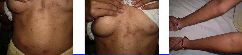
Seventeen years old female presented with multiple asymptomatic cutaneous cysts on the chest ,breast, axilla, the inguinal region and on the upper and lower extremities, sparing only head and the neck region, which had 6 months of duration. The cysts first appeared over the arms and they gradually involved other parts of the body.
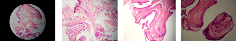
On examination, the dermal cysts were found to be round to oval, well defined and smooth surfaced, without a punctum and to vary in diameter from 2-5mm [Table/Fig-1]. The patient gave a history of similar lesions in her father too. The systemic and the laboratory findings were normal. Sonography revealed multiple nodules which were oval in shape, which were relatively well marginated and hypoechoic and with a posterior enhancement. Though the case was clinically diagnosed, FNAC and biopsy were advised to confirm the diagnosis. On FNAC, an oily material was aspirated. The smears revealed few squamous cells in an oily background. The differential diagnoses of steatocystoma and multiple epidermoid cysts were given and a biopsy was taken. On histopathological examination, the skin biopsy was found to be lined by keratinized, stratified, squamous epithelium. The dermis revealed a cyst which was lined by flattened, stratified, squamous epithelium without a granular layer and with a cellular eosinophilic cuticle over its surface. The sebaceous gland lobules were lying close to the cyst wall [Table/Fig-2]. After the histopathological confirmation, the patient was given oral isotretinoin and the cosmetic sites of the body were treated with a radiofrequency probe, but the patient declined further surgical treatment.
On examination, the dermal cysts were found to be round to oval, well defined and smooth surfaced, without a punctum and to vary in diameter from 2-5mm
Histopathological Discussion
DISCUSSION
Steatocystoma multiplex was first described by Jamieson in 1873, and the term was coined by Pringle in 1899 [5]. It is a hamartomous malformation of the pilosebaceous duct junction [2]. The cysts appear during adolescence and early adulthood and there is no sex predilection [6]. The exact origin of the cysts is still unknown but multiple theories which suggest their origin are: they result from sebaceous retention cysts of a nevoid nature or they are hamartomas or they are a variety of dermoid cysts [7]. Few authors have associated steatocystoma multiplex with ichthyosis and koilonychias, pachyonychia congenital, acrokeratosis verruciformis of hopf, hypertrophic lichen planus, hypohidrosis, hypothyroidism, hidradenitis suppurativa and hypotrichosis [2].
The familial Steatocystoma multiplex is associated with a mutation in the keratin 17 gene, in the areas which are identical to the mutations which are found in patients with pachyonychia congenita type 2 (PC-2) [8]. Uptil now, in patients with either steatocystoma multiplex or pachyonychia congenita type 2, 14 mutations have been discovered, all of which are localized to the helix initiation domain (1A domain) of the K17 gene. The KR T17 gene is located on the long(q) arm of chromosome 17, between positions 12 and 21 and the mutations in this gene interfere with the assembly of the keratin intermediate filament network [1,9].
Smith FJD have reported heterozygous missense mutations in K17(N92H and R94H) in two families who were diagnosed as steatocystoma multiplex [10].
Xiuying W studied the keratin 17 mutation in the cDNA of the cystic tissue of 5 patients of steatocystoma multiplex and in the DNA of the peripheral blood of 25 patients with steatocystoma multiplex from a steatocystoma multiplex family, by direct sequencing of the RTPCR products, nested PCR and restricted fraction length polymorphism and he revealed the R94 C mutation in the keratin 17 gene, which is one of the genetic basis of steatocystoma multiplex in the Chinese [11].
In our case, the patient had no features of Pachyonychia congenita type 2, such as hypertrophic nail dystrophy, focal keratoderma and multiple pilosebaceous cysts. The relationship of steatocystoma multiplex to the development of the sebaceous glands and its common presentation at puberty, suggest a hormonal trigger for the lesion growth [5].
Park et al., reported the appearance of steatocystoma multiplex on mammography and sonography. The finding of a well circumscribed, round, fat density nodule on mammography or an intradermal hypoechoic nodule on sonography, combined with a family history and an extensive bilateral spatial distribution of the nodules, confirm the diagnosis of steatocystoma multiplex [12].
The diagnosis should always be confirmed on histopathology, excluding its main differential diagnoses like eruptive vellus hair cysts, epidermal inclusion cysts, milia, trichilemmal cysts and tumours of the follicular infundibulum. Steatocystomas and vellus hair cysts are closely related to each other. Cases have been published with the cysts showing the features of both steatocystoma multiplex and eruptive vellus hair cysts, which are caused by a cystic change in the same pilosebaceous duct [13]. The histopathology of the Eruptive Vellus Hair Cysts (EVHCs) shows dermal cysts which are lined by stratified squamous epithelium with a granular layer and which contain many obliquely, transversely sectioned vellus hair shafts. The microscopic examination of the expressed contents in a potassium hydroxide preparation shows numerous vellus hairs in the eruptive vellus hair cysts. Generally, no sebaceous glands are present in the cyst wall [13]. The epidermoid cysts are lined with stratified squamous epithelium which contains a granular layer and they show laminated keratin inside the lumen of the cysts. Milia show very small cysts. The milium is usually located in the superficial dermis and it has an epithelial lining with a granular cell layer. It contains lamellated keratin. The common primary milia in infants and children are found in the undifferentiated sebaceous hair collar which surrounds the vellus hair follicles. Tumours of the follicular infundibulum show a platelike dermal tumour with anastomosing islands and cords, with connections to the overlying epidermis and the horn cysts. The trichilemmal cysts are lined by squamous epithelium without a granular layer and with the swelling of the cells close to the cyst cavity, which is filled with homogenous keratin. But the characteristic histopathological finding of steatocystomas is the presence of sebaceous lobules close to the cystic wall, which is lined by stratified squamous epithelium without a granular layer, though there may be scanty vellus hairs in the cystic cavity.
The patterns of keratin 10 (K10) and keratin 17 (K17) expression are also variable in the epidermoid cysts, the trichilemmal cysts, the eruptive vellus hair cysts, and in Steatocystoma multiplex. The epidermoid cysts expressed K10 and the eruptive vellus hair cysts expressed K17, whereas the trichilemmal cysts and Steatocystoma multiplex showed the expression of both K10 and K17 [14].
On electron microscopy, Steatocystoma multiplex has been found to be a nevoid sebaceous duct and a sebaceous gland tumour. There is one pilary unit which continuously produces the vellus hairs, which are trapped in the cystic cavity or in the pilary canal (trichostasis). Steatocystoma multiplex is connected to the epidermis by a straight or meandering epithelial cord, the remnant of the follicular infundibulum, which is a solid strand which contains sebocytes or sebaceous lobule like structures. A lumen which is partly present in a few areas of the cord, is filled with the cellular debris of the keratinocytes, corneocytes, sebocytes, or the trapped hairs [4]. In comparison, the trichilemmal cysts take their origin from the outer root sheath of the follicle, the epidermal cysts take their origin from the infundibulum and the vellus hair shafts take their origin from the infundibuloisthmic junction or the isthmus [12].
The various modalities in the treatment of Steatocystoma multiplex include surgery, CO2 laser therapy, oral isotretinoin and cryotherapy, but the recent procedures include the use of a radiofrequency incision probe to make mini incisions and for expressing the contents of cyst, even the cyst wall. It has the advantage of producing bloodless field which cannot be obtained by surgical blades [15] and the other advantages are the treatment with the Er:yag laser, followed by the topical tetracycline ointment, which also have shown good cosmetic results [6].
To conclude, Pachyonychia congenita type 2 and the eruptive vellus hair cysts are closely related to Steatocystoma multiplex. Therefore, a histopathological confirmation is a must before starting with any treatment. The genetic chromosomal abnormalities and mutations should be detected and a long term follow up should be taken up in such patients for further research on this disease.
[1]. Covello SP, Smith FJ, SillevisSmitt JH, Paller AS, Munro CS, Jonkman MF, Keratin 17 mutations cause either steatocystoma multiplex or pachyonychia congenita type 2Br JDermatol. 1998 139(3):475-80. [Google Scholar]
[2]. Kim SJ, Park HJ, Oh ST, Lee JY, Cho BK, A case of steatocystoma multiplex limited to scalpAnn Dermatol. 2009 21(1):106-09. [Google Scholar]
[3]. Hurley HJ, LoPresti PJ, Steatocystoma multiplexArch Dermatol. 1965 92:110-11. [Google Scholar]
[4]. Plewig G, Wolf HH, Braun-Falco O, Steatocystoma multiplex: anatomic reevaluation, electron microscopy, and autoradiographyArch Dermatol Res. 1982 272(3–4):363-80. [Google Scholar]
[5]. Davey Mathew, “Steatocystoma multiplex”Retrieved 25 may 2011 [Google Scholar]
[6]. Mumcuolu CT, Gurel MS, Kiremitci U, Erdemir AV, Karakoca Y, Huten O, Yag laser therapy for steatocystoma multiplexIndian J Dermatol. 2010 55(3):300-01. [Google Scholar]
[7]. Lima AM, Rocha SP, Batista CM, Reis CM, Leal II, Azevedo LE, What is your diagnosis. Case for diagnosisAn. Bras. Dermatol. 2011 86(1):165-66. [Google Scholar]
[8]. Chu HD, Steatocystoma multiplexDermatology Online Journal9(4):18 [Google Scholar]
[9]. Gass JK, Wilson NJ, Smith FJ, Lane EB, McLean WH, Rytina E, Steatocystoma multiplex, oligodontia and partial persistent primary dentition associated with a novel keratin 17 mutationBr J Dermatol. 2009 161(6):1396-98. [Google Scholar]
[10]. Smith FJD, Corden LD, Rugg EL, Ratnavel R, Leigh IM, Moss C, Missense mutations in keratin 17 cause either Paronychia Congenita Type 2 or a phenotype resembling steatocystoma multiplexJournal of Investigative Dermatology 1997 108:220-23. [Google Scholar]
[11]. Xiuying W, Yaozhou SHI, Yuexian YE, Keratin 17 gene mutation in patients with steatocystoma multiplexNational Medical Journal of China 2001–09 [Google Scholar]
[12]. Park KY, Oh KK, Noh TW, Steatocystoma multiplex :Mammographic and sonographic manifestationsAJR 2003 180:27 [Google Scholar]
[13]. Karen JK, Heller M, Wee SA, Mikkilineni R, Eruptive vellus hair cystsDermatology Online Journal13(1):1 [Google Scholar]
[14]. Hana T, Fujimoto Wataru, Arata Jiro, Expression of keratins (K10 and K17) in steatocystoma multiplex, eruptive vellus hair cysts, and epidermoid and trichilemmal cystsAmerican Journal of Dermatopathology 1997 19:250-53. [Google Scholar]
[15]. Choudhary S, Koley S, Salodkar A, A modified surgical technique for Steatocystoma multiplexJ Cutan Aesthet Surg. 2010 3(1):25-28. [Google Scholar]