Giant Congenital Melanocytic Nevi: A Case Report
Sudhanshu Ku. Das1, Amarendra M.2, Monalisa Subudhi3
1 Assistant Professor, Department of Pediatrics, Konaseema Institute of Medical Sciences, Amalapuram Andhra Pradesh 533201, India.
2 Associate Professor, Department of Pediatrics, Konaseema Institute of Medical Sciences, Amalapuram Andhra Pradesh 533201, India.
3 Tutor, Department of Microbiology, Konaseema Institute of Medical Sciences, Amalapuram Andhra Pradesh 533201, India.
NAME, ADDRESS, E-MAIL ID OF THE CORRESPONDING AUTHOR: Dr. Sudhanshu ku. Das, Assistant Professor, Department of Pediatrics, Konaseema Institute of Medical Sciences, Amalapuram Andhra Pradesh 533201, India.
Phone: 09441140581
E-mail: Swayam.dr007@gmail.com
Congenital melanocytic naevi which are formed by the overgrowth of melanocytes, are present at birth, which are called as bathing trunk, coat-sleeve or stocking naevi, which are commonly found over the back and the thigh areas . The giant congenital naevi of sizes which are greater than 20 cm are pigmented and are often hairy. The risk of malignant melanoma for the giant naevi is approximately 6% and 50% of the melanomas that develop by the age of two years, and 80% of those that develop by the age of seven years. So, an early removal is recommended The objective of this paper is to present a unique case of giant naevi along with a review of the literature.
Giant congenital melanocytic naevi (GCMN), Malignant melanoma, Leptomeningeal melanocytosis
INTRODUCTION
Congenital melanocytic naevi are brown to black moles which are present at birth, which are commonly found over the back and the thigh areas [1]. Giant Congenital Melanocytic Naevi (GCMN) of sizes which are larger than 20 cm diameter are rare and they occur in 1per 500,000 newborns. GCMN have irregular margins and they are dark brown to black in colour, with verrucous surfaces, with satellite lesions which are present beyond the periphery of the main lesion. They may be scattered over the entire skin surface [2,3]. These lesions are variously called as bathing trunk, coatsleeve or stocking naevi, depending on their regional distribution [4]. The giant naevi on the scalp and the neck may be associated with leptomeningeal melanocytosis and neurologic disorders which include neurofibromatosis and epilepsy [2,3].
Here, we are presenting a case of GCMN with extension to the scalp and the neck, without a leptomeningeal involvement, which has usual association with it.
CASE REPORT
A 4 months old female baby who was born out of a consanguineous marriage by a normal vaginal delivery at term to a primigravida, with an uneventful antenatal history, presented with an extensive pigmented patch over her body since birth. There was no family history of any similar lesion.
The physical examination revealed an extensive pigmented patch which covered 50% of the skin surface area over the neck, back, scalp, thigh and the legs [Table/Fig-1]. Multiple pigmented satellite lesions of size, 4-5cm were also present over the body and the extremities [Table/Fig-2].Tufts of coarse and lustreless hair were scattered all over the lesion at the back and at the right forehead, which were 2-3 cm in size [Table/Fig-3].
Extensive pigmented patch involving neck, scalp,back
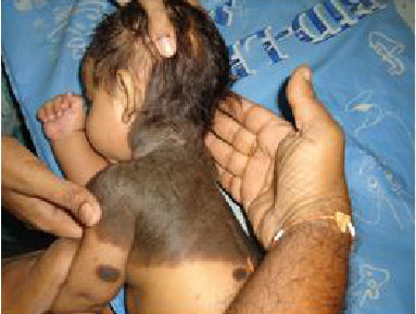
Multiple pigmented satellite lesion
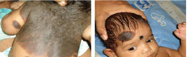
Tuft of hair over thepigmented lesion

There were no other associated congenital anomalies. A CT scan of the head showed no deep CNS extension. Ultrasound of the abdomen, an X ray of the spine and the fundus examination were normal. The biopsy of the patient was taken and the histopathological findings were consistent with those of congenital melanocytic naevi. No evidence of a malignant transformation was seen.
DISCUSSION
The congenital melanocytic nevi are pigmented cutaneous lesions which are formed by a combination of epidermally and dermally derived naevus cells, which occur in about 1% of the newborns [5]. They are classified according to their sizes as small (<1.5 cm), medium (1.5-19.9cm) and large or giant naevi (>20cm).The incidence of the small naevi is 1 in 100 births, that of the medium naevi is 6 in 1000 births, and that of GCMN which are larger than 20 cm in diameter is 1 per 500,000 newborns [6,7]. An equal prevalence exists in males and females. GCMN are thought to be caused by spontaneous mutations or during the foetal development, but in some families, the frequent appearance of these lesions suggest that they may be genetically inherited.The culture of melanocytes from such lesions showed chromosomal rearrangements which involved the chromosomal regions 1p,12p and 19p. Researchers think that a body protein which is called HGF/SF (hepatocyte growth factor /scatter factor) seems to be responsible for encouraging the neuroectodormal cells to develop,migrate and scatter. It seems that either too much or a wrong type of this protein in some cells, develop extra pigment and abnormal skin cells which are called naevus cells. These cells scatter around and so, we have naevi scattered all over the body [8] Malignancy should be suspected when there is a focal growth, pain, bleeding, ulceration, significant pigmentary changes or pruritus. The risk of malignant melanoma for the giant naevi is approximately 6% [9] and 50% of the melanomas that develop by the age of two years, and 80% of those that develop by the age of seven years. The risk of malignancy is also increased by the presence of larger naevi (>50 cm), axial locations such as on the trunk, head and neck, the presence of multiple satellite lesions and the existence of nodules, dark patches, junctional activity, deep dermal neurogenic elements or a blue naevus component [8,10,11]. Radiographic imaging, which includes MRI, is warrented to evaluate the melanocytic deposition in the CNS. The baseline MRI should be obtained when the patient is aged 4-6 months. It is impractical to prophylactically excise all the non giant congenital naevi and so, yearly examinations for the first 3 years of life are recommended, with reassessments for every 2 to 5 years. The surgical treatment of GCMN is addressed at the age of 6 months. The procedures which are used in the surgical treatment include serial excision and reconstruction with skin grafting, tissue expansion, local rotation flaps and free tissue transfer. Due to the depth of some lesions ,especially if the leptomeninges are involved, excisions may not eliminate the risk for developing melanoma [11]. The carbon dioxide laser, the Er:YAG and the Qswitched ruby laser have all been recently used for resurfacing and for selectively treating the deep pigmentations [12].
[1]. Walton RG, Jacobs AH, Cox AJ, Pigmented lesions in newborn infantsBr J Dermatol. 1996 95:289-90. [Google Scholar]
[2]. Mackie RM, Melanocytic nevi and malignant melanomaIn Rooks textbook of Dermatology,5th edition. Eds Champion RH, Burton JL, Ebling FJG 1993 OxfordBlackwell Scientific Publication:1525-60. [Google Scholar]
[3]. Arons MS, Management of giant congenital neviPlastReconster. Surg. 2002 110:352-53. [Google Scholar]
[4]. Caro William A, Tumours skinIn :Dermatology, Edited by Moschello SL, Pillshuy DM,HJ 1978 PhiladelphiaWB Saunders Company:1323-407. [Google Scholar]
[5]. Rhodes AR, Benign neoplasias and hyperplasias of melanocytesIn: Fitz Patricks Dermatology in General medicine year 1999 5th:1026-32. [Google Scholar]
[6]. Rhodes AR, Melanocytic precursors of cutaneous melanoma. Estimated risks and guidelines for managementMed Clin North Am. 1986 70(1):3-37. [Google Scholar]
[7]. Rhodes AR, Weinstock MA, Fitzpatrick TB, Risk factors for cutaneous melanoma: a practical method of recognising predisposed individualsJAMA 1987 258:3146-54. [Google Scholar]
[8]. Nevi and Malignant melanomaIn: Habit TP .Clinical Dermatology: A colour guide to Diagnosis and Therapy 2004 4thEdinburghMosby:776-77. [Google Scholar]
[9]. Rhodes AR, Melski JW, Small congenital nevocellular nevi and the risk of cutaneous melanomaJ Pediatric. 1982 100:219-24. [Google Scholar]
[10]. Foster RD, William ML, Barkovich AJ, Hoffman WY, Mathes SJ, Friden IJ, Giantcongenital melanocytic nevi: The significance of neurocutaneous melanosis in neurologically asymptomatic childrenPlast Reconstr. Surg. 2001 107:933-41. [Google Scholar]
[11]. Mark GJ, Mihm MC, Liteplo MG, Reed RJ, Clark WH, Clinical.histologic and ultrastructural studiesHum Pathol. 1973 4:395-18. [Google Scholar]
[12]. Park SH, Koo SH, Choi EO, Combined laser therapy for difficult dermal pigmentation: resurfacing and selective photothermolysisAnn Plast.Surg. 2001 47:31-6.1. [Google Scholar]