Acoustic Radiation Force Impulse Imaging of the Liver: Measurement of the Normal Mean Values of the Shearing Wave Velocity in a Healthy Liver
Rajneesh Madhok1, Chaitanya Tapasvi2, Umakant Prasad3, Ashish Kr Gupta4, Abhinav Aggarwal5
1 Associate Professor, Shri Ram Murti Smarak Institute of Medical Sciences, Bareillly, U.P., India.
2 Assistant Professor, Shri Ram Murti Smarak Institute of Medical Sciences, Bareillly, U.P., India.
3 Assistant Professor, Shri Ram Murti Smarak Institute of Medical Sciences, Bareillly, U.P., India.
4 Assistant Professor, Shri Ram Murti Smarak Institute of Medical Sciences, Bareillly, U.P., India.
5 Junior Resident –II, Shri Ram Murti Smarak Institute of Medical Sciences, Bareillly, U.P., India.
NAME, ADDRESS, E-MAIL ID OF THE CORRESPONDING AUTHOR: Dr. Rajneesh Madhok, Associate Professor, Department of Radiodiagnosis, Shri Ram Murti Smarak Institute of Medical Sciences, Bhojipura, Bareilly -243202, U.P., India.
Phone: 9897606172
E-mail: drmadhok@rediffmail.com
AIM: To find the normal mean values of the liver elasticity/stiffness by Acoustic Radiation Force Impulse (ARFI) Elastography in healthy subjects.
Material and Method: This study was conducted on 137 healthy subjects without any known liver pathology or a history of jaundice by using the Siemens Acuson S2000TM Ultrasound machine with a convex probe . A routine ultrasound was also performed in each case, and the subjects with fatty liver changes or any other signs of a chronic liver pathology were excluded. In each subject, 10 measurements were taken and the median value of the 10 measurements was calculated, which was expressed in metres/sec (m/s). The inter quartile range method was used for the interpretation of the data. Only the measurements with an IQR of < 30% and a Success Rate (SR) of >60% were considered. The measurements were taken at 1-2 cm depth from the liver capsule through an intercostal approach, with the subject lying in the decubitus position. The mean ARFI values and the mean values according to the age and gender of the subjects were evaluated.
Result: Valid ARFI measurements were taken in 108/137 patients (78.83%). The mean value of the ARFI measurements in the normal individuals was 1.197±0.25 m/s. There were no significant differences between the mean ARFI values in men vs women (1.195±.25 vs. 1.199±0.26m/s, p = 0.939), and also among the different age groups (p>0.05).
Conclusion: In our study, the mean liver elasticity value (shearing wave velocity) which was obtained by ARFI in the healthy subjects was 1.197±0.25 m/s.
ARFI, Liver tissue elasticity, Shearing wave velocity
INTRODUCTION
ARFI is an elastography method which is used to measure the liver elasticity /stiffness for fibrosis. There are various elastography methods which are used for assessing liver fibrosis. They are, Transient Elastography (Fibro Scan) [1], Real-Time Tissue Elastography, Acoustic Radiation Force Impulse (ARFI) [2] and MRI elastography [3]. Some of these techniques have already been well established, while others still have to prove their usefulness. ARFI is an ultrasound based, new non-invasive technique which is used to measure the tissue stiffness, it is the most reliable measurement procedure for assessing the liver elasticity/ stiffness and it has reproducibility [4].
It is an acoustic radiation force-based imaging method that is provided by the conventional B-mode ultrasonography. It offers elastography with a flexible metering box at a variable depth, thus allowing the examination of a specific area. A focused ultrasound is used to apply a localized radiation force to small volumes of tissue (2mm3) [5]. The ARFI imaging involves the transmission of an initial ultrasonic pulse at diagnostic intensity levels to obtain a baseline signal for a later comparison. A short-duration (approximately 0.3 s), high-intensity, acoustic ‘pushing pulse’ is then transmitted by the same transducer, followed by a series of diagnostic intensity pulses, which are used to track the displacement of the tissue which is caused by the pushing pulse. The response of the tissue to the radiation force is observed by using the conventional B-mode imaging pulses, and it is possible to display the quantitative shear-wave velocity (SWV; meter/sec) of the ARFI displacement. This velocity (m/s) is proportional to the square root of the tissue elasticity. Because the velocity of the shear wave depends on the tissue stiffness, it is possible to apply the ARFI technology to elastography, in which the wave propagation speed is evaluated to study the viscoelastic properties of the target tissues [6]. This technology was named as “Virtual Touch Tissue Quantification” by SIEMENS. Previous reports have also referred to it as ARFI elastography or ARFI elastometry. The measured Shear-Wave Velocity (SWV) is an intrinsic calculation and a reproducible property of the tissue [7] and also, the procedure can be performed during a routine ultrasound examination.
Acoustic Radiation Force Impulse Elastography is used for the assessment of liver fibrosis, thyroid gland nodules, breast nodules and, liver and kidney tumours, for the characterization of atherosclerotic plaques, as well as for monitoring the results of radiofrequency ablations.
ARFI is a non-invasive alternative to a liver biopsy for the evaluation of Liver fibrosis. A liver biopsy is still recommended as the gold standard method for determining the fibrosis stage, the prognosis and the therapeutic indications in patients with chronic liver disease. However, a liver biopsy is an invasive procedure which is associated with a risk of serious complications. Approximately 1–3% of the patients require hospitalization for the complications, and a quarter of them report pain after a percutaneous liver biopsy. The diagnostic accuracy of liver biopsy for assessing liver fibrosis is influenced by the quality of the biopsy samples. In addition, there are several absolute or relative contraindications to the liver biopsy, which include severe coagulopathy [8]. In order to differentiate a normal liver from a fibrotic liver, the normal range of the liver elasticity measurements must be established. The aim of this study was to define the normal shearing wave velocity values in a healthy liver.
MATERIALS AND METHODS
One hundred and thirty seven subjects without any known liver pathology or a history of liver disease, underwent the acoustic radiation force impulse imaging tissue quantification. The subjects with fatty liver or an enlarged spleen at ultrasound were excluded. This study was approved by the local ethics committee and the patients’ informed consent was obtained for the investigations.
In each subject, the liver elasticity was measured by means of ARFI in the right liver lobe, 1-2 cm below the liver capsule, by an intercostal approach, with the patient lying in the left lateral decubitus, with minimal scanning pressure being applied by the operator, while the patients were asked to stop breathing for a moment, in order to minimize the breathing motion. Care was taken not to include any blood vessels or billiary structures. The ARFI measurements were performed by using the Virtual Touch issue Quantification software. In each patient, valid ARFI measurements were made in the liver and the median values were calculated, the results being expressed in metres/second (m/s).
Our data analysis was performed by the hospital’s statistician by using the Inter Quartile Range (IQR) [9]. The IQR is used in statistics for finding the difference between the biggest and the smallest values in the middle 50% of the data. It is the measure of the variability, which is based on dividing a data set into quartiles, where the quartiles divide a rank order data set into four equal parts. The values which divide each part are called the first, second and the third quartiles and they are denoted as Q1, Q2 and Q3. IQR is difference between Q3 and Q1.
For the normally distributed data, the arithmetic mean and the standard deviation are used. For the data which is not normally distributed, the median with the data range are used. Therefore, the range is an informative tool which is used as a supplement to the other measures such as standard deviation.
The ARFI measurements were numeric variables and so the mean and the standard variations were calculated. The mean ARFI values and the mean values according to the age and gender were evaluated.
RESULTS
Our study group comprised of 137 subjects and their mean age was 34.139±10.674 years. There were 40 females and 97 males (the ARFI values ranged between 0.23 and 2.98 m/s). One hundered eight patients (78.83%) had valid ARFI measurements with an IQR of <30% and an SR of ≥60% (35 women and 73 men, whose mean age was 34.5±9.53 years). Only in 8 subjects (5.83%), we could not obtain 10 valid measurements, and in 21 (15.32%) patients, the IQR and the SR criteria were not fulfilled [Table/Fig-1].
Distribution of Median of ARFI values in Healthy Population-According to the Age Group.
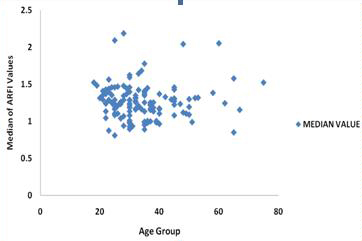
The mean shearing wave velocity value which was determined by ARFI in the healthy subjects with an IQR of <30% and an SR of ≥ 60% was 1.197 ±0.25 m/s. The ARFI values ranged between 0.23 and 2.98m/s.
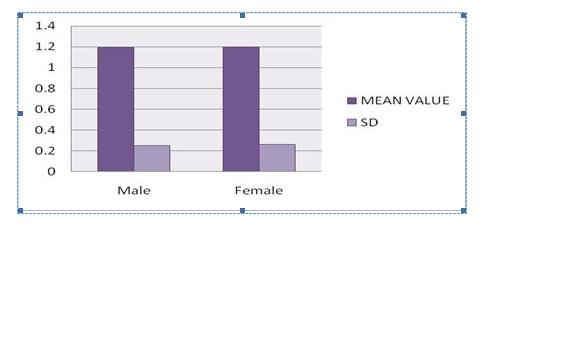
We did not find significant differences between the mean ARFI values in men vs. women (1.195±0.25 vs. 1.99± 0.26 m/s, p=0.939) [Table/Fig-2], [Table/Fig-3] and also among the different age groups [Table/Fig-4], [Table/Fig-5].
Distribution of Mean/SD ARFI values in Healthy Population-According to the Gender
Distribution of Mean/SD ARFI values in Healthy Population-According to the Age Group
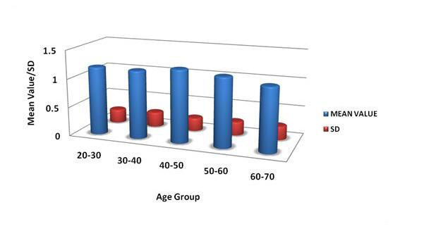
Age group wise distribution of mean ARFI values
| Age Group | Frequency | Mean value |
| 20-30 | 45 | 1.192±0.24 |
| 30-40 | 42 | 1.184±0.26 |
| 40-50 | 15 | 1.262±0.23 |
| 50-60 | 3 | 1.20±0.24 |
| 60-70 | 3 | 1.106±0.24 |
Age group wise distribution of mean ARFI values
| Gender | Frequency | Mean value | SD |
| Male | 73 | 1.195 | 0.25 |
| Female | 35 | 1.199 | 0.26 |
In our group, 21 subjects (15.32%) had ARFI values which were higher than the mean value of 1.195±0.25m/s and measured 1.252 to 1.276±0.24m/s.
DISCUSSION
There are only few published data regarding the normal range of the shearing wave velocity in healthy subjects.
A recent study which was published by Alina Popescu et al., [10] used the interquartile range for the data evaluation in 82 subjects with a mean age group of 34 years and it showed that the normal mean Liver Stiffness (LS) /elasticity by the ARFI Shear Wave Velocity(SWV) in the healthy subjects was 1.15 m/s(range 0.69 -1.6m/s). A study which was published by Horster et al., [11] showed that the mean ARFI shearing wave velocity in a group of 68 healthy volunteers, with a mean age of 28 years was 1.19 m/s (range 0.77-1.63). This was similar to our study results, 1.195m/s (range 0.23 and 2.98 m/s). Both the authors also showed that the age and gender did not influence the ARFI shearing wave velocity. The same was also observed in our study.
The intercostals approach had the highest success rates (97.2%), while the skin-liver distance significantly influenced the ARFI SWS .The best correlation with fibrosis (which was obtained by a liver biopsy), was obtained for the measurements which were made at 1-2cm (with a success rate of 95.5%) and at 2-3 cm ( with a success rate of 85.6%) below the capsule and so the best place for the ARFI determination would be 1-2 cm below the capsule [12] . Goertz et al obtained a mean LS value of 1.09 m/s (range 0.79-1.32 m/s) in a group of 20 subjects without any liver pathology and Fierbinteanu-Braticevici [13] et al., obtained a mean value of 1.18 m/s. Friedrich-Rust et al., [14] had performed the ARFI measurements in 20 healthy volunteers in the right liver lobe, through the intercostals space at 2cm below the liver capsule, with 10 valid values for each patient. The median SWV which was measured was 1.10 m/sec, with a mean value of 1.13±0.23 m/ sec and a range of 0.85-1.42 m/sec.
Gallotti et al., [15] measured the elasticity of various abdominal organs by ARFI elastography in 35 healthy subjects and obtained a mean value of 1.59 m/s for the liver, which was higher than the values which were obtained in the other studies. Grgurevic et al., [16] established a SWV cut-off value of 1.3 m/s for the healthy liver, that could reliably differentiate between the healthy and the non-cirrhotic HCV patients.
We also considered like Alina Popescu et al, that only the measurements with an IQR of <30% and an SR of ≥60% should be validated.
The published data regarding the value of ARFI in predicting the severity of liver fibrosis tried to established cut-off values for the different stages of fibrosis. In the study of Lupsor et al., [17], the cut-off values (m/s) which were predictive for each fibrosis stage were 1.19 (F≥1), 1.34(F≥2), 1.6((F≥3) and 2.00 (F4). Friedrich-Rust et al showed the following mean values for the different fibrosis stages: F0-1.16 ± 0.17 m/sec, F1-1.18±0.18 m/sec, F2 -1.34±0.34 m/sec, F3-1.75±0.51 m/sec and F4 -2.38±0.74 m/ sec. From these data, we observed that the mean SWV value which was obtained by us for the normal subjects was close to the cut-off values for F1 . Further large studies on healthy subjects are required to establish the normal ARFI measurements and to establish their place in the diagnosis of patients with chronic diffuse liver diseases.
CONCLUSION
In our study, the mean LS value which was obtained by ARFI in the healthy subjects was 1.197 m/s, and we did not find significant differences between the mean ARFI values in men vs. women and among the different age groups.
Conflict of interest: None.
[1]. Talwalkar JA, Kurtz DM, Schoenleber SJ, West CP, Montori VM, An ultrasound-based, transient elastography for the detection of hepatic fibrosis: a systematic review and a meta-analysisClin Gastroenterol Hepatol. 2007 5:1214-20. [Google Scholar]
[2]. Boursier J, Isselin G, Fouchard-Hubert I, The acoustic radiation force impulse: a new ultrasonographic technology for the widespread noninvasive diagnosis of liver fibrosisEur J Gastroenterol Hepatol. 2010 22:1074-84. [Google Scholar]
[3]. Huwart L, Sempoux C, Vicaut E, Magnetic resonance elastography for the noninvasive staging of liver fibrosisGastroenterology 2008 135:32-40. [Google Scholar]
[4]. Shan B, Pelegri AA, Maleke C, A mechanical model for computing the elastic modulus of the tissue for harmonic motion imagingJ Biomech 2008 41:2150-58. [Google Scholar]
[5]. Zhai L, Palmeri ML, Bouchard RR, Nightingale RW, Nightingale KR, An integrated indenter-ARFI imaging system for the tissue stiffness quantificationUltrason Imaging 2008 30:95-111. [Google Scholar]
[6]. Sporea I, Popescu A, Sirli R, How to improve the quality of the tissue sample which is obtained by doing a percutaneous liver biopsyCEJ Med 2010 6:103-06. [Google Scholar]
[7]. D’Onofrio M, Gallotti A, Mucelli RP, Tissue quantification with acoustic radiation force impulse imaging: the measurement repeatability and the normal values in the healthy liverAmerican Journal of Roentgenology 2010 [Google Scholar]
[8]. Haque M, Robinson C, Owen D, Yoshida EM, Harris A, Comparison of acoustic radiation force impulse imaging (ARFI) to the liver biopsy histologic scores in the evaluation of chronic liver disease: A pilot studyAnn Hepatol 2010 9:289-93. [Google Scholar]
[9]. Popescu A, Bota S, Sporea I, Do the success rate and the interquartile range influence the liver stiffness which is measured by the acoustic radiation force impulse technique?Euroson 2010 Abstract BookPE38:116 [Google Scholar]
[10]. Popescu A, Sporea I, Sirli R, Bota S, Focsa M, Danila M, The mean values of the liver stiffness which were assessed by acoustic radiation force Impulse elastography in normal subjectsMedical Ultrasonography 2011 13(1):33-37. [Google Scholar]
[11]. Horster S, Mandel P, Zachoval R, Clevert DA, Comparing acoustic radiation force impulse imaging to transient elastography in assessing the liver stiffness in healthy volunteers with and without the Valsalva maneuverClin Hemorheol Microcirc 2010 46:159-68. [Google Scholar]
[12]. Goertz RS, Zopf Y, Jugl V, Measurement of the liver elasticity by using the acoustic radiation force impulse (ARFI) technology: an alternative noninvasive method for staging the liver fibrosis in viral hepatitisUltraschall Med 2010 31:151-55. [Google Scholar]
[13]. Fierbinteanu-Braticevici C, Andronescu D, Usvat R, Cretoiu D, Baicus C, Marinoschi G, Acoustic radiation force imaging sonoelastography for the noninvasive staging of liver fibrosisWorld J Gastroenterol 2009 15:5525-32. [Google Scholar]
[14]. Friedrich-Rust M, Wunder K, Kriener S, Liver fibrosis in viral hepatitis. A noninvasive assessment with acoustic radiation force impulse imaging versus transient elastographyRadiology 2009 252:595-604. [Google Scholar]
[15]. Gallotti A, D’Onofrio M, Pozzi Mucelli R, The Acoustic Radiation Force Impulse (ARFI) ultrasound technique in virtual touch with the quantification of the upper abdomenRadiol Med 2010 115:889-97. [Google Scholar]
[16]. Grgurevic I, Cikara I, Horvat J, The noninvasive assessment of liver fibrosis with acoustic radiation force impulse imaging: the increased liver and splenic stiffness in patients with liver fibrosis and cirrhosisUltraschall Med 2010 Nov 23 [Epub ahead of print] [Google Scholar]
[17]. Lupsor M, Badea R, Stefanescu H, The performance of a new elastographic method (ARFI technology) which was compared to unidimensional transient elastography in the noninvasive assessment of chronic hepatitis C. The preliminary resultsJ Gastrointestine Liver Dis 2009 18:303-10. [Google Scholar]