The knee joint is one of the most important joints of the human body. The knowledge of the anatomy of this joint is fundamental for understanding any pathology and for thus augmenting an accurate diagnosis and treatment [1]. A well preserved knee region specimen thus becomes a necessity, not only for anatomists, but also for radiologists and orthopaedicians. During routine dissections of the knee joints, due to repeated handling and inappropriate preservation and aftercare, the boundaries and attachments of the synovial capsule and other minute details of the structures in the joint cavity, such as the cruciate ligaments and the relationship of the neurovascular bundle, may remain indistinct. This may also be attributed to the high concentration of formalin which is used during the embalming and the storage of these specimens. Unfortunately, these formalin fixed specimens are not user friendly, due to the objectionable odour of the fumes, the health hazards which are caused by the excessive exposure to formalin and the cost and time which are spent on their maintenance [2, 3]. Moreover, their repeated handling reduces their instructional value, as the muscles, vessels and the neurovascular bundle turn dark, rigid and dry and present with disturbed altered relations of the structures. All these drawbacks of the formalin preservation led us to explore the technique of plastination: a better alternative for the preservation of gross specimens [4]. The plastinated specimens offer several advantages over other methods, as they are dry, odourless, durable, life-like, maintenance-free and nontoxic, with good colour retention [5-7]. They provide a long term structural preservation of the tissue details, with minimal shrinkage and they are soft, supple and flexible [8, 9]. Their aesthetic superiority in terms of their morphological features and their easy handling makes them student friendly and also popular among the staff. So, the reliance on plastinated specimens in education is certainly on the rise.
Thus, the present study was undertaken to prepare plastinated specimens of old embalmed and freshly fixed knee regions and also to compare the aesthetic value of the two, which have not been reported in the literature so far.
MATERIALS AND METHODS
Group I: A total of 10 knee specimens from fresh cadavers were obtained from the Department of Forensic Medicine. Each specimen was thoroughly washed with running water through the femoral artery, in order to empty the vessels and this was followed by a heparin injection (25,000 I.U. in 5ml) which was diluted with distilled water in the proportion of 0.2 in 5ml. 5-8% formalin was injected through the femoral artery for fixation and the joint capsule was filled with 120-250 ml of fixative. The further specimens were submerged in 5% formalin for 1-2 weeks for a better fixation.
GROUP II: A total of 5 knee specimens were collected from the cadavers which were being used in the Department of Anatomy. They were fixed by using the standard embalming procedure. The embalming fluid was composed of formalin, glycerin, methanol and phenol. These cadavers were then stored in formalin (30-40%) tanks till they were used further.
All the collected specimens were washed in running tap water for 1-2 days to remove the excess formalin, cooled to 4°C and transferred to 100% cold acetone at-20°C. The dilated joint capsule was flushed with acetone several times. The acetone concentration was measured daily with an acetonometer and the acetone was replaced when its concentration reached around 90%. The dehydration was considered to be complete when the water content was below 1% for three consecutive days. The specimens were then immersed in the silicone resin, S10 with the S3 Catalyst [10] for 24 hours. In the joint cavity, the acetone was drained out and the cavity was refilled with the silicone polymer. The specimens were infiltrated with the polymer reaction mixture, just before their impregnation. They were then subjected to a forced impregnation where the pressure was reduced gradually by using a vacuum pump. The rate of impregnation was monitored by the rate of acetone bubbles which came on the surface. The specimens were kept immersed in the polymer for one day before taking them out. They were finally gas cured in a curing chamber by passing S6 vapour. After the specimens were dry on touch, they were enclosed in polythene bags for the final curing. The finally plastinated fresh and old formalin fixed specimens were compared for their morphologies.
RESULTS
The method of plastination was standardized, the specimens of both the groups were well plastinated and the following features were observed:
1. demonstration of the morphological features of the knee
The knee joint is the largest synovial and the most complicated joint in the body, which consists of an extensive network of ligaments and muscles. The following structures were observed:
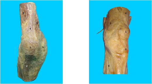
The muscle groups which surround the knee joint [Table/Fig-1] and [Table/Fig-2]
Morphological features of plastinated right knee region.
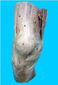
The two main muscle groups of the knee joint, the quadriceps and the hamstrings, were seen on the ventral and the dorsal aspects of the thigh. The proximal and the distal attachments of all the muscles were clearly visible in both the experimental groups, but the direction of the fibres were better maintained in group I. Popliteus originated from the capsule of the knee joint by a strong tendon. The attachment of Popliteus to the lateral meniscus was better observed in the fresh formalin fixed, plastinated (group I) specimens, as compared to that in the old embalmed (group II) plastinated specimens.
The popliteal fossa: The following muscles which formed the boundaries of the popliteal fossa were observed ie. semimembranosus, semitendinosus, tendon of the biceps femoris, the medial and lateral heads of the gastrocnemius and the plantaris. In group I, the middle genicular nerve and vessels were clearly visible. They pierced the floor which was opposite the oblique popliteal ligament.
The popliteal vessels were lying on the floor of the popliteal fossa. The popliteal artery extended obliquely as a continuation of the femoral artery and at the lower border of the popliteus muscle, it divided into the anterior and posterior tibial arteries. The popliteal vein ascended through the popliteal fossa. Its relationship to the popliteal artery changed as the vein ascended. In the upper part, the vein was seen posterolateral to the artery. It then crossed superficial to the artery and in the lower part, it ran medial to the artery. Its tributaries, the short saphenous vein and the veins which corresponded to the branches of the popliteal artery, were also observed in both the groups [Table/Fig-2].
Posterior view of plastinated right knee region showing muscles, nerves and vessels.
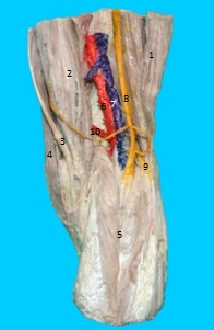
The overall morphology with respect to the muscles and vessels were superior in the group I plastinated knee specimens as compared to that in the group II specimens.
The knee joint capsule
The thick ligamentous joint capsule which surrounded the entire knee was well preserved in both the groups. The infrapatellar padoffat and the infrapatellar bursa were well preserved in group I.
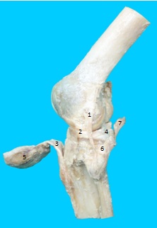
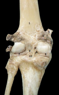
The ligaments and the menisci of the knee joint [Table/Fig-3] and [Table/Fig-4]
Lateral view of plastinated left knee joint showing intracapsular structures.
Posterior view of plastinated left knee joint showing ligaments and tendons.
The following ligaments and menisci were observed: the ligamentum patellae, the Medial Collateral Ligament (MCL), the lateral collateral ligament (LCL), the Anterior Cruciate Ligament (ACL), the Posterior Cruciate Ligament (PCL), the transverse ligament and the menisco-femoral ligament – The posterior horn of the lateral meniscus was connected to the medial condyle of the femur by anterior (ligament of Humphery) and posterior (ligament of Wrisberg) menisco-femoral ligaments. Their colour preservation was inferior in group II as compared to that in group I.
The medial meniscus: The attachments of the semilunar medial meniscus were better visualized in the group I knee joints as compared to those in the group II knee joints. The anterior horn was narrower than the posterior horn and both the horns were attached to the intercondylar area on the tibia [Table/Fig-4].
The lateral meniscus: Similarly, the circular lateral meniscus which had two horns and was attached closer on the intercondylar area of the tibia and was grooved posteriorly by the tendon of popliteus, was better observed in group I [Table/Fig-4].
Thus, the gross morphological features of the knee region and the internal structures of the knee joint were well preserved in both the groups but they were aesthetically superior in the freshly fixed, group I plastinated specimens, as they exhibited a life-like look, as compared to the corresponding old embalmed group II plastinated specimens.
2. Evaluation of the morphological features of the plastinated knee specimens
External features [Table/Fig-5] and [Table/Fig-6]
Comparison of morphological features of plastinated old embalmed and freshly fixed knee joints.
Comparison of morphological features of Group I (freshly fixed) and Group II (old embalmed) plastinated specimens.
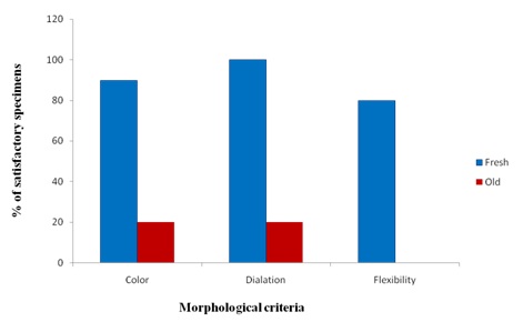
Each specimen of both the groups was evaluated on the basis of its colour, dilatation of its joint cavity, its flexibility and the preservation of its morphology. The grading of these qualitative data was expressed as the overall percentage of the satisfactory specimens out of the total specimens in the group. The responses were recorded as Yes/No(Y/N) as regards to the criteria which were under evaluation. A response of ‘Yes’ was considered as satisfactory.
Colour: Did the specimens show a life-like colour? -Y/N
Dilatation: Did the joint cavity get dilated satisfactorily?? -Y/N
Flexibility: Was the knee joint flexible?? -Y/N
Colour
The specimens with a life-like colour were marked as satisfactory, while those with any discolouration were marked as unsatisfactory. All the specimens of group I had a good colour preservation except one specimen. On the other hand, only one of the specimens from group II showed a satisfactory colour, as all the other specimens had turned dark.
Dilatation
Dilatation of the joint cavity was evaluated by the amount of fluid which was required for its infiltration. All the specimens from group I showed a satisfactory dilatation, which required 200-250 ml of fluid for their infiltration. Only one specimen from group II showed a satisfactory dilatation, while the other 4 showed an unsatisfactory dilatation, which required only 50-100 ml of fluid. Thus, the success rate for the dilatation in group I (fresh) was 100% as compared to 20% in the group II (old) specimens.
Flexibility
Most of the specimens of group II lost their flexibility, while those of group I remained flexible.
DISCUSSION
Anatomy is a fundamental educational science in medical universities. In the study of anatomy, the use of gross specimens is mandatory. The decay of this material is an impediment to all morphological studies, teaching and research. Thus, the preservation of biological materials becomes essential for them to be used as an educational tool. Their preservation is most commonly achieved by using liquids such as formaldehyde, alcohol and glycerin. A prolonged exposure to high concentrations of formalin can not only discolour the specimens, but it also makes them toxic, hazardous, fragile and unpleasant to use. The tissues which are fixed in formalin require periodic wetting to prevent them from drying. Inhaled formaldehyde is proven to cause carcinoma in rats and mice [3]. Moreover, the repeated handling of these specimens may lead to a disturbance in the relationship of the structures, making them inappropriate for anatomy teaching. Due to the ease of the procedure, if the preservation of the formalin-based solutions have gained acceptance globally among the staff for anatomical teachings, they have limited use in hands-on experiences in cadaveric workshops. During one such workshop on knee joints, it was observed that the knee joints which had been fixed in higher concentrations of formalin for a longer duration, had turned harder and non-flexible and that the cavity had not remained fully dilated. So a hands -on experience for arthroscopic procedures was not possible. Also, the relationship of the cruciate ligaments with the neurovascular bundle had remained unclear, which is very crucial in order to prevent iatrogenic injuries during the procedure of arthroscopy. Thus, in the Department of Anatomy, we planned to preserve the knee joint by plastination, in order to overcome the disadvantages of formalin preservation.
We observed that plastination had resulted in specimens that were clean, dry, odourless, user friendly, life-like and nontoxic [11,12]. They required minimal aftercare and could be stored on shelves or in display cases for a longer duration. As the fully cured specimens are dry and transportable to classrooms, they can be rotated to each student without gloves, to appreciate the features which lead to a higher effectiveness of the lectures, as well as for maintaining a student/specimen interaction.
The comparison among the 2 groups showed a gross difference in terms of the colour, dilation and the flexibility of the specimens. The discolouration and the non-flexibility in group II could be attributed to the high concentration of formalin (>30%) which was used for the preservation of the specimens. These old, embalmed, plastinated knee specimens could also be used for teaching, but with some limitations. On the other hand, the freshly fixed (Group I), plastinated knee specimens revealed all the structures, that could be well compared with the living anatomy, thus making them superior for teaching anatomy as well as for hands-on cadaveric practices. These knee joints were flexible and they could be bent at different angles for an arthroscopic examination. As their cavities remained dilated, they offered a better visualization of the intracapsular structures.
CONCLUSION
We infer that the plastinated knee specimens that were fixed in lower concentrations of formalin for an adequate period of time were superior to the ones which were fixed in higher concentrations of formalin for a prolonged period. The preservation of knee joints by plastination could provide an excellent teaching tool for studying anatomy. A well plastinated specimen will not only increase its acceptability among students, the faculty and clinicians, but it can also increase its instructional potential.