Footdrop can be caused by several pathologies at various levels of the central and peripheral nervous systems, which include lesions of the spinal cord, nerve roots or the peripheral nerves. Most vulnerable to trauma is the common peroneal nerve, as it is closest to the surface and the least protected [1].
After taking its origin from the sacral plexus, the sciatic nerve divides into the common peroneal (fibular) and the tibial nerves, which are proximal to the popliteal fossa [2]. The common peroneal nerve then becomes superficial, as it winds laterally around the head of the fibula and it bifurcates into the superficial and the deep peroneal nerves. The superficial peroneal nerve supplies the everters of the foot. The deep peroneal nerve innervates the dorsiflexors of the foot and it provides sensations to the web space between the first and the second toes [3]. The several peculiarities of the common peroneal nerve make it vulnerable to injuries. The peroneal nerve is fixed at the fibular head, which leads to more mechanical stretching [2], as the nerve has a limited longitudinal mobility [4]. The most common mechanism of the injury is acute compression, traction or laceration at the fibular head. Also being known as the foot drop and strawberry picker’s palsy, this palsy occurs with bed positioning, squatting, casting and tight boots. The nerve may also be entrapped, as it passes through a fibro-osseous tunnel [5]. The postoperative palsies occur especially after operations which are performed with the patients in the lateral decubitus position or in the lithotomy position. The other causes of the compression are chronic squatting and prolonged sitting in the cross-legged position [3].
The most usual cause of a unilateral drop foot is the common peroneal nerve palsy of a spontaneous, traumatic, or a pressure type. Large studies which were done on this condition in military casualties, are available [6]. A recent study which was done on a civilian population described a relatively large number of cases which were due to gunshot wounds [7]. The other published reports dealt with small numbers of patients with palsy which were due to single causes. A therapeutic weight loss, on itself, can cause a foot-drop [8]. With lesions at the fibular head, especially in farmers, the deep branch of the peroneal nerve is affected more commonly than the whole nerve [9]. The reports on more unusual cases are also available.
A patient with the peroneal nerve entrapment presents as a footdrop, with an unstable ankle and a gait difficulty. The sensory symptoms include numbness and decreased sensations along the anterolateral leg and the dorsum of the foot, between the first and the second toes [5]. The superficial peroneal nerve is commonly involved in the lesions of the common peroneal nerve or it may be seen in isolation to involve the paresis and atrophy of the peronei muscles (foot evertors) and the sensory disturbance which affects the skin of the lateral distal portion of the lower leg and the dorsum of the foot. Nerve conduction studies which are done across the entrapment site reveal slowing and small action potential amplitudes. Needle electromyography may reveal denervation of the peroneally innervated muscles [5]. Only a few electrodiagnostic studies had been carried out in the past, to study the cases of footdrop in the farmers [3].
Materials and methods
50 male patients who were aged between 20 to 50 years, who were involved in all types of farming activities, were recruited as the subjects. These patients presented to the Orthopaedics OPD with a clinical history of a unilateral footdrop and a gait difficulty. The patients had been harvesting (43 cases) and weeding (7 cases) 4 to 5 months before suffering from the unilateral footdrop. 50 normal, healthy, age matched subjects who were not involved in farming activities were selected as the controls. The presence of Diabetes mellitus and polyneuropathy was ruled out by taking the clinical history and by doing urine and blood sugar level tests. The subjects were examined clinically to rule out the causes of the foot drop which were other than a peroneal nerve injury at the fibular head.
Electrophysiological studies were carried out to compare the sensory as well as the motor parameters of the deep and the superficial peroneal nerve and the tibial and the sural nerves. These were stimulated distally as well as proximally.
The electodiagnostic studies
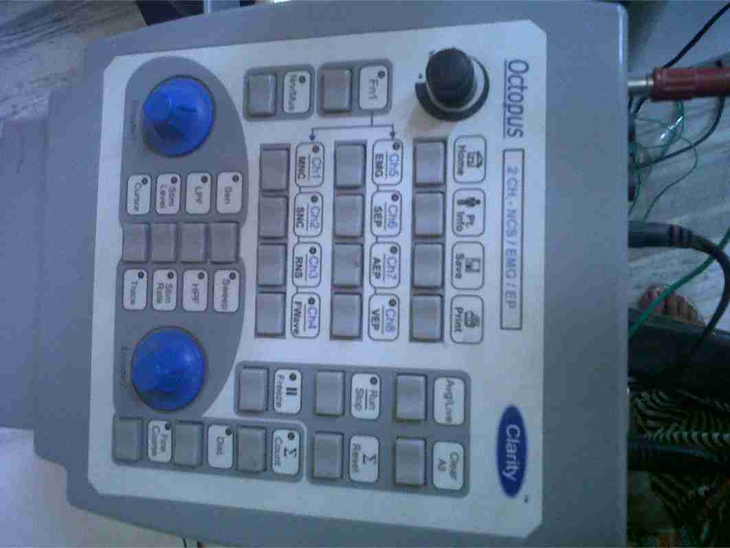
These studies were performed by using the Clarity vision NCS/EMG machine [Table/Fig-1], in the Department of Physiology, Adesh Institute of Medical Sciences and Research, Bathinda Punjab,India. The subjects were examined while they were lying comfortably in the supine position. The room temperature was kept at 25-28°C. The filters were set at 2 Hz to 5 kHz for the motor studies and at 20 Hz to 2 kHz for the sensory studies. The sweep speed was set at 5ms/division for the motor studies and 2 ms/division for the sensory studies. A stimulus duration of 50 -1000 μs and a current of 0–100 mA were required for an effective nerve stimulation. Supramaximal stimuli were delivered in order to get adequate responses. The surface fixed bar stimulating electrodes and the surface.
1-cm disc recording electrodes were used for the motor studies as well as for the sensory studies. The data was collected for the following parameters:
For the motor nerves
The Distal Motor Latency (DML), the Motor Nerve Conduction Velocity (MNCV) and the amplitude of the Compound Muscle Action Potential (CMAPA).
For the sensory nerves
The Sensory Latency (SL), the Sensory Nerve Conduction Velocity (SNCV) and the amplitude of the Sensory Nerve Action Potential (SNAPA), which were measured from the peak of the negative potential to the peak of the positive potential. For the peroneal motor nerve, the below fibular head-to-ankle conduction velocity, for the tibial motor nerve, the knee-to-ankle conduction velocity and for the sural nerve, the antidromic calf-to-ankle conduction velocity were recorded.
A standardized technique was used to obtain and to record the action potentials for the motor and the sensory studies.
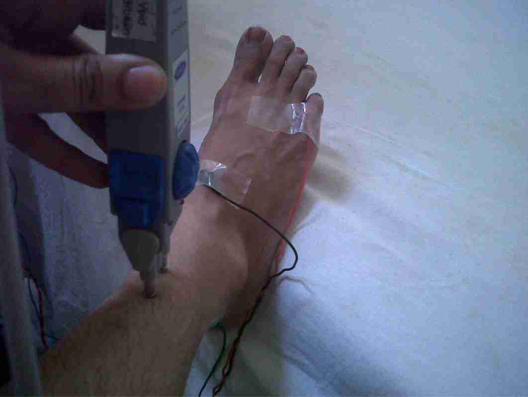
A. The motor nerve conduction studies [Table/Fig-2a & 2b]:
The Deep Peroneal Nerve:
The stimulation was done in three regions:
The ankle: The dorsal aspect of the distal lower leg between the tendons of the tibialis anterior (medially) and the extensor hallucis (laterally), at 8cm to the active recording electrode.
below the fibular head: 3-4cm distal to the proximal tip of the fibular head
The popliteal fossa: Medial to the biceps femoris tendon, 10 cm below the fibular head.
Recording from E1 (over the extensor digitorum brevis: EDB) and E2 (over the fifth toe)
The ground electrode (GND) was placed on the lateral malleolus
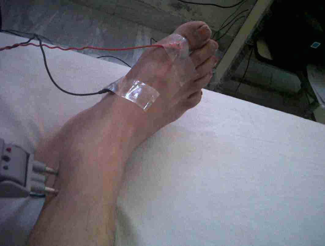
The Tibial Nerve:
The stimulation was done in two regions:
The ankle: 4-5cm below and lateral to the medial malleolus.
The popliteal fossa: Medially along the flexor crease of the knee.
Recording from E1 (over the abductor hallucis) and E2 (over the first toe).
The ground electrode (GND) was placed on the lateral malleolus.
B. The sensory nerve conduction studies:
The sensory nerve conduction was evaluated in the superficial peroneal nerve and in the sural nerves:
The superficial peroneal nerve:
The stimulation site was in the lateral calf, 14 cm proximal to E1.
The recording electrodes: E1 was placed between the tibialis anterior tendon and the lateral malleolus, and E2 was placed 3-4 cm distally.
The sural nerve:
The stimulation site was in the lateral leg, 14 cm proximal to E1.
The recording electrodes: E1 was placed between the Achilles tendon and the lateral malleolus and E2 was placed 3-4 cm distally.
Ethics
An informed consent was taken from all the subjects. The procedures which were followed were in accordance with the ethical standards of the committee which was responsible for human experimentation.
Statistical methods
The statistical analysis was carried out by using the Student’s paired ‘t’-test. The data was expressed as mean ± SD and the p values which were <0.05 were taken as significant.
Results
[Table/Fig-3] shows the comparison of the nerve conduction parameters of the motor and the sensory peroneal nerves in both the limbs of the farmers with those of the controls. The CMAPA and the MNCV were significantly decreased, while the DML and the F-wave (min) latency were significantly increased in the deep peroneal nerve of the affected limbs of the farmers as compared to those in the controls. The DML was increased and the CMAPA was reduced significantly in the contralateral limbs of the farmers, while there was no significant variation in the MNCV and the F-wave (min) latency as compared to those in the controls.
On studying the parameters of the superficial peroneal nerve, it was observed that the SL was significantly increased, while the SNAPA and the SNCV values were significantly lowered in the affected limbs of the farmers as compared to those in the controls. For the contralateral limb, the SNAPA and the SNCV values were significantly lowered in the farmers, while the SL values were not significantly variable from those of the controls.
Clarity Vision 2 Ch NCS/EP/EMG Machine
Electrode placement for motor peroneal nerve study
Electrode placement for motor tibial nerve study
Comparison of Nerve conduction parameters of motor and sensory peroneal nerve in both limbs of farmers and with those of controls
| (N=50) Farmers Mean ± SD | (N=50) Controls Mean ± SD |
|---|
| Deep Peroneal nerve (motor) | Affected limb | Contralateral limb | Dominant limb |
| DML (msec) | 4.62 ± 0.20*** | 4.18 ± 0.85††† | 3.43 ± 0.60 |
| CMAPA (mv) | 2.10 ± 0.85*** | 3.42 ± 0.36††† | 10.91 ± 1.16 |
| MNCV (m/s) | 20.31 ± 1.54*** | 40.80 ± 2.64† | 41.56 ± 2.23 |
| F-wave (min) latency (msec) | 60.79 ± 2.25*** | 50.15 ± 2.19† | 49.9 ± 2.97 |
| Superficial Peroneal nerve (sensory) | | | |
| SL (msec) | 4.78 ± 0.45*** | 2.59 ± 0.42† | 2.54 ± 0.13 |
| SNAPA (μv) | 3.29 ± 0.71*** | 4.96 ± 0.52†† | 5.59 ± 1.19 |
| SNCV (m/s) | 35 .81 ± 1.13*** | 45.73 ± 2.93††† | 54.78 ± 4.13 |
p>0.05,
p<0.01,
p<0.001, when affected limb of farmers compared with controls
p>0.05,
p<0.01,
p<0.001, when contralateral limb of farmers compared with controls
Comparison of Nerve conduction parameters of tibial motor nerve and sural nerve in both limbs of farmers and with those of controls
| (N=50) Farmers Mean ± SD | (N=50) controls Mean ± SD |
|---|
| Tibial nerve (motor) | Affected limb | Unaffected limb | Dominant limb |
| DML (msec) | 4.02 ± 0.36* | 4.10 ± 0.70† | 3.94 ± 0.25 |
| CMAPA (mv) | 11.29 ± 1.14* | 12.3 ± 1.71† | 12.22 ± 0.91 |
| MNCV (m/s) | 46.09 ± 2.89* | 48.93 ± 1.52† | 47.19 ± 2.32 |
| F-wave (min) latency (msec) | 48.19 ± 2.82* | 49.18 ± 2.63† | 48.74 ± 1.87 |
| Sural nerve (sensory) | | | |
| SL (msec) | 3.12 ± 0.23* | 2.89 ± 0.22† | 2.94 ± 0.13 |
| SNAPA (μv) | 17.84 ± 0.86* | 16.4 ± 1.35† | 18.4 ± 1.93 |
| SNCV (m/s) | 52.83 ± 1.97* | 53.1 ± 0.89† | 51.3 ± 2.54 |
p>0.05 when affected limb of farmers compared with controls
p>0.05 when contralateral limb of farmers compared with controls
[Table/Fig-4]shows the comparison of the nerve conduction parameters of the tibial motor nerve and the sural nerve in both the limbs of the farmers with those of the controls. It was observed that none of the above mentioned parameters showed any statistically significant variations in the affected as well as the contralateral limbs of the farmers when they were compared with those of the controls.
Discussion
Footdrop has many causes. Various pathologic lesions at different anatomic levels in the nervous system can lead to it. The technique of the motor nerve conduction velocity measurement in man was demonstrated and this was applied to the common peroneal nerve [10,11]. The conduction studies of the common peroneal nerve have been used chiefly for detecting peripheral neuropathy. Electrodiagnosis and electromyography are extremely helpful in establishing the diagnosis and the location of the nerve lesion and in determining the pathophysiology and in explaining the prognosis [13]. Peroneal neuropathy can be diagnosed if the electrodiagnostic findings fulfil one of the following criteria: i) An absent or a low CMAPA-6mV with fibular neck stimulation, ii) a low MNCV of < 40 m/s, and iii) an absent or low F-wave (min latency) [12].
In our study, we observed that CMAPA and MNCV were significantly decreased, while DML and the F-wave (min) latency were significantly increased in the deep peroneal nerve on the side of the foot drop in farmers, which indicated peroneal motor neuropathy as a cause of the foot drop. But we also observed an increased DML and decreased values of CMAPA in the contralateral limbs, which were asymptomatic and showed no signs of the foot drop. These findings indicated that some pathology appeared in the contralateral peroneal nerve, also once the farmers developed foot drop on the affected side. Similar findings were observed for the superficial peroneal nerve where the SNAPA and the SNCV values were significantly lowered on both the affected and the contralateral limbs in farmers. Recording the CMAPA of the peroneal nerve at the fibular neck may help in determining the precise location of the peroneal nerve lesion in the knee. In our study, the CMAPA was decreased at the ankle as well as at the fibular head in the knee, which indicated that the site of the peroneal nerve damage in the farmers was at the level of the knee joint.
Foot drop can also occur due to a lesion of the sciatic nerve or the lumbosacral plexus, thus indicating a proximal neuropathy in which the tibial motor and the sural nerve conduction parameters are also affected. In our study, there were no significant variations in the electrodiagnostic studies on the tibial motor and the sural nerves on the affected and the contralateral limbs as compared to those in the controls [Table/Fig-4]. This finding clarified that the cause of the foot drop in farmers was mainly the involvement of the peroneal nerve. In another study which was done on 103 patients, 52 lesions of peroneal neuropathy were localized at the fibular head [13]. In a retrospective study, the results of the electrophysiologic examinations in patients with paresis or paralysis of the foot dorsiflexors were analyzed. It was observed that 76.5% of the sciatic nerve lesions and 67.0% of the common peroneal nerve lesions were traumatic in origin [14].
In another study, 25 patients with commom peroneal mononeuropathy in the knee were retrospectively reviewed. The findings of the CNAPs of the CPN which were recorded at the fibular neck and the sural SNAPs were analyzed [15]. Seven patients were diagnosed to have a lesion at or distal to the fibular neck (normal CNAPs), and eighteen cases were diagnosed to have fibular head lesions (abnormal CNAPs). The sural SNAPs were normal in all the cases of the lesions which were at or distal to the fibular neck. Among the eighteen cases of the fibular head lesions, the sural SNAPs were normal in seven patients.
In the patients who present with a unilateral footdrop,bilateral common peroneal neuropathy might occur due to the nerve compression. This may be due to a repeated trauma to the peroneal nerve at the fibular head or it may be due to a particular sitting position that the patient assumes. As a matter of fact, during harvesting or weeding, a farmer squats, he bends the dominant knee completely and the other knee partially, and then makes to and fro movements of the dominant hand to cut the plants or weeds. Whether it is the complete flexion of the knee or the repeated trauma to the fibular head which are made by the forearm, the injures to the peroneal nerve are not fully understood
Conclusion
In our study, we observed that the most common cause of the foot drop in farmers was peroneal neuropathy. The peroneal neuropathy can occur on the asymptomatic side as well. We suggest that further investigations be carried out, in order to find out the mechanisms of the bilateral common peroneal nerve injury and that prophylactic measures be taken to prevent it in the farming population.
*p>0.05,
**p<0.01,
***p<0.001, when affected limb of farmers compared with controls
†p>0.05,
††p<0.01,
†††p<0.001, when contralateral limb of farmers compared with controls
*p>0.05 when affected limb of farmers compared with controls
†p>0.05 when contralateral limb of farmers compared with controls