Developmental dental anomalies are marked deviations from the normal colour, contour, size, number, and the degree of the development of teeth. Local as well as systemic factors may be responsible for these developmental disturbances. Such influences may begin before or after birth and hence the deciduous or the permanent teeth may be affected. These anomalies not only affect the aesthetic appearance of the teeth but also pose difficulties during the dental treatment and sometimes are the cause of dental problems.
A rare case of dental developmental fusion of the permanent right lateral incisor with two supernumerary teeth, with dens invagination, dilacerations, talon’s cusp and a periradicular lesion reported to the Department of Oral Medicine and Radiology, NHDC.
The clinical and the radiographical findings led to the diagnosis of fusion of the permanent lateral incisor with two supplementary teeth. All the 3 involved teeth showed dens invaginatus, dilacerations of the roots and a talons cusp with a chronic periapical lesion.
The rarity with which this entity appears, along with its complex characteristics, often makes it difficult to treat. A multidisciplinary approach, with different specialists working together, can contribute to the success of a treatment plan.
Introduction
Developmental dental anomalies [1] are marked deviations from the normal colour, contour, size, number, and the degree of development of teeth. Local and systemic factors may be responsible for these developmental disturbances and hence the deciduous or the permanent teeth may be affected. These anomalies affect the aesthetic appearance of the teeth and pose difficulties during the dental treatment.
A fusion is a developmental anomaly which is characterized by the union of two adjacent teeth. The fused teeth usually present asymptomatically. The terminologies, ’dental fusion’ and ’germination’ are used to define two different morphological dental anomalies which are characterized by the formation of a clinically wide tooth [2]. Despite the considerable number of cases which have been reported in the literature, making the differential diagnoses of these abnormalities is difficult.
Supernumerary teeth are a developmental disturbance [3]. They are expressed by an increased number of teeth over that which has been described by the normal dental formula, I2, C1, PM2, M3 [4].
Dens invaginatus [5] is known as the ’pregnant woman anomaly’, extensive compound odontoma, and dens in dente, that occurs as a consequence of an invagination on the external surface of the tooth crown before calcification occurs [6].
A talon’s cusp which is known as an “eagle’s talon”, is an extra cusp on an anterior tooth [7, 8]. This term refers to the same condition as dens evaginatus, but it is the manifestation of dens evaginatus on the anterior teeth [9].
Dilaceration is a developmental disturbance that refers to an angulation, or a sharp bend or a curve in the root or the crown of a formed tooth. These arise secondary to trauma during the tooth formation, altering the angle between the tooth germ and the portion of the tooth which had already developed. Occasionally, the bend is created due to the pressure from the adjacent cysts, tumours or odontogenic hamartomas [10].
The case histories and the clinical and the radiographic examinations can provide the information which is required for the diagnosis of all the above mentioned five abnormalities.
This paper is reporting a rare case of dental developmental anomalies in a single patient; the fusion of the permanent right maxillary lateral incisor with two supernumerary teeth, along with dens invaginatus, dilacerations and the talon’s cusp with a periradicular lesion.
The dental anomalies are clinically evident abnormalities which cause various dental problems. A careful observation and the appropriate investigations are required to diagnose the condition and to institute the treatment.
Case Report
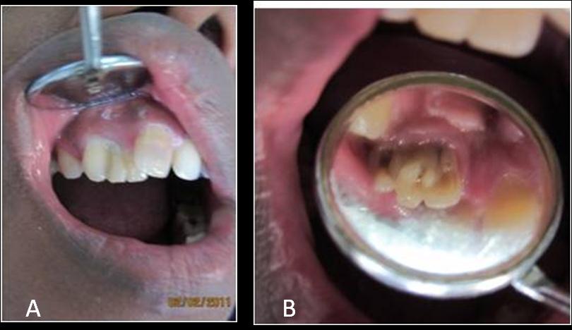
A 19-year old male patient came to the department with the complaint of pain in the lower left back region. On clinical examination, the lower left molar was found to be carious. The patient had poor oral hygiene and malaligned teeth. On inspection, the number of teeth which were present in the upper arch were found to be sixteen, and on the right side, the central incisor was rotated outwards, overlapping a large tooth which consisted of fused teeth, which included two supernumeraries which were fused with each other, which were slightly palatally placed and not in alignment and the lateral incisor which was slightly rotated mesially and fused to the supernumeraries. On palatal inspection, it was noted the two teeth which were mesial to the canine had deep lingual pits and that the tooth which was distal to the central incisor had an enamel elevation which mimicked a talon’s cusp [Table/Fig-1a and 1b]. The teeth were non tender to percussion. The vitality test gave no response.
(a) Intra oral photograph showing rotated right upper central incisor tooth, over lapping the fused supernumeraries and lateral incisor which are palatally placed. (b) Intra oral photograph showing palatal aspect; deep lingual pits and talons cusp on the tooth distal to the central incisor.
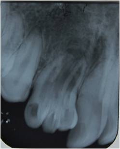
An intraoral radiograph showed [Table/Fig-2]; 3 teeth; the anatomy of the crown of the tooth which was distal to the central tooth appeared like a lateral tooth and there was an overlapping radiopacity in the cervical third, due to the deposition of enamel;-talon’s cusp. Dilacerations were present, with wide root canals which appeared to be fused. The tooth which was mesial to the permanent canine and the centre tooth appeared to be invaginated, with deep lingual pits. There was widening of the periodontal ligament space around the fused teeth, with loss of the lamina dura and a periapical radiolucency which could have been a chronic granuloma, as the patient had no pain or discomfort with respect to the same.
Intra oral Radiograph showing fused teeth
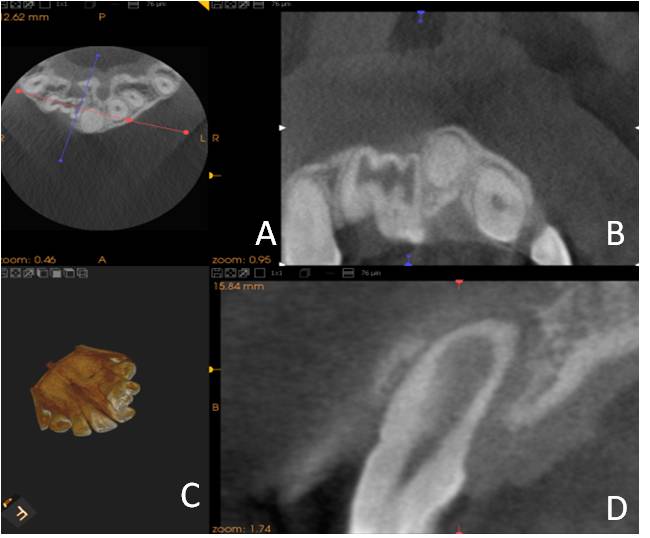
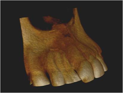
Cone Beam Computed Tomography (CBCT) demonstrated that in the axial plane [Table/Fig-3a], there was continuity of the pulp canals. The coronal plane [Table/Fig-3b] and the pulp canals of all the 3 teeth were continuous. The lingual view [Table/Fig-3c] showed a deep lingual pit. The saggital section [Table/Fig-3d] of the tooth which was mesial to the central incisor showed the presence of a dilated pulp canal. The three dimensional view [Table/Fig-4] showed the lateral incisor to be fused to two supernumeraries with bone loss in relation to the three teeth.
CBCT view showing (a) Axial view; (b) Coronal view (c:) 3D view and (d) Saggital view
Diagnosis
There was fusion of the permanent lateral incisor with the 2 supplementary teeth. All the 3 teeth showed dilacerations with dens invaginatus in two teeth and a chronic periapical lesion granuloma).
It was difficult to decide as to which was the permanent lateral incisor; as clinically the tooth which was mesial to the canine appeared to be anatomically bigger and more similar to a lateral incisor in morphology, but the presence of a rudimentary talon’s cusp and the radiographic anatomy of the tooth which was distal to the central incisor ruled more in favour of this tooth being the lateral incisor.
Discussion
The aetiology of the fusion between teeth is unknown, but the influence of pressure or physical forces which produce a close contact between two developing teeth has been reported as one possible cause [10]. A fusion can occur between the teeth of the same dentition or of mixed dentitions, and between normal and supernumerary teeth [11-17]. The number of teeth in the dental arch is also normal and their differentiation from the gemination is clinically difficult or impossible.
In the anterior region, this anomaly causes an unpleasant aesthetic tooth shape due to the irregular morphology. These teeth are greatly predisposed to caries and periodontal disease and the endodontic treatment is complicated [18]. Furthermore, the fusion may have an adverse effect on the occlusion, thus causing a deviation and delaying the eruption [2].
The supernumerary teeth are detected during the clinical examination or incidentally in the radiographs. In patients with nonsyndromic supernumerary teeth, a heredity factor has been proposed in several reports and therefore the family history should be carefully explored.
Dens Invaginatus (DI) [5] presents clinically as a pit or a fissure on the lingual surfaces of the anterior teeth. A majority of the cases are located in the maxilla and in the lateral incisors. The possibility of the pulp being affected without a clinically detectable lesion as a result of a tortuous lingual anatomy, makes DI clinically significant [19].
The talon’s cusp has a higher frequency in males than in females. This anomaly has a greater predilection for the maxilla, and the maxillary lateral incisors are commonly affected in the permanent dentition, followed by the central incisors and the canines [7,8].
An early diagnosis and management of the talon’s cusp is important, in order to prevent complications such as an occlusal interference, compromised aesthetics, caries and periapical pathologies, and periodontal problems. The prevention of an accidental cusp fracture and attrition has also been stressed on.
In the cases of dilacerations, the affected teeth are the maxillary incisors, followed by the mandible anteriors. They have been noted more in males than in females. This may produce a delayed eruption or difficulties during a root canal therapy or a tooth extraction. Their early recognition on the preoperative radiographs will minimize these problems.
In this case, there was a traumatic occlusion which resulted from the fused teeth which were out of alignment. As the patient had no complaint regarding the fused teeth, no treatment, therapeutic or aesthetic was given on a routine basis; but the patient was informed as regards the pathology and the treatment options.
Conclusion
The different cases require a variety of knowledge about the alternative operative techniques and abilities. A multidisciplinary approach with different specialists working together, can contribute to the success of a treatment plan, especially in the cases where there are multiple dental anomalies [20,21].
[1]. Ezoddini AF, Sheikhha MH, Ahmadi H, The prevalence of dental developmental anomalies: a radiographic studyCommunity Dent Health 2007 Sep 24(3):140-44. [Google Scholar]
[2]. Yang Y, Xia X, Wang W, Qin M, The uncommon fusion of teeth and a lateral periodontal cyst in a Chinese girl: a case reportOral Surg Oral Med Oral Pathol Oral Radiol Endod 2011 112:e18-e20. [Google Scholar]
[3]. White S, Pharoah M, O’Connor D, Oral Radiology: Principles and Interpretation 2000 2nd edLondonSt. Mosby Louis, [Google Scholar]
[4]. Arathi R, Ashwini R, Supernumerary teeth: A case reportJournal of Indian Society of Pedodontics and Preventive Dentistry 2005 23(2):103 [Google Scholar]
[5]. Vardhan TH, Subramanyam S, Dens evaginatus and dens invaginatus in all the maxillary incisors: the report of a caseQuintessence Int 2010 41(2):105-07. [Google Scholar]
[6]. Atkinson SR, “The permanent maxillary lateral incisor.”American Journal of Orthodontics and Oral Surgery 1943 29(12):685-98. [Google Scholar]
[7]. Hegde KV, Poonacha KS, Sujan SG, Bilateral labial talon cusps on the permanent maxillary central incisors: the report of a rare caseActa Stomatol Croat 2010 44(2):120-22. [Google Scholar]
[8]. Sarraf-shirazi A, Rezaiefar M, Forghani M, A rare case of multiple talon cusps in three siblingsBraz Dent J 2010 21(5):463-66. [Google Scholar]
[9]. Neville BW, Damm D, Allen C, Bouquot J, Oral and Maxillofacial Pathology 2002 Second edition:78 [Google Scholar]
[10]. Neville DW, Damm DD, Allen CM, Bouquot JE, Color Atlas of Clinical Oral Pathology 1991 2ndBaltimore, MDWilliams and Wilkins:62-64. [Google Scholar]
[11]. Shafer WG, Hine MK, Levy BM, A Textbook of Pathology 1983 4th edPhiladelphiaWB Saunders Company [Google Scholar]
[12]. Turell IL, Zmener O, Endodontic therapy in a fused mandibular molarJ Endod 1999 25:208-09. [Google Scholar]
[13]. Peyrano A, Zmener O, The endodontic management of a mandibular lateral incisor which was fused with a supernumerary toothEndod Dent Traumatol 1995 11:196-98. [Google Scholar]
[14]. Hülsmann M, Bahr R, Grohmann U, Hemisection and vital treatment of a fused tooth – a literature review and case reportEndod Dent Traumatol 1997 13:253-58. [Google Scholar]
[15]. Velasco LF de, Araujo FB, Ferreira ES, Velasco LE, The esthetic and the functional treatment of a fused permanent tooth: a case reportQuintessence Int 1997 28:677-80. [Google Scholar]
[16]. Camm HJ, Wood JA, Gemination, fusion and a supernumerary tooth in the primary dentition: the report of a caseJ Dent Child 1989 56:60-61. [Google Scholar]
[17]. Spatafore CM, The endodontic treatment of fused teethJ Endod 1992 18:628-31. [Google Scholar]
[18]. Pereira AJA, Fidel RAS, Fidel SR, A maxillary lateral incisor with two root canals: fusion, gemination or dens invaginatus?Braz Dent J 2000 11:141-46. [Google Scholar]
[19]. Mupparapu M, Singer SR, A rare presentation of dens invaginatus in a mandibular lateral incisor, which occurred concurrently with bilateral maxillary dens invaginatus: A case report and review of the literatureAust Dent J 2004 49:90-93. [Google Scholar]
[20]. Suprabha BS, Sumanth KN, Boaz K, George T, “An unusual case of the non-syndromic occurrence of multiple dental anomalies.”Indian Journal of Dental Research 2009 20(3):385-87. [Google Scholar]
[21]. Nagaveni NB, An unusual occurrence of multiple dental anomalies in a single nonsyndromic patient: A case reportCase Reports in Dentistry Volume 2012 2012 Article ID 426091:4pages doi:10.1155/2012/426091 [Google Scholar]