Cysticercus of the Breast which Mimicked a Fibroadenoma: A Rare Presentation
Karthikeyan T.M.1, Manimaran D.2, Mrinalini V.R.3
1. Associate Professor, Department of Pathology, Melmaruvathur Adhiparasakthi Medical College, Melmaruvathur, Tamil Nadu, India – 603319.
2. Associate Professor, Department of Pathology, Melmaruvathur Adhiparasakthi Medical College, Melmaruvathur, Tamil Nadu, India – 603319.
3. Professor & HOD, Department of Pathology, Melmaruvathur Adhiparasakthi Medical College, Melmaruvathur, Tamil Nadu, India – 603319.
NAME, ADDRESS, E-MAIL ID OF THE CORRESPONDING AUTHOR: Dr. Karthikeyan T.M., Associate Professor, Department of Pathology, Melmaruvathur Adhiparasakthi Medical College, Melmaruvathur, Tamil Nadu, India – 603319.
Phone: 9994743277
E-mail: karthikeyan.tmariappan@gmail.com
Human cysticercosis is an infection which is caused by the larvae of the pork tapeworm, Taenia solium. They can affect any part of the body, the most common sites being the muscle, the CNS and the subcutaneous tissues. In this report , we are presenting the case of a 32-year old woman who came with a history of a painless, freely mobile lump in the left breast. A clinical diagnosis of a fibroadenoma was made and an excision biopsy was done, which revealed the presence of cysticercus lavae, along with a foreign body giant cell reaction. A diagnosis of cysticercus at the atypical sites is rare and it depends mainly on the histopathological examination. Although it is rare, cysticercus should be considered as a differential diagnosis for a mass in the breast and in the areas of a greater prevalence.
Cysticercus, Breast, fibroadenoma
Introduction
Human cysticercosis is a parasitic infestation which is caused by Cysticercus cellulosae, the larvae of the pork tape worm, Taenia solium [1–3]. It is a major public health problem in the developing countries, where open air defaecation and food contamination is rampant. The common sites of occurrence of cysticercus are the skeletal muscle, the subcutaneous tissues , the brain and the eye, in the decreasing order of frequency. The breast is an uncommon site for cysticercosis, with only a few cases having been reported in the literature [2–5]. The patients commonly present with a cyst or a lump in the breast.
Case discussion
A 32-year old married female presented with a painless lump in the left breast, which was there over a period of 1 year. On examination, a freely mobile lump which measured 2x1cm was found in the left upper outer quadrant of the breast, which was non-tender and soft to firm in consistency. FNAC (Fine Needle Aspiration Cytology) yielded scanty material and it was inconclusive, following which an excision biopsy was done and the lump was sent for a histopathological examination.
Pathological findings
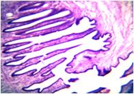
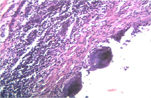
Grossly, the lump consisted of a grey white, cystic, nodular swelling which measured 1.5x0.75 cm. The external surface was smooth and glistening. The cut section showed a cyst which contained a white mural nodule which was attached to the wall, with a clear serous fluid. The nodule measured 2x1mm. The entire specimen was submitted in toto. Its microscopic sections showed a cyst which was composed of 3 layers [Table/Fig-1]. The outer cuticular layer, the middle cellular layer and the inner fibrillary layer formed a racemose pattern. The scolex was not identified. The surrounding breast tissue showed a foreign body giant cell granulomatous reaction [Table/Fig-2].
Section shows a cyst composed of 3 layers, outer cuticular layer, middle cellular layer and inner fibrillary layer thrown in racemose pattern
Surrounding stroma shows foreign body giant cell reaction
In view of the histological diagnosis of cysticercus, an extensive search was made, to exclude the infestation at the other sites.
The physical and the radiological examinations of the whole body was normal. The stool examination did not show any eggs or proglottids. The patient was given a course of Albendazole, following a surgical excision.
Discussion
Cysticercosis is caused by the larval stage of the human tape worm, Taenia solium. It continues to be a major public health problem in the developing countries, where open air defaecation and a lack of hygiene are rampant.
Human cysticerosis, a potentially deadly infestation, is the conse-quence of the ingestion of the eggs of Taenia solium which is present in contaminated food or water or on unwashed hands, or by means of autoinoculation which results from reverse peristalsis. The cysticerci are commonly found in the CNS, the eye, the subcutaneous tissues and the striated muscle and rarely in the heart, lung and the bone. The breast is an unusual site for the cysts to form and only few such cases have been reported in the literature [2,4–6].
Amatya and Kimula reported 62 cases of histologically diagnosed cysticercosis, five of which were found in the breast substance [4]. In this case, an initial diagnosis of fibroadenoma of the breast was made, due to its typical feature of a painless, firm and a freely mobile mass [4–6]. Thus, a diagnosis of cysticercosis in the unusual sites may be clinically difficult. It can be diagnosed only by the histological demonstration of the parasite in surgically removed tissues or by radiological means, which may give a clue. Radiologically it can be detected by X-ray visualization of the calcifying cysticerci and by the use of computerized tomography. In the present case, X-ray and CT scan of the other parts of the body were normal.
FNAC also plays an important role in diagnosing cysticercosis, but it is limited by the varying cytomorphological features of cysticercosis [7]. The host tissue response is extremely variable and it ranges from an insignificant response to the markedly cellular response, which consists of epitheloid cell granulomas and histiocytes. In a study which was done by K Sahai et al., the presence of palisading histiocytes and eosinophils was found consistently in the patients with a cysticercosis breast. The hooklets and the scolex were occasionally seen. In the present case, because of a scanty cellularity, the FNAC was nondiagnostic.
Serological tests such as the Indirect Haemagglutination Test and the Indirect Fluorescent Antibody Test ELISA (Enzyme Linked Immunosorbent Assay)can be used to diagnose cysticercosis in the suspected cases, but these are limited by a low sensitivity and specificity [8].
This case report emphasizes the fact that cysticercosis of the breast should be considered as a differential diagnosis for a mass in the breast, especially in the areas of greater prevalence.
[1]. Ouedrago EG, Hydatid cyst of the breast: 20 casesJ Gynecol Obstet Biol Reprod (Paris) 1986 15:187-94. [Google Scholar]
[2]. Kunkel JM, Hawksley CA, Cysticercosis presenting as a solitary dominant breast massHum Pathol 1987 18:1190-91. [Google Scholar]
[3]. Alagaratnam TT, Wing YK, Tuen H, Cysticercosis of the breastAm J Trop Med Hyg 1988 38:601-02. [Google Scholar]
[4]. Amatya BM, Kimula Y, Cysticercosis in Nepal; A histopathologic study on sixty two casesAm J Surg Pathol 1999 23:1276-79. [Google Scholar]
[5]. Sah SP, Jha PC, Gupta AK, An incidental case of breast cysticercosis which was associated with a fibroadenomaIJPM 2001 44(1):59-61. [Google Scholar]
[6]. Geetha TV, Krishnanand BR, Pai CG, Cysticercosis of the breast: A rare presentationJ. Nep Med Assoc 2000 39:184-85. [Google Scholar]
[7]. Sahai K, Kapila K, Verma K, Parasites in fine needle breast aspirates – Assessment of the host tissue responsePostgrad Med J 2002 78:165-67. [Google Scholar]
[8]. Hubert K, Andriantsimahavandy Al, Michault A, Frosch M, Muhlschlegel FA, The serological diagnosis of human cysticercosis by the use of recombinant antigens from the Taenia solium cysticerciClinical and Diagnostic Laboratory Immunology 1999 July6(4):479-82. [Google Scholar]