Introduction
The swim up and the density-gradient centrifugation are the two main techniques which are used to separate the viable motile sperm fraction in the assisted reproductive technology. However, there are several published studies about these methods, but there is no sufficient evidence for recommending the superiority of one of them. This study was designed to study the efficiency of the swim-up and the density gradient techniques to recover the spermatozoa with a high degree of motility, a normal morphology and a low level of DNA fragmentation.
Material and Methods
A total of 35 semen samples were included in the study. The semen samples were collected, one part of the semen was spread on a slide and the remainder was prepared by using the swim-up or the density gradient techniques. The recovered spermatozoa were evaluated for concentration, motility, and normal morphology. A comet assay was carried out to assay the DNA fragmentation in all the samples.
Results
There were significant differences in the sperm parameters between the density gradient and the swim up techniques. Also, the swim-up technique showed a significantly higher level of DNA fragmentation as compare to the density gradient technique.
Conclusion
The results of this study demonstrated several benefits of the gradients method in the separation of normal and motile spermatozoa with healthy DNA, in comparison to the swim up method.
Sperm, Gradients sperm separation, Swim up, Comet assay
Introduction
In the assisted reproductive technology, swim up and density-gradient centrifugation are the two main techniques which are used to separate the viable motile sperm fraction from the other semen components [1–3]. Although several studies have been published on the effectiveness of these methods, there is insufficient evidence for recommending the superiority of one of them. Over the past years, the comparative studies on the sperm preparation methods have essentially investigated the outcomes such as the recovery rates and the conventional semen parameters [3–5]. And more recently, researchers have focused on the evaluation of the molecular parameters such as sperm DNA damage or apoptosis for the comparison of these different separation methods [6–12]. Spano et al., [7] in 1999, have been shown that the swim-up sperm separation may improve some of the sperm chromatin structure assay–related parameters. Younglai et al., [8] in 2001, have reported that the swim up method does not induce sperm DNA damage. Furthermore, Zini et al., [5] in 2000, found that the percentage of sperm with denaturated DNA was reduced significantly among the swim-up–treated but not among the density-gradient centrifugation–treated sperm, as compared to the whole semen. In contrast, it has been reported that the motile sperms which were obtained by density-gradient centrifugation had a higher mitochondrial membrane potential and a lower DNA fragmentation, they generated a lower ROS and they were more viable than those which were among the whole semen [9,10]. Also, according to Sakkas et al., [11] results, there was a significant decrease in the percentage of sperm with DNA damage on using the density-gradient centrifugation technique, whereas on using the swim-up method, the recovered sperm showed no significant improvement. On the other hand, some studies have investigated apoptosis in the prepared sperm by swim-up [12–14] and density-gradient centrifugation [15,16] and they have reported contradictory results. Because of these results, there was no consensus about which method was superior for isolating the “functionally normal” sperm. The aim of the present study was to compare the effects of the density-gradient centrifugation and the swim-up methods on the sperm parameters and the DNA fragmentation.
Materials and methods
Semen Analysis
Semen samples were obtained from 35 men who underwent a semen analysis. This study was approved by the Research Committee of Hamadan University of Medical Sciences (Iran). A routine semen analysis was performed according to the World Health Organization (WHO) guidelines [17]. The sperm morphology was assessed by using the WHO criteria at a cut-off point of 30% normal sperm. From each ejaculate, two aliquots were taken for density-gradient centrifugation and the swim-up preparation.
Swim-Up
An aliquot of 0.5ml of whole semen was washed with 4ml of medium (Hams F10,Sigma) which was supplemented with 10% human serum albumin in a 15ml Falcon conical tube and it was then centrifuged at 300g for 10 minutes. The supernatant was discarded and 0.5ml of medium was gently layered on the pellet. Then, the tube was inclined at an angle of 45 degrees and incubated at 37°C for at least 45 minutes. The tube was then gently set upright and the upper interface was gently aspirated with a Pasteur pipette. An aliquot was examined for the sperm concentration and motility, and another aliquot was used for the comet assay analysis.
Density-Gradient Centrifugation
Silane-coated silica particles were used for the gradient separation (PureSperm 40/80; Nidacon International, Goteborg, Sweden). A 2 layer gradient was prepared by using ready-to-use solutions of 80% and 40% PureSperm. The media were warmed to 37°C. By using a sterile pipette, 0.5ml of liquefied semen sample was placed on top of the upper layer in a conical 5ml centrifuge tube. The tube was centrifuged at 300g for 20 minutes. The supernatant was then removed and the pellet was suspended in a volume of 1ml of medium. It was again centrifuged at 500g for 10 minutes. The pellet was resuspended in a volume of 0.5ml of medium. An aliquot was examined for the sperm concentration and motility, and another aliquot was used for the comet assay analysis.
The Single Cell Gel Electrophoresis (comet) Assay
In this study, the comet assay was performed by using a modification of the Angelis method [18] in order to detect both the single and the double stranded breaks.
Pre-cleaned slides (ROTH, Germany) were dipped in a solution of 1.5% ( w/v) normal melting point agarose (NMPA) in PBS, coverslips was then placed on top of them, and the agarose was allowed to solidify at room temperature overnight. The next day, the coverslips were removed and 100 micro litter suspensions of spermatozoa in 1% (w/v) low melting point agarose (LMPA), at a concentration of 1×10⁴ cell/ml, were pipetted onto the slides and they were covered with coverslips. The slides were allowed to solidify at 4ºC for 5 minutes and then the coverslips were gently removed, 1% LMPA was used to form a third layer and the slides were allowed to solidify at 4°C for at least 1 hour. Then, the slides with the coverslips were removed and they were placed in cold lysis buffer (2.5 M NaCL, 100 mM EDTA, 10 mM Tris, 1% Triton X-100, 1% DMSO, and 10 mM Dithiothreitol [DDT] at a PH of 10 for 30 min at 4°C. They were protected from light. The slides were then incubated at 37°C in 10µg/ml of Proteinase K (Sigma) in lysis buffer for 2.5 hours.
Following the cell lysis, all the slides were washed through three changes of distilled water at 5 min intervals to remove salt and detergent from the microgels. The slides were placed in a horizontal electrophoresis tank which was filled with electrophoresis buffer (10 mM Tris containing 0.08 M boric acid and 0.5 M EDTA pH=8.2) and they were kept for 20 minutes to allow the DNA to unwind. The electrophoresis buffer was adjusted at a level of ~0.25 cm above the surfaces of the slides. The electrophoresis was performed for 20 minutes at 25V which was adjusted to 300 mA, by either raising or lowering the buffer level in the tank. When the electrophoresis was completed, the slides were dried and flooded with three changes of neutralization buffer (0.4 mol/l Tris; PH 7.5), each for 5 minutes. After a neutralization step, the slides were stained with ethidium bromide (20µg/ml dissolved in distilled water) and they were mounted with cover slips. The cells were visualized at 200X by using a fluorescent microscope (Nikon).
Each cell with fragmented DNA had the appearance of a comet [Table/Fig-1], with a bright fluorescent head and a tail on one side, which was formed by the DNA, which contained strand breaks that were drowned away during the electrophoresis. The samples were run in duplicate, and 50 cells were randomly analyzed per slide for a total of 100 cells per sample. The percentage of the sperms with a comet appearance was considered as the comet index on each slide.
Comet assay, The figure A shows sperm without DNA fragmentation and the figure B shows sperm with high degree of DNA fragmentation

Statistical Analysis
The statistical analysis was performed by using the SPSS, version 11 software. The normal distribution of the data was checked by using the Kolmogrov-Smirnov test. The independent sample t-test was used to compare the mean differences between the test and the control samples. The data were represented as mean ± S.D. and a p value of < 0.05 was considered as statistically significant.
Results
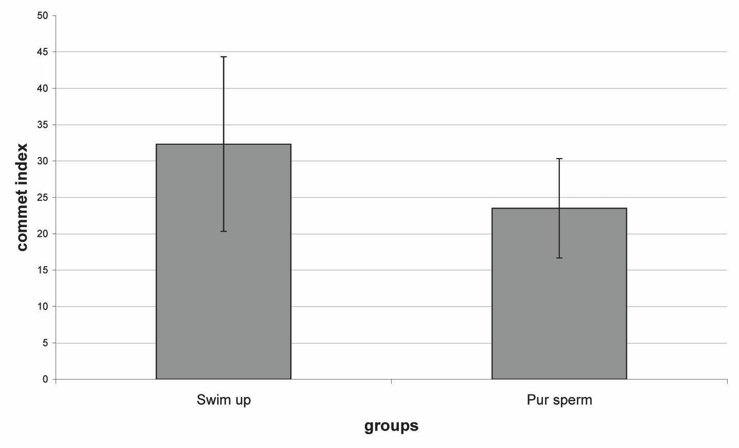
The recovery rate of the total count, total motility, and the sperm with a normal morphology were significantly higher on using the density-gradient centrifugation as compared to that which was obtained on using the swim-up preparation [Table/Fig-2] and [Table/Fig-3]. The means of the sperm concentrations in the density-gradient centrifugation method were significantly higher than those in the swim-up method (47.3±13.9 million vs. 50.6±31.1 million). The total sperm motility in the density-gradient centrifugation method was also significantly higher as compared to that in the swim-up method (75±15.1% vs. 52.3±10.2%). The normal sperm morphology in the density-gradient centrifugation method was also significantly higher as compared to that in the swim-up method (28±13.11% vs. 11.4 ±8.4%). The mean of the comet index in the sperms after the density-gradient centrifugation was significantly lower than that in the swim-up method (23.51±7.59 vs. 32.33±12, p< 0.001).
Comparsion of comet index after density-gradient centrifugation and swim-up methods. P< 0.05 was considered significant.

Comparsion of Sperm parameters and comet index after density-gradient centrifugation and swim-up methods.
| Swim-up | Pur Sperm | P value |
|---|
| Concentration (×106)/ml | 50.3 ± 10.2 | 74.3± 13.9 | <.0003 |
| Total motility (%) | 52.3 ± 10.2 | 75.5 ± 15.1 | <.0001 |
| Normal morphology (%) | 14 ± 8.4 | 28 ± 13.11 | <.0001 |
| Comet index | 32.33 ± 12 | 23.51 ± 6.83 | <.0001 |
Note: Values are mean ± SD, P< 0.05 was considered significant.
Discussion
In the present study, the comet assay data showed that the mean percentage of the DNA fragmented sperms in the swim-up–processed samples was significantly higher than that in the gradient-density–processed samples. Our results were inconsistent with the findings of Jayaraman and colleagues (2012) [19]. Their results showed no significant differences in the rates of the apoptotic sperm, which were indicated by the Tunnel technique, which were recovered by the density-gradient and the swim-up processing methods. This inconsistency may be have been due to the different technique (comet assay) which was used for investigating the DNA fragmentation in our study. According to our results, the lower percentage of the DNA fragmentation which was found in the density-gradient fractions, suggested that this method allowed the removal of most of the sperms with fragmented DNA. The use of sperms with DNA fragmentation during ART may be one of the causes for the suboptimal results. The negative association between the sperm DNA fragmentation and the fertilization rate has been documented in clinical and experimental studies [16,19]. The sperm DNA fragmentation seems to have a negative impact on the sperm oocyte penetration. Therefore, the selection of sperms with normal DNA should be one of the prerequisites for achieving optimal conception rates after ART [19] and to obtain this goal, the sperm processing method is important. In the present study, we compared the two routine sperm separation methods. The lower percentage of the apoptotic sperm which was found in the density-gradient fractions suggested that this method allowed the removal of most of the apoptotic sperm. So, it can be hypothesized that in comparison to the swim up method, the density-gradient method induces less DNA fragmentation. Therefore, the risk of selecting the DNA fragmented sperm during the clinical ART seems to be low. In agreement with our results, a meta analysis [1] showed that the density-gradient technique seemed to result in a higher sperm concentration and in a higher progressive motile sperm recovery rate than the swim-up technique.
In conclusion, the sperms which are obtained through density gradient centrifugation provide spermatozoa with a higher quality in terms of the motility, viability and low DNA fragmented as compared to those which are obtained by the other conventional sperm preparation methods.
Note: Values are mean ± SD,
P< 0.05 was considered significant.
[1]. Henkel RR, Schill WB, The sperm preparation for ARTReprod Biol Endocrinol 2003 14:108-29. [Google Scholar]
[2]. Boomsma CM, Heineman MJ, Cohlen BJ, Farquhar C, Semen preparation techniques for intrauterine inseminationCochrane Database Syst Rev 2004 3:CD004507 [Google Scholar]
[3]. Aitken RJ, Clarkson JS, The significance of the reactive oxygen species and the antioxidants in defining the efficacy of the sperm preparation techniquesJ Androl 1988 9:367-76. [Google Scholar]
[4]. Mortimer D, The sperm preparation techniques and the iatrogenic failures of in-vitro fertilizationHum Reprod 1991 6:173-76. [Google Scholar]
[5]. Zini A, Mak V, Phang D, Jarvi K, The potential adverse effects of semen processing on the human sperm deoxyribonucleic acid integrityFertil Steril 1999 72:496-99. [Google Scholar]
[6]. Henkel RR, Franken DR, Lombard CJ, Schill WB, The selective capacity of glass-wool filtration for the separation of human spermatozoa with condensed chromatin: a possible therapeutic modality for male-factor casesJ Assist Reprod Genet 1994 11:395-400. [Google Scholar]
[7]. Spanò M, Cordelli E, Leter G, Lombardo F, Lenzi A, Gandini L, Nuclear chromatin variations in the human spermatozoa which underwent swim-up and cryopreservation, which were evaluated by the flow cytometric sperm chromatin structure assayMol Hum Reprod 1999 5:29-37. [Google Scholar]
[8]. Younglai EV, Holt D, Brown P, Jurisicova A, Casper RF, Sperm swim-up techniques and DNA fragmentationHum Reprod 2001 16:1950-53. [Google Scholar]
[9]. Donnelly ET, ‘Connell MO, McClure N, Lewis SE, The differences in the nuclear DNA fragmentation and the mitochondrial integrity of semen and the prepared human spermatozoaHum Reprod 2000 1:1552-61. [Google Scholar]
[10]. Marchetti C, Obert G, Deffosez A, Formstecher P, Marchetti P, A study on the mitochondrial membrane potential, the reactive oxygen species, the DNA fragmentation and the cell viability which was done by using flow cytometry on human spermHum Reprod 2002 17:1257-65. [Google Scholar]
[11]. Sakkas D, Manicardi GC, Tomlinson M, Mandrioli M, Bizzaro D, The use of two density gradient centrifugation techniques and the swim-up method to separate the spermatozoa with chromatin and nuclear DNA anomaliesHum Reprod 15 2000 15:1112-26. [Google Scholar]
[12]. Muratori M, Piomboni P, Baldi E, Filimberti E, Pecchioli P, Moretti E, The functional and the ultrastructural features of DNA-fragmented human spermsJ Androl 2000 21:903-12. [Google Scholar]
[13]. Almeida C, Cardoso MF, Sousa M, Viana P, Goncalves A, Silva J, A quantitative study which was done on the caspase-3 activity in semen and after the swim-up preparation in relation to the sperm qualityHum Reprod 2005 20:1307-13. [Google Scholar]
[14]. Paasch U, Grunewald S, Fitzl G, Glander HJ, The deterioration of the plasma membrane is associated with the activated caspases in human spermatozoaJ Androl 2003 24:246-52. [Google Scholar]
[15]. Marchetti C, Gallego MA, Deffosez A, Formstecher P, Marchetti P, Staining of human sperm with the fluorochrome-labeled inhibitor of caspases to detect the activated caspases: correlation with apoptosis and the sperm parametersHum Reprod 2004 19:1127-134. [Google Scholar]
[16]. The WHO, Laboratory manual for the examination of human semen and the sperm-cervical mucus interaction 1999 4th [Google Scholar]
[17]. Angelis KJ, McGuffie M, Menke M, Schubert I, The adaptation to the alkylation damage in DNA which was measured by the comet assayEnviron Mol Mutagen 2000 36(2):146-50. [Google Scholar]
[18]. Said T, Agarwal T, Grunewald S, Rasch M, Baumann T, Kriegel C, Selection of nonapoptotic spermatozoa as a new tool for enhancing the assisted reproduction outcomes: an in vitro modelBiol Reprod 2006 74:530-37. [Google Scholar]
[19]. Jayaraman V, Upadhya D, Narayan PK, Adiga SK, The sperm processing by swim-up and density gradient is effective in the elimination of the sperm with DNA damageJ Assist Reprod Genet 2012 Jun29(6):557-63. [Google Scholar]