Fine Needle Aspiration Cytology of Adrenal Oncocytic Neoplasm of Uncertain Malignant Potential
Hilda Fernandes1, Maria Bukelo2
1 Professor of Pathology, Fr Muller Medical College, Mangalore, Karnataka, India
2 Resident, Fr Muller Medical College, Mangalore, Karnataka, India
NAME, ADDRESS, E-MAIL ID OF THE CORRESPONDING AUTHOR: Dr. Hilda Fernandes, Dept of Pathology, Fr Muller Medical College, Mangalore, Karnataka, India -575002.
E-mail: hilda67@rediffmail.com
Sir,
Adrenocortical oncocytic neoplasms (AONs) are rare tumors with only about 55 cases reported in the English language literature [1]. Histologic features that predict the biologic behaviour of AONs appear to differ from those of conventional adrenocortical tumors. In 2004, the modified Weiss system was proposed for AONs [2].
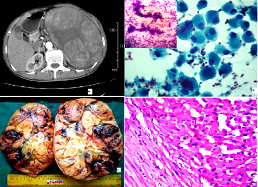
A 72-years old male was admitted for evaluation of a palpable abdominal mass. Computer Tomography scan of the abdomen revealed a large heterogeneously enhancing mass lesion arising from the left upper retroperitoneum having clear fat planes with pancreas, spleen and left kidney [Table/Fig-1A]. Possibility of malignant adrenal neoplasm was considered. Image guided Fine Needle Aspiration [FNA] smears were cellular showing singly dispersed polygonal cells with eccentric nucleus and dense granular cytoplasm [Table/Fig-1B]. A few cells were clinging to delicate capillaries in a clean background [Table/Fig-1B] (inset) suggesting a diagnosis of adrenal oncocytic neoplasm. The left adrenalectomy specimen consisted of a 2005 gm solid, encapsulated, globular mass measuring 20 × 15 × 12 cm. Cut surface of the tumor was solid, fleshy tan brown with lobulations of haemorrhage and necrosis [Table/Fig-1C]. The tumor was encapsulated and showed neoplastic cells arranged in a diffuse, solid pattern with multiple foci of haemorrhage, microcystic degeneration and necrosis [Table/Fig-1D]. Tumor cells were uniform with abundant brightly eosinophilic granular cytoplasm, round to oval vesicular nucleus. Mitotic figures, capsular and venous invasion were absent. A final diagnosis of adrenal oncocytic tumor of uncertain potential was given based on modified Weiss Criteria. Electron Microscopy confirmed the oncocytic nature of cells.
[Table/Fig-1]: (1A) - CT scan showing a large well defined mass with heterogenous echotexture; (1B) - Smear showing singly dispersed cells with eccentric nucleus and granular cytoplasm (PAP 40X); (1B inset) - Smear showing tumor cells attached to delicate capillaries.(MGG 20X); (1C) - Cut surface shows encapsulated tumor with lobulated areas and necrosis. (1D): Section shows capsule and oncocytic cells (H&E 20X).
AONs have the potential to behave in a benign fashion, recur or extensively infiltrate into the adjacent organs. Average age at diagnosis is 47 years (range 6-77) with a slight female predilection [3]. Size varies from 2.2 to 20 cm and weight from 8 to 1900 g [1]. In our case the tumor weighed 2005 gm which is the highest so far reported. Imaging characteristics of adrenal oncocytic tumors are described as single case reports . Fibrous encapsulation and heterogeneous areas do not differentiate between benign and malignant oncocytic tumors [4]. Cytopathologic diagnosis of AONs by FNA is described by some authors.[5] Some of the criteria of Modified Weiss system like necrosis and atypical mitosis can be evaluated on cytology. Unfortunately accurate mitotic count, venous invasion and capsular invasion can not be assessed in smears. In the absence of major criteria, it is best to designate the tumor as an oncocytic adrenal neoplasm with a note for a detailed histopathologic assessment. If any minor criteria are present, designation AON of uncertain malignant potential may be rendered.
In our case there was no necrosis or atypical mitosis on smears. However due to the large size and heterogeneous areas on CT scan, possibility of malignancy was still considered preoperatively. Only after detailed histopathologic examination, it was designated AON of uncertain malignant potential. Awareness of Modified Weiss criteria will help in averting over diagnosis of malignancy while dealing with FNA of AON.
[1]. Cham E, Watkin W, Goldschmidt R, Lin Lin, Fine Needle Aspiration Cytology of Adrenocortical Oncocytic Neoplasm. A case reportActa Cytol 2010 54:627-34. [Google Scholar]
[2]. Argyaion P, Zisis C, Alevizopoulos N, Kefaloyannis EM, Gennatas C, Petraki CD, Adrenocortical oncocytic carcinoma with recurrent metastasis: a case report and review of literatureWorld J Surg Oncol. 2008 16:234-42. [Google Scholar]
[3]. Tahar GT, Nejib KN, Sadok SS, Rachid LM, Adrenocortical oncocytoma: a case report and review of literatureJ. Pediatric Surg. 2008 43:E1-3. [Google Scholar]
[4]. Shah RK, Oto A, Ozkan OS, Ernst RD, Fernandez JA, Chaudhary HB, Adrenal oncocytoma: US and CT findingsJBR-BTR 2004 87:180-82. [Google Scholar]
[5]. Wang T, Nguyen GK, Cytopathology of Adrenal cortical OncocytomaDiagn Cytopathol 2001 24:222-23. [Google Scholar]