Bilateral Type-I Duane Syndrome with Multiple Anamolies: A Case Report
Chaitanya Varma1, Shrikiran Aroor2, Suneel C Mundkur3, Karthick Annamalai4
1 Assistant Professor, Department of Paediatrics, Kasturba Medical College, Manipal, Karnataka, India
2 Professor, Department of Paediatrics, Kasturba Medical College, Manipal, Karnataka, India
3 Associate Professor, Department of Paediatrics, Kasturba Medical College, Manipal, Karnataka, India
4 Senior Resident, Department of Paediatrics, Kasturba Medical College, Manipal, Karnataka, India
NAME, ADDRESS, E-MAIL ID OF THE CORRESPONDING AUTHOR: Dr. Shrikiran Aroor, Professor, Department of Paediatrics, Kasturba Medical College, Manipal, Karnataka, India.
Phone: +919448177671,
E-mail: kiranaroor@yahoo.com
The Duane syndrome is a strabismus syndrome which is characterized by congenital non-progressive horizontal ophthalmoplegia which primarily affects the abducens nerve. Approximately 70% of the individuals with the Duane syndrome have an isolated disease. We have described here, a case of bilateral Duane syndrome with associated anamolies.
Border line intelligence, Crocodile tears, Duane Retraction Syndrome, Stilling-Turk-Duane Syndrome, Spina bifida occulta
Introduction
The Duane Syndrome (DS) or the Duane Retraction Syndrome is a congenital form of strabismus which is characterized by horizontal eye movement limitation and globe retraction with palpebral fissure narrowing during an attempted adduction. It was first described by Sinclair, Turk and Stilling in 1895. In 1905, Duane summarized the clinical presentations and postulated the theory for its pathogenesis and treatment [1]. It constitutes approximately 1% of the total cases of strabismus. It usually presents as an isolated unilateral condition and it is only rarely associated with systemic anomalies. Here, we have reported a case of bilateral Duane syndrome with crocodile tears, mental retardation, and spina bifida occulta.
Case Report
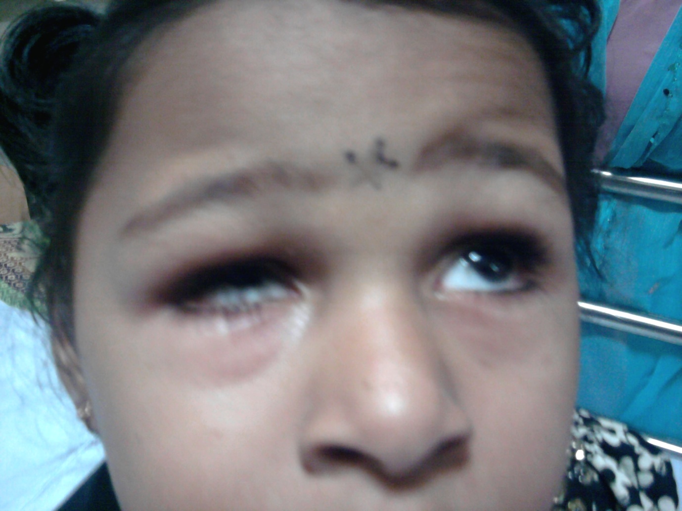
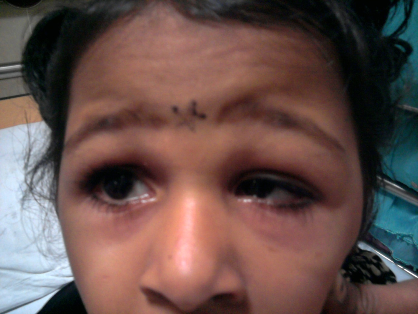
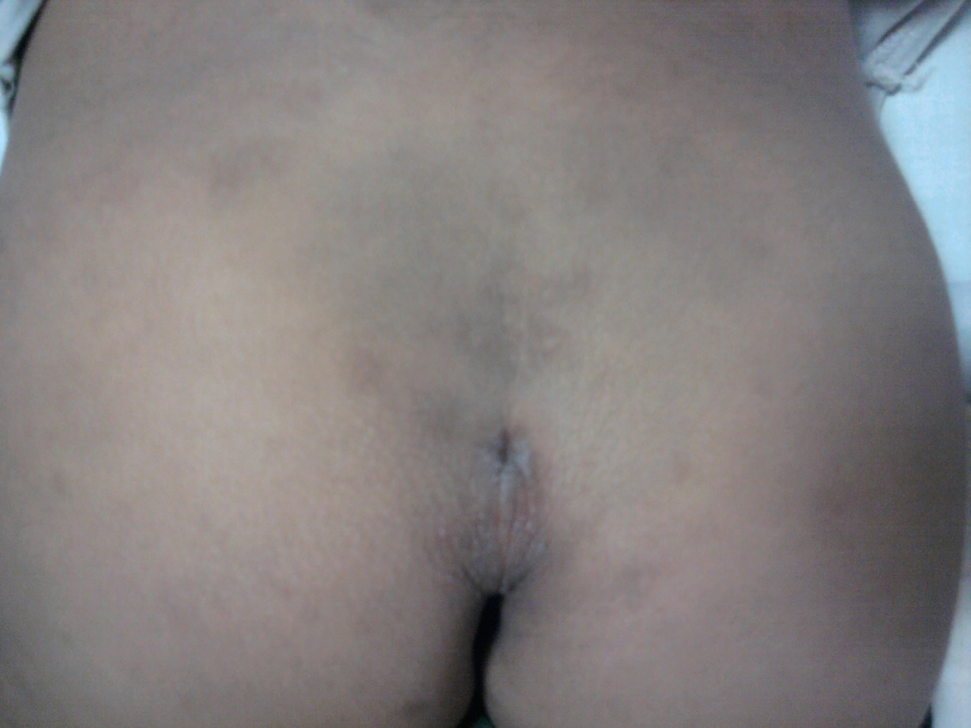
A 6-year old girl who was born out of a non-consanguineous marriage with an uneventful perinatal period was brought with a history of delay in attaining age specific milestones and excessive watering from the eyes since the time she started chewing solid food. The parents also noticed that there was a decrease in the size of the eye and an increased tendency for the eyes to move independently while she looked to the sides. Her developmental age corresponded to around 3 years. The child was born to non-consanguineous parents at full term, with an uneventful perinatal period. On examination, her height was found to be 102cm, with a weight of 15kg. She had limited abduction movements in both her eyes, along with globe retraction and narrowing of the palprebral fissure while adduction was attempted [Table/Fig-1(a) and (b)]. The child was found to have epiphora from both the eyes while she chewed food. A sacral dimple and hyper extensibility of the joints was also noticed [Table/Fig-2]. Her cardiovascular and neurological examinations were unremarkable. An ophthalmology consultation confirmed the finding as bilateral Duane's syndrome along with “crocodile” tears. An x-ray of the lumbosacral spine revealed a spina bifida occulta. Her IQ was 70, which put her at borderline intelligence. Her hearing evaluation and ultrasonography of her abdomen were normal. Her haemogram, renal function tests and thyroid profile were normal.
Limited abduction movements in both her eyes along with globe retraction and narrowing of the palprebral fissure while attempting adduction
Discussion
Duane's Retraction Syndrome (DRS), also known as the Stilling-Turk-Duane syndrome, is a congenital oculomotor anomaly that presents as globe retraction with simultaneous narrowing of the palpebral fissure on attempted adduction. It is caused by an absence or hypoplasia of both the abducens nucleus and the nerve with an anomalous innervation of its target, the lateral rectus muscle, by a branch of the oculomotor nerve [2]. The incidence of DRS is approximately 1% of the total cases of strabismus. It commonly occurs unilaterally and sporadically, with a predominant tendency to affect females and the left eye. 10% of the Duane syndrome cases are inherited in an autosomal dominant pattern and are usually bilateral. Disorders which are similar in presentation to the Duane syndrome can be acquired as a result of trauma, or following a localized infection of the orbit, leading to inflammation and consequent mechanical restrictions of the eye movement. A complete case history will usually help in distinguishing between these conditions. Sixth nerve paralysis, a tight medial rectus, crossed fixation, pseudo duanes or inverse duanes are some of the other differential diagnosis for this condition. The primary lesion in pseudo-Duane's retraction syndrome is suspected to be due to entrapment of the medial rectus muscle within the medial orbital wall due to trauma [3].
In 1974, based on electromyography studies, Huber classified the DRS into 3 types; Type 1 with a marked limitation of abduction, Type 2 with a limitation of adduction, and Type 3 showing limitation of both adduction and abduction [4].
Esotropia is the most common type of strabismus which is encountered and characteristic up shoots and down shoots occur in adduction. The other associated ocular anomalies which can occur in subjects with the Duane syndrome can include nystagmus, anisocoria, ptosis, optic nerve colobomas, epibulbar dermoids, crocodile tears and aniridia. This syndrome is also associated with skeletal (limb hypoplasia, polydactyly and hypoplastic or absent radius and/or thumb), vertebral (scoliosis, spina bifida, “butterfly” vertebrae and Klippel-Feil anomaly), genitourinary (renal agenesis and vesicoureteral reflux), and cardiac (patent ductus arteriosus and auricular septal defect) defects. The Duane syndrome can occur with other syndromes like the Okihiro (Duane syndrome and radial ray defects), the Wildervanck (Duane syndrome, Klippel-Feil anomaly, and deafness), the Moebius (congenital paresis of facial and abducens cranial nerves) and the Townes-Brocks (ear, limb, anal, renal and heart anomalies) syndromes [5,6]. The treatment of the DS may involve correction of the refractory error and squint and surgical procedures like muscle recession procedures, vertical transposition of the rectus muscle, or a combination of the two, for improving or eliminating the head turns and misalignment of the eyes [7]. Our child presented with Type-1, bilateral DS and “crocodile” tears- a rare combination which has been scarcely reported in the Indian medical literature [8]. In addition to the above features, the child also had sacral dimpling, spina bifida occulta and borderline intelligence, a combination which has not been associated with the Duane syndrome earlier.
Conclusion
The DS is a rare syndrome and bilateral DS is all the more so. Any child with bilateral DS should be completely evaluated to rule out other syndromic associations. Immediate and early ophthalmological interventions with refractory error treatment, use of prisms and surgical corrections, will go a long way in helping such children to lead a normal life.
[1]. Duane A, A congenital deficiency of abduction which was associated with the impairment of adduction, contraction of the palprebral fissure and oblique movements of the eyeArch Opth. 1905 34:133-59. [Google Scholar]
[2]. Andali D, Javadzadeh A, Lateral rectus muscle disinsertion and reattachment to the lateral orbital wall in exotropic Duane syndrome: a case reportJ Med Case Reports 2008 2:253 [Google Scholar]
[3]. Lee SH, Lee JH, Lee SY, Kim SY, A case of pseudo-Duane's retraction syndrome with old medial orbital wall fractureKorean J Ophthalmol. 2009 December23(4):329-31. [Google Scholar]
[4]. Tomi A, Preda C, Poenaru O, Zamfiroiu F, The Duane's syndrome-etiopathogenesis, clinical features and diagnosisOftalmologia 2005 49(2):10-4. [Google Scholar]
[5]. Gutowski MJ, The Duane syndrome. A reviewEuropean J Neurology 2000 7:145-49. [Google Scholar]
[6]. Yüksel D, Orban de Xivry J-J, Lefèvre P, Review of the major findings about the Duane retraction syndrome (DRS), leading to an updated form of classificationVision Research 2010 50(23):2334-47. [Google Scholar]
[7]. Wang KM, Liu LJ, Zhang FH, Treatment of Duane's retraction syndrome by recession of the medial and the lateral rectus muscles, combined with a Y-splitting procedureChinese Journal of Ophthalmology 2007 Nov43(11):972-76. [Google Scholar]
[8]. Mugundhan K, Thiruvarutchelvan K, Sivakumar S, Congenital crocodile tears with the Duane's syndrome—the congenital cranial dysinnervation syndromeJ Assoc Physicians India 2011 May59:316 [Google Scholar]