The Abnormal Origin, Course and the Distribution of the Arteries of the Upper Limb: A Case Report
Surekha D Shetty1, Satheesha Nayak B2, Venu Madhav N2, Srinivasa Rao Sirasanagandla2, Abhinitha P2
2 Corresponding Author
1,3-5Department of Anatomy, Melaka Manipal Medical College (Manipal Campus), Manipal University, Madhav Nagar, Manipal, Karnataka State, India. 576104
NAME, ADDRESS, E-MAIL ID OF THE CORRESPONDING AUTHOR: Dr. Satheesha Nayak B, Professor and Head, Department of Anatomy, Melaka Manipal Medical College (Manipal Campus), International Centre for Health Sciences, Manipal University, Madhav Nagar, Manipal, Udupi District, Karnataka State, 576104, India.
Phone: +91 820 2922519, 91 9844009059, Fax: 91 820 2571905,
E-mail: nayaksathish@yahoo.com
The knowledge on the arterial variations in the arm is of importance for a clinician, as it is a frequent site of injury and as it is also involved in many surgical and invasive procedures. During the routine dissection classes for medical students, we came across the multiple arterial variations in the right upper limb of an approximately 45-year-old male cadaver.
The brachial artery was very short, and it terminated by dividing into the radial and the ulnar arteries in the upper part of the arm. The radial collateral, the middle collateral and the superior ulnar collateral arteries arose from a common trunk. This common trunk originated from the proximal part of the brachial artery. The ulnar artery was the lateral branch and the radial artery was the medial branch of the brachial artery at their point of origin. The radial artery had a tortuous course, and it crossed the ulnar artery from the medial to the lateral side in the middle third of the arm. The ulnar artery gave anterior and posterior interosseous arteries and a common trunk that divided into the anterior and the posterior ulnar recurrent arteries in the cubital fossa.
The knowledge on these variations is very useful for radiologists and surgeons.
Brachial artery, Radial artery, Ulnar artery, Profunda brachii artery, Superior ulnar collateral artery, Variation
Introduction
The brachial artery is the main artery of the arm. It is a continuation of the axillary artery, at the lower border of the teres major muscle. It usually terminates at the level of the neck of the radius, in the cubital fossa, by dividing into the radial and the ulnar arteries. The radial artery runs along the lateral part of the front of the forearm, with the superficial branch of the radial nerve. The ulnar artery passes medially deep to the pronator teres muscle and it then runs to the distal part of the forearm together with the ulnar nerve. The profunda brachii artery is a large branch from the brachial artery and it follows the radial nerve closely, between the long and the medial heads of the triceps and then in the radial groove. It divides into the radial collateral and the middle collateral arteries. The superior ulnar collateral artery is the branch of the brachial artery and it runs along with the ulnar nerve. In the present case, we are reporting the observed high bifurcation of the brachial artery.
Case Report
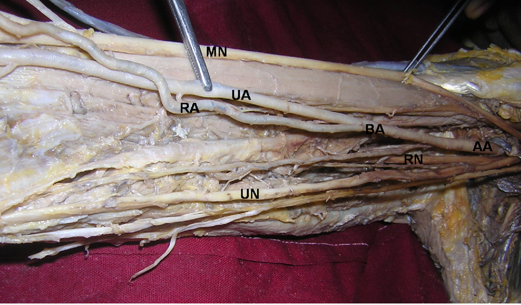
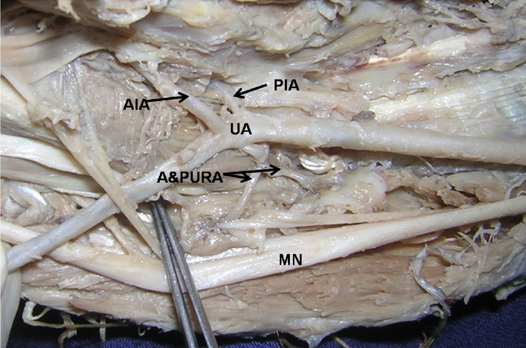
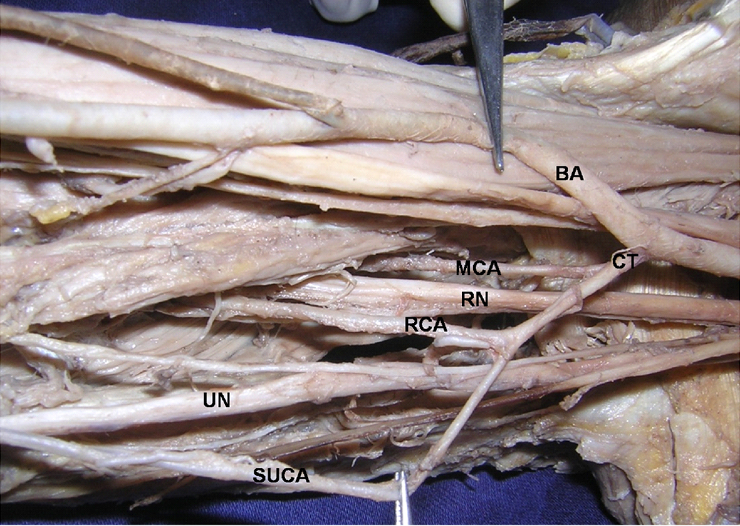
During the regular dissections which were done for undergraduate medical students at Melaka Manipal Medical College (Manipal Campus), we found arterial variations in a formalin embalmed male cadaver who was aged approximately 45 years. In the right upper extremity, the axillary artery continued as the brachial artery, at the lower border of the teres major muscle. The brachial artery bifurcated into the radial and the ulnar arteries, just an inch below the lower border of the teres major muscle [Table/Fig-1]. The radial artery was superficial, throughout the course from its origin. It was very torturous and it arose from the medial side of the brachial artery. It then crossed the ulnar artery and the median nerve superficially and continued down on the lateral side of the cubital fossa. In the cubital fossa, it gave the radial recurrent artery and numerous muscular branches and passed superficial to the bicipital aponeurosis. In the forearm, the artery was superficial to the forearm muscles, but it was lying deep in the antebrachial fascia. The ulnar artery coursed on the medial side of the biceps brachii, till the cubital fossa. In the cubital fossa, it gave the anterior interosseous, the posterior interosseous, the anterior ulnar recurrent and the posterior ulnar recurrent arteries [Table/Fig-2]. The two branches of the profunda brachii; the radial collateral and the middle collateral arteries originated from a common trunk, which also gave origin to the superior ulnar collateral artery [Table/Fig-3].
Photograph of high bifurcation of the brachial artery (AA - axillary artery; BA - brachial artery; UA - ulnar artery; RA - radial artery; MN - median nerve; UN - ulnar nerve; RN - radial nerve.)
Photograph of ulnar artery and its branches in the cubital fossa.(UA - ulnar artery; AIA – anterior interosseous artery; PIA – posterior interosseous artery; A&PURA – anterior and posterior ulnar recurrent arteries; MN – median nerve.)
Photograph of common trunk of the profunda brachii and superior ulnar collateral arteries.(BA - brachial artery; CT – common trunk; MCA - middle collateral artery; RN - radial nerve; RCA - radial collateral artery; UN - ulnar nerve; SUCA- superior ulnar collateral artery.)
Discussion
The variations in the branching patterns of the arteries of the limbs have clinical and surgical significance [1]. Anomalies of the arteries of the upper limb are common, as their development is dependent upon many sources, as well as on a precise sequential pattern of the formation and the regression of some of the arteries [2]. Some of the earliest study reports on the variations in the arterial system have been made by Senior [3] and Singer [4]. According to the Bergman et al., the high origin of the radial artery is seen in 15% cases and that of the ulnar artery is seen in 2% cases. This high division may occur at any point in the normal course of the vessels, but it is more common in the middle third portion of the course [5]. In the present case, the brachial artery bifurcated into the radial and the ulnar arteries. The radial artery was very torturous and it arose from the medial side of the brachial artery. It was superficial, throughout the course from its origin. The ulnar artery coursed on the medial side of the biceps brachii, till the cubital fossa. In the cubital fossa, it gave the anterior interosseous, the posterior interosseous, the anterior ulnar recurrent and the posterior ulnar recurrent arteries. Celink et al., [6] have reported the early division of the brachial artery into the radial and the ulnar arteries after giving origin to the profunda brachii artery.
The variations of the superficial brachial artery have been reported by Yang et al., [7]. Rodriguez-Niedenfuhr et al., [8] named the radial artery which arose in the arm as the brachio-radial artery and they reported 12 upper limbs which showed the radial artery to arise from the axillary artery, among 192 upper limbs. Harrington [9] reported a case of high bifurcation of the brachial artery with a reunion at the elbow. A case of the high origin of the radial artery has been also reported by Clemente [10]. The incidence of the superficial radial artery is one of the frequent variations and it has been observed in 14.26% cases in the cadaveric studies and in 9.75% cases in the angiographic studies [11].
The profunda brachii is largest branch of the brachial artery and it shows considerable variation in its origin. In 55% of the cases, it arises as a single trunk at the level of the tendon of the teres major muscle [12]. It may arise from the axillary artery (in 22% cases), as a common trunk with the superior ulnar collateral artery (in 22% cases) or as a branch of the circumflex humeral artery (in 7% cases). In the current case, the two branches of the profunda brachii and the superior ulnar collateral artery arose from the brachial artery as a common trunk. Singal and Lalit [13] have reported the high origin of the profunda brachii and the common interosseous arteries. The superficial brachio-ulnar and the superficial brachio-radial arteries have also been reported [14].
The knowledge on the variation which has been reported here, is important in procedures like cardiac catheterizations, arterial grafting and other angiographic procedures. This type of a variation may cause a misinterpretation of the angiographic images. Accidental punctures of the superficially placed arteries may occur while venipunctures are attempted, which are susceptible to damage in orthopaedic and plastic surgery operations. The presence of a tortuous radial artery has not been reported as yet. The origin of the radial artery from the medial side of the brachial artery, its tortuous course and its crossing superficial to the ulnar artery makes this variation special and unique. This course and nature of the radial artery might cause problems in invasive procedures.
[1]. Jurjus A, Sleir R, Bezirdjian R, An unusual variation of the arterial pattern of the human upper limbAnat Rec 1986 215:82-3. [Google Scholar]
[2]. Standring S, Gray’s Anatomy 2006 39thElsevier Churchill Livingstone Publishers:941 [Google Scholar]
[3]. Senior HD, A note on the development of the radial arteryAnat Rec 1926 32:220-1. [Google Scholar]
[4]. Singer E, An embryological pattern which persists in the arteries of the armAnat Rec 1933 55:403-9. [Google Scholar]
[5]. Bergman RA, Thompson SA, Afifi AK, Saadeh FA, A Compendium of the Human Anatomic VariationsBaltimore: Urban Schwarzenberg 1988 :65 [Google Scholar]
[6]. Celik HH, Gormus G, Aldur MM, Ozcelik M, Origin of the radial and the ulnar arteriesMorphologie 2001 85:25-7. [Google Scholar]
[7]. Yang HJ, Gil YC, Jung WS, Lee HY, Variations of the superficial brachial artery in Korean cadaversJ Korean Med Sci 2008 23:884-7. [Google Scholar]
[8]. Rodriguez-Niedenfuhr M, Vazquez T, Parkin IG, Sanudo JR, The arterial patterns of the human upper limb: an update on the anatomical variations and the embryological developmentEur J Anat 2003 7(1):21-8. [Google Scholar]
[9]. Harrington A high bifurcation of the brachial artery with a reunion at the elbowJohns Hospkins Hosp. Bull 1905 16:65-6. [Google Scholar]
[10]. Clemente CD, Anatomy of the Human Body 1985 30thPhiladelphiaLea and Febiger:829 [Google Scholar]
[11]. Karlsson S, Niechajev IA, The arterial anatomy of the upper extremityActa Radiol Diagn (Stockh) 1982 23:115-23. [Google Scholar]
[12]. Tountans CP, Bergman RA, Anatomical variations of the upper exterimitesNew York, Churchill Livingstone 1993 :197-210. [Google Scholar]
[13]. Singal KR, Lalit M, A superficial brachial artery with a high origin of the profunda brachii and the common interosseous arteryJournal of Clinical and Dignostic Reserch 2011 5(3):628-80. [Google Scholar]
[14]. Fatah MF, Nancarrow JD, Murray DS, Raising the radial artery forearm flap; the superficial ulnar artery trapBr. J Plast Surg 1985 38(3):394-5. [Google Scholar]