Body-Painting: A Tool Which Can Be Used to Teach Surface Anatomy
Komala Nanjundaiah1, Sheshgiri Chowdapurkar2
1 Assistant Professor, Department of Anatomy, MS Ramaiah Medical College, Bangalore, Karnataka, India - 560054
2 Senior Professor and Head of Department, Department of Anatomy, MS Ramaiah Medical College, Bangalore, Karnataka, India - 560054
NAME, ADDRESS, E-MAIL ID OF THE CORRESPONDING AUTHOR: Dr. Komala Nanjundaiah, Assistant Professor, Department of Anatomy, M.S. Ramaiah Medical College, M.S.R.I.T Post, Bangalore, Karnataka, India - 560054,
Phone: 9480259177, 080-23421593,
E-mail: komas2001@yahoo.com, komalananjundaiah@gmail.com
Introduction
The primary method of learning the surface anatomy is by making the students mark structures on mummified bodies. The students feel that learning the surface anatomy on mummified cadavers is not interesting. The present project on learning the surface anatomy through the body painting method was undertaken to evoke interest among the students.
Materials and Methods
Physiotherapy and dental undergraduate students who volunteered were involved in this study. A few surface anatomy classes were conducted by using the traditional method and a few more by using the body painting exercise. Non toxic body paints of various colours and brushes of different sizes were used for the body painting.
Results
A feedback was obtained from the students by using a structured questionnaire. The students opined that the body painting method was advantageous to them in learning the human anatomy. They also felt that they could have more practice sessions in any setting other than in the classroom and that they did not need to rely upon the mummified bodies. They described the body painting method as self explanatory, which gave them the feel of live structures.
Conclusion
This project was successful in achieving its objectives as the students felt that the method was exciting, with lots of fun during the learning. The body painting method was well accepted by the students as an effective method for learning the surface and the clinical anatomy.
Living anatomy, Body painting, Medical education, Visual images, Memory
Introduction
In many medical schools, the lack of cadavers is driving changes to the novel learning and the teaching methodologies [1]. The living anatomy, which is defined as the anatomy which is revealed on the living humans, is gaining importance in the modern anatomy education, and it has even been considered to replace the cadaver-based anatomy study [2]. As educators, it is necessary for us to innovate the teaching practices to help the students in understanding the anatomy. The traditional method of learning the surface anatomy on cadavers requires the student to put in lot of efforts on memorizing the landmarks. Their recall of the landmarks during the assessments is also poor. With this in mind, we tried body painting exercises for the students to learn the surface anatomy of certain regions. The students were well engaged during the sessions and they had fun while learning. Their active involvement made the students learn. The colourful visual images of various structures which were obtained after the painting exercises, increased their memory.
Aims of the Study
To ensure active participation of students during the learning
To help the students enlist the landmarks of the surface marking easily
To facilitate peer to peer learning
To make the learning of the surface and the clinically relevant anatomy more interesting and enjoyable for the students and to thus enable them to correlate the anatomy with the clinical diagnoses and to excel during their clinical years.
Methodology
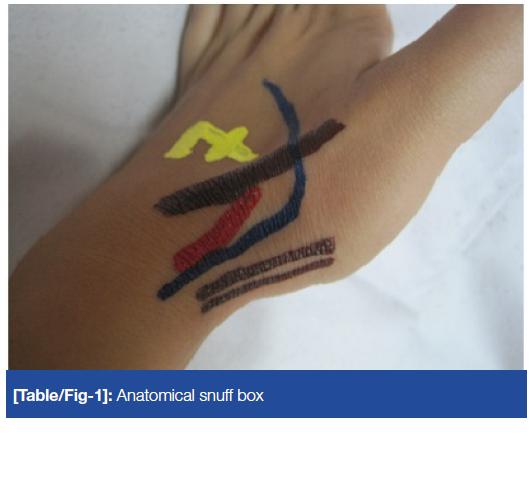
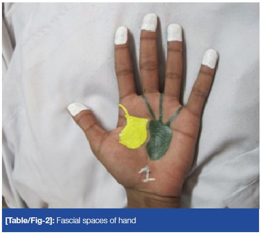
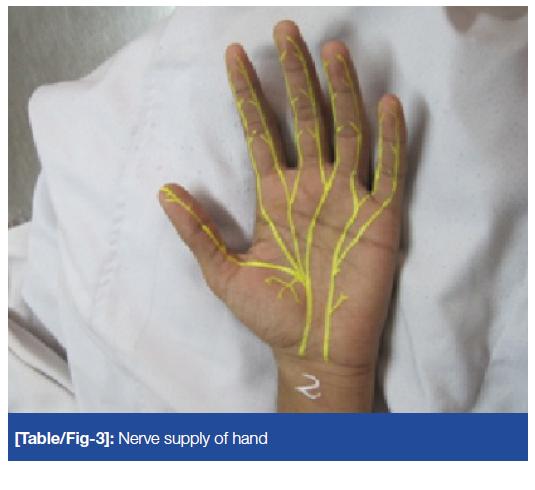
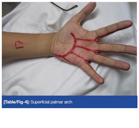
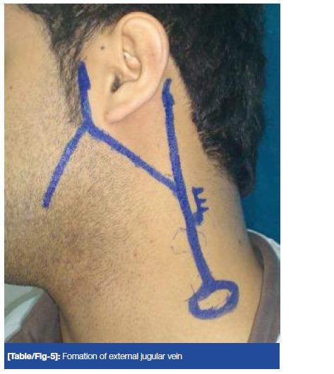
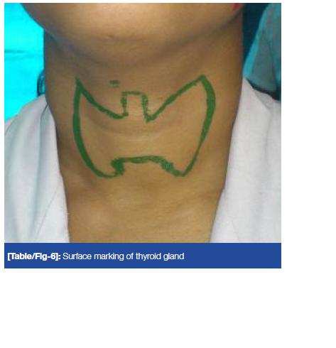
All the Ist year physiotherapy (25) and the Ist year dental (60) students (2011-2012 batch) volunteered for the body painting programme. For the physiotherapy students, the surface anatomy classes were conducted by using the traditional method for the lower limb. The surface anatomy classes for the upper limb were conducted by using body painting exercises. For the dental students, the surface anatomy of certain parts of the head and neck were taught by the traditional method, while certain other parts were taught by using body painting. Non toxic body paints of various colours (which were easily washable with water) and brushes of different sizes were used for the body painting. The students, on the previous day, were asked to do a sensitivity test for the colours which were used. The students were given 20 min to paint on their peer partners and another 20 min to get themselves painted [Table/Fig-1,2,3,4,5 and 6].
Fomation of external jugular vein
Surface marking of thyroid gland
A written consent from the volunteer students and ethical clearance for the project from the institutional review board were obtained.
A feedback was obtained from the students by using a structured questionnaire. The respondents' agreements/disagreements were noted with a set of statements by using a six point Likert scale.
Results
Seventy-seven students (percentage) answered the feedback questionnaire. The data was analyzed by using the SPSS software. 98.7% of the students agreed that the body painting method of learning the surface marking was interesting. 98.7% agreed that they actively participated in the body painting sessions. 96.1% agreed that it was easy to list out the landmarks after the body painting exercise. 96.1% felt that painting on each other facilitated a peer to peer learning. 90.9% agreed that body painting gave them the feel of the landmarks and various structures. 98.7% felt that the retainment of knowledge was better when it was learnt through body painting [Table/Fig-7]. The varying degrees of agreements and disagreements for the closed questions for the body painting exercise have been tabulated in [Table/Fig-8].
Graphical representation of the feedback from the students
Feed back analysis of varying degrees of aggrement and disaggrement for closed questions for body painting exercise as per 0-5 Liken Scale in Percentages, Chronbach's alpha-78.4%
| Parameters | Is interesting | Actively participated | Easy to list out landmarks | Peer to peer learning | Live feel of landmarks | Retainment is good |
|---|
| Very Strongly Disagree | – | – | – | – | 13 | – |
| Strongly Disagiee | – | 13 | – | – | – | – |
| Disagree | 1.3 | – | 3.9 | 3.9 | 7.8 | 1.3 |
| Agree | 54.5 | 40.3 | 59.7 | 45.5 | 41.6 | 42.9 |
| Strongly Agree | 28.6 | 36.4 | 22.1 | 37.7 | 32.5 | 42.9 |
| Veiy Strongly Agree | 15.6 | 22.1 | 14.3 | 13.0 | 16.9 | 13.0 |
The internal consistency (Cronbach's Alpha) for the questions which were asked was 0.784.
57 students were assessed for the surface marking of the structures which was learnt by either method. Standardized checklists were used for the assessments. The students' learning by both the methods was quantified by comparing the scores which were obtained by them. Their score did not follow the normal distribution (Shapiro-Wilk test = 0.953, p-value = 0.01). So, the Man-Whitney U test was used and it was found to be insignificant [Table/Fig-9].
Scores obtained by the students
| Method | N | Median | IQR | p-value (exact) |
|---|
| Body painting | 57 | 4.00 | 27.42 | 0.07 |
| Traditional | 57 | 4.00 | 27.09 | |
Discussion
The body painting method which was used to learn the surface and the clinical anatomy was well accepted by our students as an effective method to learn the surface and the clinical anatomy. The visual images increased their memory of the landmarks.
The students who used drawing, commented that the use of drawing while they learned from a text, required a lot of concentration (57%), lateral thinking (31%), making an alignment between what they understood from the text (60%) and transferring the knowledge into drawings (63%). This experience helped them in deepening their understanding (92%) [3].
While a cadaver is still the best study material for the construction of a three-dimensional image of human anatomy, considering the pressure which is required for reducing the hours which are geared towards the anatomy education, the education in anatomy in the 21st century must be revolutionized to utilize the state-of-the-art modalities to formulate a contemporary anatomy course. Such modalities allow the students to carry on self –directed learning, leading to a positive outcome in the anatomy education. The problem arises if we have to incorporate more living anatomy, for which the time which is necessary for the dissection needs to be minimized or compromised [2].
The approaches to the learning, correlate positively with the quality of the learning. A successful learning of the anatomy requires a balance between the memorization with understanding and the visualization [4].
One hundred and thirty-three preclinical medical students participated in 24 focus groups over the period from 2007-2009 at Durham University. Focus groups were conducted to ascertain whether or not the medical students found the body painting of the anatomical structures to be an educationally beneficial learning activity. The data were analyzed by using a grounded theory approach. Five principal themes emerged: body painting as a fun learning activity, body painting as promoting the retention of knowledge, the factors which contributed to the memorability of the body painting, removal from the comfort zone, and the impact of the body painting on the students' future clinical practice [5].
Op Den Akker JW et al., designed a course in which the students familiarized themselves with the surface markings and subsequently painted the full organ at the site of its projection on the body surface. They concluded that the course was a successful and enjoyable means of teaching various aspects of the anatomy in relation to the physical examination [6].
Body painting was introduced into the integrated clinical skills teaching sessions which included the clinically important aspects of the respiratory system, the musculoskeletal system, and topics in the regional anatomy, including the head and neck. The kinaesthetic nature and the active participation, together with the powerful visual images of the underlying anatomy, appear to contribute to the value of body painting as a teaching exercise. In addition, it may have the added bonus of helping break down the apprehension regarding the peer-peer examination [1].
Conclusion
The project was successful in achieving its objectives. The students felt that the body painting method was fun and that lots of peer learning happened. The students felt that they need not wait for the mummified bodies any more. They could paint on their friends outside the classroom setting also. While doing body painting, they could actually get the feel of and the relationships between the various anatomical structures. The feeling of the students regarding the method was, “Body painting ROCKZ!!!”. At the same time, the students also expressed a view that body painting was time consuming and that not all the structures could be marked at a time. However, this can be overcome by arranging proper preplanned sessions.
[1]. McMenamin PG, Body painting as a tool in the clinical anatomy teachingAnatomical Sciences Education 2008 1(4):139-44. [Google Scholar]
[2]. Ganguly PK, Chanz LK, The living anatomy in the 21st century: how far can we go?South East Asian Journal of Medical Education 2008 2(2):52-57. [Google Scholar]
[3]. Azer SA, Learning the surface anatomy: Which learning approach is effective in an integrated PBL curriculum?Medical Teacher 2011 33:78-80. [Google Scholar]
[4]. Pandey P, Zimitat C, The medical students' learning of the human anatomy: memorisation, understanding and visualizationMedical education 2007 Jan41(1):7-14. [Google Scholar]
[5]. Finn GM, Mclachlan JC, A qualitative study on the student responses to body paintingAnatomical Sciences Education 2010 3(1):33-38. [Google Scholar]
[6]. Op Den Akker JW, Bohnen A, Oudegeest WJ, Hillen B, Giving color to a new curriculum: Body painting as a tool in medical educationClin Anat 2002 15(5):356-62. [Google Scholar]