Dermatofibrosarcoma Protuberans of Inguinal Region: A Case Report
Priyanka Suknandan Hande1, Suresh Vasant Phatak2, Prashant Onkar3, Deepali Mohan Trimukhe4
1 Junior Resident, Department of Radiodiagnosis, N.K.P. Salve Institute of Medical Science and Research Centre and Lata Mangeshkar Hospital, Nagpur, Maharashtra, India.
2 Professor, Department of Radiodiagnosis, N.K.P. Salve Institute of Medical Science and Research Centre and Lata Mangeshkar Hospital, Nagpur, Maharashtra, India.
3 Professor, Department of Radiodiagnosis, N.K.P. Salve Institute of Medical Science and Research Centre and Lata Mangeshkar Hospital, Nagpur, Maharashtra, India.
4 Junior Resident, Department of Radiodiagnosis, N.K.P. Salve Institute of Medical Science and Research Centre and Lata Mangeshkar Hospital, Nagpur, Maharashtra, India.
NAME, ADDRESS, E-MAIL ID OF THE CORRESPONDING AUTHOR: Dr. Suresh Vasant Phatak, Professor, Department of Radiodiagnosis, N.K.P. Salve Institute of Medical Science and Research Centre and Lata Mangeshkar Hospital, Nagpur-440019, Maharashtra, India.
E-mail: suresh_phatak@yahoo.com
Dermatofibrosarcoma is a rare tumour involving the skin. It is a low-grade soft-tissue sarcoma that is commonly seen in middle-aged individuals, usually in the third and fourth decades of life, with males showing a higher prevalence than females. The authors reported a case of a 48-year-old female patient who presented to the hospital with complaints of multiple right inguinal swellings for three months, along with a reported recent increase in size over the last month. On examination, multiple papulomatous lesions, approximately 5×3 cm in size, were observed in the right inguinal region. Ultrasonography (USG) revealed well-defined lesions with a relatively mixed pattern and a few cystic areas noted in the subcutaneous plane, measuring approximately 40×25 mm. These lesions exhibited raised vascularity on colour Doppler imaging. The histopathological examination revealed myxoid tissue showing spindle or satellite cells with oval elongated nuclei and indistinct cytoplasm, suggestive of dermatofibrosarcoma. Dermatofibrosarcoma protuberans is an unusual soft-tissue tumour of the skin, and ultrasonography can significantly help in its diagnosis and management.
Sarcoma, Skin tumour, Soft-tissue tumour
Case Report
A 48-year-old female patient came to the hospital with complaints of multiple right inguinal swellings for three months, with a reported recent increase in size over the last month. There was no history of chills, fever, cold, night sweats, pain, trauma, previous similar complaints, pus discharge, or bowel/bladder complaints. There was no history of allergies. The patient had taken antitubercular medication five years ago for pulmonary tuberculosis and had no other drug intake in the past.
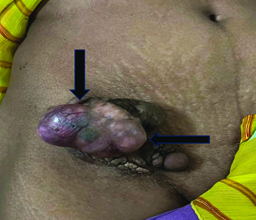
On examination, multiple papulomatous lesions, approximately 5×3 cm in size, were present in the right inguinal region. These lesions were irreducible when pressure was applied. They were pinkish-blue in colour, soft, non tender, and showed no local rise in temperature. There was no history of discharge or bleeding [Table/Fig-1]. A probable clinical diagnosis of a papillosquamous lesion was made. The patient was referred to the Radiodiagnosis Department for ultrasonography of the local right inguinal region.
Multiple, papulomatous lesions (black arrow) over right inguinal region.
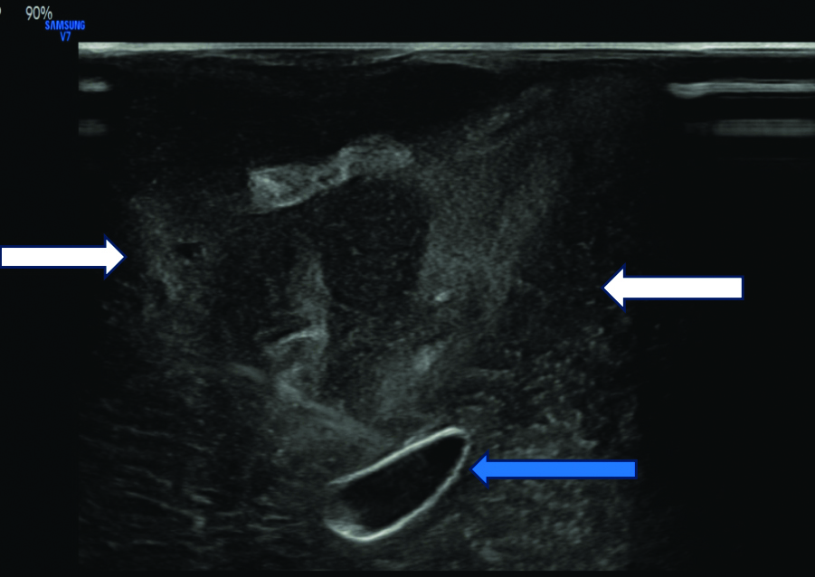
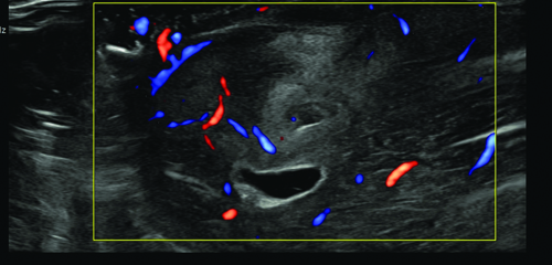
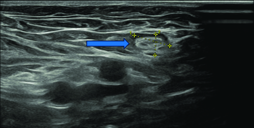
On ultrasonography, a well-defined, relatively mixed-pattern lesion with a few cystic areas was noted in the subcutaneous plane, approximately 40×25 mm in size [Table/Fig-2a]. It showed raised vascularity on colour Doppler [Table/Fig-2b] and no evidence of calcification. A probable radiological diagnosis of a soft-tissue tumour was made, and a few subcentimetric lymph nodes were noted in the right inguinal region [Table/Fig-2c].
USG image showing heterogenous lesions (white arrows) with few cystic areas (blue arrow) in the subcutaneous plane in right inguinal region.
Colour Doppler image showing high vascularity in the lesion.
USG showing lymph node (blue arrow) in right inguinal region.
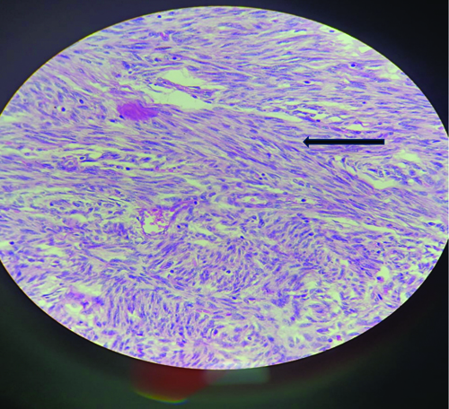
The patient underwent surgical excision, and the mass was sent for histopathology, which revealed myxoid tissue showing spindle cells with oval elongated nuclei and indistinct cytoplasm, features suggestive of dermatofibrosarcoma protuberans [Table/Fig-3]. The patient did not return for follow-up.
Myxoid tissue showing spindle cells (black arrow) with oval elongated nuclei and indistinct cytoplasm suggestive of dermatofibrosarcoma (H&E, 40x).
Discussion
Dermatofibrosarcoma protuberans most commonly occurs on the trunk, head, neck and proximal extremities, with unusual and rare presentations occurring on the head and neck, breast and vulva [1]. It accounts for less than 0.1% of cases amongst sarcomas of cutaneous origin and approximately 1% of all soft-tissue sarcomas. The annual incidence of dermatofibrosarcoma protuberans is 0.8-4.5 cases per million individuals [1]. Multiple factors, such as oncogenes, tumour suppressor genes, immunodeficiency, and, in some patients, a history of trauma, are involved in the development of dermatofibrosarcoma protuberans [1,2]. Clinically, most of these tumours present as asymptomatic, bluish or brownish, multinodular, raised lesions that are superficial and movable over deeper structures [3].
In a study by Gong X et al., these lesions commonly show a hypoechoic pattern with occasional hyperechoic dots within and lobulated lateral borders. A few lesions show hyperechoic areas with occasional hypoechoic bands or hypoechoic areas in the dermis; however, a mixed pattern (hyperechoic and hypoechoic areas) was also found in seven cases, which were similar to the appearance in the present case [4]. Elastography helps differentiate benign from malignant lesions. This method is more accurate and reliable than palpation. The red-green-blue colour scale represents soft, moderate and hard tissue consistency [5]. Magnetic Resonance Imaging (MRI) imaging is not routinely performed because these tumours have a typical clinical appearance and superficial location. It is used only in cases involving larger, atypical lesions or recurrent disease. The largest MRI study of dermatofibrosarcoma protuberans tumours was carried out by Torreggiani WC et al., in which T1-weighted images showed low signal intensity and T2-weighted sequences showed higher intensity than fat [6]. Fluorodeoxyglucose-Positron Emission Tomography/Computed Tomography (FDG-PET/CT) is useful in imaging dermatofibrosarcoma protuberans for assessing the extent of disease, staging, and recurrence, which mainly helps in planning surgery. A few cases have shown dermatofibrosarcoma protuberans’ response to imatinib on FDG-PET but not on CT. Therefore, FDG-PET/CT plays an important role in the patient’s treatment plan [7].
The differential diagnosis includes hypertrophic scar, keloid, and skin manifestations of myofibroblastoma [3]. These conditions were ruled out with the help of the histopathological features of dermatofibrosarcoma protuberans. A precise diagnosis will reduce the morbidity and mortality associated with this entity.
Conclusion(s)
The present case report aids in studying the salient features of a rare tumour in a middle-aged female. The patient’s history, clinical features and radiological investigations help in differentiating dermatofibrosarcoma protuberans from other pathologies; however, confirmation is achieved through histopathology.
Author Declaration:
Financial or Other Competing Interests: None
Was informed consent obtained from the subjects involved in the study? Yes
For any images presented appropriate consent has been obtained from the subjects. Yes
Plagiarism Checking Methods: [Jain H et al.]
Plagiarism X-checker: Jan 18, 2024
Manual Googling: Apr 20, 2024
iThenticate Software: Apr 23, 2024 (4%)
[1]. Chan IL, Carneiro S, Menezes M, Ramos-e-Silva S, Magalhães T, Cuzzi T, Dermatofibrosarcoma protuberans: A case reportCase Rep Dermatol 2014 6(2):134-39.10.1159/00036290024926255PMC4035680 [Google Scholar] [CrossRef] [PubMed]
[2]. Hao X, Billings SD, Wu F, Stultz TW, Procop GW, Mirkin G, Dermatofibrosarcoma protuberans: Update on the diagnosis and treatmentJ Clin Med 2020 9(6):175210.3390/jcm906175232516921PMC7355835 [Google Scholar] [CrossRef] [PubMed]
[3]. Dragoumis DM, Katsohi LAK, Amplianitis IK, Tsiftsoglou AP, Late local recurrence of dermatofibrosarcoma protuberans in the skin of female breastWorld J Surg Oncol 2010 8(1):4810.1186/1477-7819-8-4820525288PMC2892497 [Google Scholar] [CrossRef] [PubMed]
[4]. Gong X, Li J, Ding A, Chen J, Tao X, Xiong P, Multimodal ultrasound for preoperative evaluation of dermatofibrosarcoma protuberans: A series of 40 casesBMC Cancer 2022 22:113710.1186/s12885-022-10211-436335314PMC9637320 [Google Scholar] [CrossRef] [PubMed]
[5]. Zou MH, Huang Q, Yang T, Jiang Y, Zhang LJ, Xie Y, Role of ultrasound in the diagnosis of primary and recurrent dermatofibrosarcoma protuberansBMC Cancer 2021 21(1):90910.1186/s12885-021-08476-234376150PMC8356448 [Google Scholar] [CrossRef] [PubMed]
[6]. Torreggiani WC, Al-Ismail K, Munk PL, Nicolaou S, O’Connell JX, Knowling MA, Dermatofibrosarcoma protuberansAm J Roentgenol 2002 178(4):989-93.10.2214/ajr.178.4.178098911906888 [Google Scholar] [CrossRef] [PubMed]
[7]. Al-Tamimi A, Zaheer S, Pierce CKH, Osmany S, Sittampalam K, Recurrent dermatofibrosarcoma protuberans of the shoulder with rare distant abdominal metastasis detected by Fluorodeoxyglucose-Positron Emission Tomography/Computed Tomography (FDG-PET/CT)Sultan Qaboos Univ Med J 2012 12(3):371-74.10.18295/2075-0528.139223269951PMC3529665 [Google Scholar] [CrossRef] [PubMed]