The OA (OA: 3β-hydroxyolean-12-en-28-oic acid) is a naturally active pentacyclic triterpenoid molecule found in over 2,000 plant species, including many food and medicinal plants [1]. The traditional uses of plants containing OA are multiple, encompassing anti-inflammatory, hepatoprotective, analgesic, cardiotonic and sedative properties, among others [2]. Many of these therapeutic effects, such as antihypertensive activity, analgesic effects and anti-inflammatory effects in rat models, have been demonstrated in contemporary scientific research [3,4]. The most prominent sources of OA in the human diet are olives (Olea europaeaL.). Olive fruit, apple peel, ginseng, papaya fruit and black plums can all contain up to 1% OA [5].
Alzheimer’s Disease (AD) is a neurodegenerative condition characterised by amyloid accumulation in the brain’s neuropil and vasculature [6,7]. AD is marked by the build-up of abnormal proteins in the brain (amyloid plaques and neurofibrillary tangles) that disrupt communication between brain cells and lead to their eventual death [8]. The symptoms progressively worsen over time, causing cognitive decline and functional impairment. Jack CR et al., 2018 grouped the biomarkers into A (amyloid), T (phosphorylated tau) and N (neurodegeneration, measured by total tau where applicable): the ATN framework [9]. The hyperphosphorylation of tau proteins and deposition of amyloid proteins was found to result from oxidative stress caused by an imbalance in the redox state, due to the generation of excess Reactive Oxygen Species (ROS). Impaired synaptic plasticity, neuroinflammation, disruptions in vascular reactivity, cholinergic denervation, neurotransmitter imbalances, neuronal degeneration, dendritic changes and significant synaptic loss are all factors influenced by oxidative stress [10]. Therefore, it is essential for an anti-Alzheimer drug to possess the ability to reduce oxidative stress in the brain.
Therapeutic approaches to AD can be classified into three types: symptomatic, disease-modifying and regenerative. Current treatments for AD mainly focus on controlling symptoms rather than stopping the disease from progressing. There has also been a significant failure rate in recent clinical trials for new drugs for AD treatments [11]. Medications such as AChE inhibitors (donepezil, rivastigmine, galantamine) and N-methyl-D-aspartate (NMDA) receptor antagonists (memantine) provide symptomatic relief but are not very effective at targeting the core neurodegenerative processes [12,13]. Ongoing research is primarily focused on finding a drug that has a disease-modifying effect on AD. OA has been found to possess antioxidant and neuroprotective potential [14]. Hence, the present study was conducted to investigate the antioxidant and anti-Alzheimer’s effects of OA.
Materials and Methods
An in-vitro study was conducted in the Centre for Global Health Research, Saveetha Medical College and Hospital, Chennai, Tamil Nadu, India, from May 2024 to June 2024. The study commenced after receiving approval from the Scientific Review Board (SMC/R02/2024). OA (CAS No. 508-02-1), with a purity of 98%, was procured commercially from TCI Chemicals, India, for the study.
Study Procedure
In-vitro Antioxidant Activity
2,2-Diphenyl-1-Picrylhydrazyl (DPPH) free radical scavenging activity assay: The 2,2-DPPH assay was used to investigate the free radical scavenging activity of the compound, following previously described methods [15,16]. A 0.004% DPPH solution in methanol was prepared and 10 μL of OA at concentrations ranging from 5 μM to 320 μM was mixed with 190 μL of the DPPH solution. The mixture was vortexed and incubated at 37°C for 20 minutes. A blank control without the test compound was included. Absorbance was measured at 517 nm and the Half-maximal Inhibitory Concentration (IC50) value was calculated as the concentration required to reduce free radical scavenging activity by 50%. The experiments were conducted in triplicate and the percentage inhibition was determined using the formula:
% scavenging effect=(Absorbance of control-Absorbance of sample)×100/Absorbance of control.
2,2′-azino-bis (3-ethylbenzothiazoline-6-sulphonic acid) (ABTS) radical scavenging assay: The ABTS radical scavenging activity was evaluated using the method described by Gonzalez-Palma I et al., 2016. ABTS was dissolved in water to a concentration of 7 mM [17]. The ABTS radical cation (ABTS·+) was generated by reacting ABTS (7 mM) with 2.45 mM potassium persulfate and incubating the mixture in the dark at room temperature for 12-16 hours. The resulting solution was diluted with water to achieve an absorbance of 0.70 (±0.02) at 734 nm. In the assay, 0.07 mL of OA at concentrations ranging from 5 μM to 320 μM was mixed with 3 mL of the ABTS+solution. After 6 minutes of incubation, absorbance was recorded at 734 nm. Ascorbic acid was used as the standard and antioxidant activity was calculated using the formula:
% inhibition={(control-test)/control}*100
Control=Absorbance of negative control at the moment of solution preparation
Sample=Absorbance of sample after six minutes
In-vitro Lipid Peroxidation (LPO) Inhibition Assay
The in-vitro lipid peroxidation inhibition assay was carried out using the method described by Ohkawa H et al., 1979 [18]. To initiate lipid peroxidation, 1 mL of tissue homogenate was added to test tubes containing 0.5 mL of OA, followed by 0.1 mL each of FeSO4 (25 μM), ascorbate (100 μM) and KH2PO4 (10 mM). The final volume was adjusted to 3 mL with distilled water and the mixture was incubated at 37°C for one hour. Subsequently, 1 mL of 5% TCA and 1 mL of TBA were added and the tubes were boiled for 30 minutes. The reaction mixture was then centrifuged at 3,500 rpm for 10 minutes. Lipid peroxidation inhibition was assessed by measuring the TBARS level through absorbance at 532 nm. Experiments were conducted in triplicate and the IC50 value was determined as the concentration that reduced lipid peroxidation by 50%. The percentage inhibition was calculated using the formula:
(%) Inhibition={(Abs. of control-Abs. of test)/Abs. of control}×100.
In-vitro Acetylcholinesterase (AChE) Inhibition Assay
Oleanolic acid and donepezil hydrochloride (the standard) were tested for AChE inhibitory activity at concentrations of 5, 10, 20, 40, 80, 160 and 320 μM using 0.05 M Tris base. The assay involved mixing 200 μL of acetylthiocholine iodide (15 mM), 1,000 μL of DTNB (3 mM) and 200 μL of OA or Donepezil, followed by a 15-minute incubation at 30°C. The reaction was initiated by adding 200 μL of AChE (0.3 U/mL) solution to the mixture. Absorbance was recorded at 412 nm for 10 consecutive 13-second intervals. A control sample included all reagents except the test compounds and standard [19]. The percentage of AChE inhibition (% IA) was calculated using the formula:
IA (%)=(activity of control-activity of test)/activity of control×100.
OA was compared with Donepezil as a reference standard because Donepezil is a well-established and widely used drug for the treatment of Alzheimer’s disease (AD), recognised for its potent neuroprotective and antioxidative effects. Given its proven efficacy, Donepezil serves as an ideal benchmark to evaluate the therapeutic potential of OA in both antioxidant and neuroprotective activities. Furthermore, focusing on a single, well-validated standard allows for a clearer and more precise assessment of OA’s comparative efficacy in these specific activities [20].
Assessment of Aβ (1-42) Concentration
Preparation of Aβ Solution: The Aβ solution was produced using the method of Miyazaki H et al., 2019 [21]. To prevent preaggregation, synthetic β-amyloid peptide 1-42 (Aβ1-42) (PP69, Sigma Merck, USA) was dissolved in a 0.1% ammonia solution at a final concentration of 250 μM and sonicated in ice-cold water for five minutes (1 min×5 times). To prepare the Aβ solution, aliquots were diluted to 25 μM in 50 mM phosphate buffer (pH 7.5) and 100 mM NaCl.
Thioflavin T fluorescence assay: The Thioflavin T (ThT) fluorescence assay was performed as described by Miyazaki H et al., 2019 [21]. An 8 μL Aβ solution was mixed with varying concentrations of OA and standard Donepezil hydrochloride, then combined with 1.6 mL of ThT solution (5 μM ThT in 50 mM NaOH-glycine buffer, pH 8.5). The samples were incubated at 37°C and fibrillogenesis was monitored using ThT fluorescence. Fluorescence levels were measured with a Biotek Synergy H4 Multi-Mode reader at excitation and emission wavelengths of 446 nm and 490 nm, respectively.
In-vitro Inhibition Study on β-Secretase (BACE1) Enzyme
The β-Secretase enzyme inhibitory assessment was carried out using the BACE1 fluorescence assay. In brief, 10 μL of BACE1 enzyme solution at a concentration of 1.0 unit/mL (Thermo Fisher Scientific, USA), 10 μL of the OA compound (5 μM, 10 μM, 20 μM, 40 μM, 80 μM, 160 μM and 320 μM) along with standard Donepezil hydrochloride and 10 μL of 750 nM β-secretase substrate IV (Calbiochem, Merck, Darmstadt, Germany) were mixed in the reaction wells. The reaction mixture was allowed to incubate for one hour at ambient temperature. Fluorescence readings were recorded at 380 nm (excitation) and 510 nm (emission) using the Biotek Synergy H4 Multi-Mode reader (Molecular Devices, USA) [22].
Statistical Analysis
The data were analysed using GraphPad Prism (version 7.0). The results were expressed as Mean±Standard Error of the Mean (SEM), and the IC50 values were obtained from the linear regression plots. A two-way ANOVA was used to assess differences between means at a significance level of p-value <0.001. The means were compared with standard groups using the Holm-Sidak test.
Results
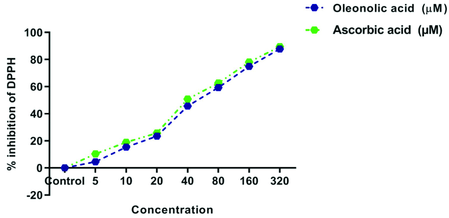
The DPPH free radical scavenging assay: The DPPH assay was conducted for OA, and it was found to exhibit a good antioxidant effect when compared with ascorbic acid. It demonstrated a concentration-dependent inhibitory effect. At a concentration of 20 μM, oleanolic acid showed 23.47±0.21% inhibition, whereas ascorbic acid showed around 25.77±0.38% inhibition. Even at a concentration of 320 μM, both OA and ascorbic acid exhibited almost the same inhibitory effect, with 87.83±0.22% inhibition on DPPH, indicating that OA has good antioxidant properties [Table/Fig-1,2].
Effect of Oleanolic Acid (OA) on DPPH, ABTS and LPO.
| Concentration (μM) | % inhibition of DPPH | % inhibition of ABTS | % inhibition of LPO |
|---|
| Oleanolic acid | Ascorbic acid | Oleanolic acid | Ascorbic acid | Oleanolic Acid (OA) | Ascorbic acid |
|---|
| Control | 0.00±0.54 | 0.00±0.54 | -0.27±0.58 | -0.27±0.58 | -0.48±0.45 | -0.48±0.45 |
| 5 | 4.50±0.08*** | 10.39±0.67 | 2.86±0.05*** | 6.93±0.01 | 1.56±0.07*** | 4.86±0.04 |
| 10 | 15.39±0.04*** | 18.92±0.19 | 9.28±0.06*** | 14.88±0.02 | 4.67±0.02*** | 8.27±0.08 |
| 20 | 23.47±0.21** | 25.77±0.38 | 18.38±0.04** | 21.02±0.07 | 12.55±0.17*** | 17.78±0.04 |
| 40 | 45.60±0.13*** | 50.89±0.55 | 28.38±0.04*** | 32.30±0.03 | 27.78±0.02* | 29.88±0.11 |
| 80 | 59.39±0.04** | 62.63±0.12 | 42.30±0.03*** | 54.50±0.06 | 44.78±0.06** | 47.87±0.06 |
| 160 | 74.94±0.21** | 78.16±0.19 | 65.39±0.04*** | 71.39±0.05 | 60.98±0.04** | 62.38±0.08 |
| 320 | 87.83±0.22* | 89.53±0.43 | 82.33±0.22* | 84.30±0.06 | 84.82±0.08*** | 89.08±0.06 |
Results are presented as mean±SEM (n=3); Differences between means were evaluated using two-way ANOVA, followed by Holm-Sidak post-hoc analysis. Statistical significance was indicated as ***p-value <0.001, **p-value <0.01 and *p-value <0.05, compared to the standard (ascorbic acid) group
Graph showing free radical scavenging activity of Oleanolic Acid (OA) by DPPH assay.
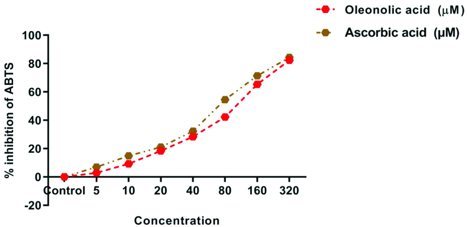
The ABTS radical cation decolorisation assay: OA demonstrated good antioxidant activity. At a concentration of 20 μM, it showed 18.38±0.04% inhibition, while ascorbic acid showed 21.02±0.07% inhibition. At a concentration of 320 μM, both showed around 82.33±0.22% and 84.30±0.06% inhibition of ABTS radical cations, respectively [Table/Fig-1,3].
Graph showing free radical scavenging activity of Oleanolic Acid (OA) by ABTS assay.
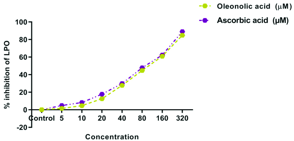
Lipid Peroxidation (LPO) assay: OA exhibited potent inhibitory effects on LPO, a major enzyme responsible for causing oxidative stress in the brain, which can lead to AD. At a concentration of 40 μM, both substances showed an inhibition of approximately 20%, with values of 12.55±0.17% and 17.78±0.04% inhibition of the LPO enzyme. At 320 μM, the inhibition was recorded as 84.82±0.08% for OA and 89.08±0.06% for ascorbic acid, respectively [Table/Fig-1,4].
Graph showing inhibition of lipid peroxidation of Oleanolic Acid (OA).
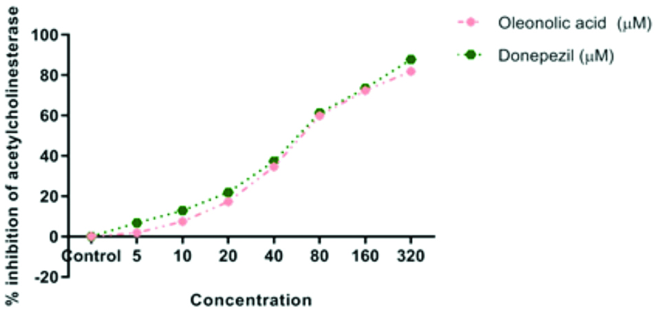
Acetylcholinesterase (AChE) inhibition assay: OA demonstrated a potent inhibitory effect on AChE, similar to donepezil, at a concentration of 20 μM, where it showed inhibition of 17.27±0.05% and 21.83±0.08%, respectively. Both OA and donepezil showed 81.78±0.07% and 87.76±0.07% inhibition at a concentration of 320 μM [Table/Fig-5,6].
Effect of Oleanolic Acid (OA) on in-vitro Acetylcholinesterase (AChE) activity, amyloid aggregation and β-secretase activity.
| Concentration (μM) | In-vitro Acetylcholinesterase (AChE) | % inhibition of amyloid aggregation | % inhibition of β-secretase activity |
|---|
| Oleanolic acid | Donepezil | Oleanolic acid | Donepezil | Oleanolic Acid (OA) | Donepezil |
|---|
| Control | 0.00±0.09 | 0.00±0.09 | 99.36±0.35 | 99.36±0.35 | -0.23±0.18 | -0.23±0.18 |
| 5 | 1.88±0.04*** | 6.73±0.06 | 99.03±0.02** | 96.72±0.06 | 3.67±0.06** | 5.37±0.05 |
| 10 | 7.48±0.03*** | 12.89±0.09 | 87.27±0.08** | 85.70±0.51 | 7.86±0.02*** | 10.93±0.06 |
| 20 | 17.27±0.05** | 21.83±0.08 | 68.36±0.02 | 67.89±0.29 | 14.77±0.09*** | 19.28±0.08 |
| 40 | 34.58±0.10** | 37.28±0.07 | 59.28±0.05** | 57.87±0.15 | 32.29±0.05** | 34.39±0.09 |
| 80 | 59.83±0.08** | 61.29±0.03 | 45.39±0.05** | 47.01±0.08 | 48.39±0.06*** | 58.82±0.02 |
| 160 | 72.37±0.08* | 73.40±0.05 | 29.84±0.04** | 27.66±0.15 | 65.92±0.08*** | 72.29±0.05 |
| 320 | 81.78±0.07*** | 87.76±0.07 | 14.63±0.04** | 12.33±0.24 | 81.28±0.06*** | 88.09±0.08 |
Results are presented as mean±SEM (n=3); Differences between means were evaluated using two-way ANOVA, followed by Holm-Sidak post-hoc analysis. Statistical significance was indicated as ***p-value <0.001, **p-value <0.01 and *p-value <0.05, compared to the standard (Donepezil) group
Graph showing inhibition of AChE by Oleanolic Acid (OA).
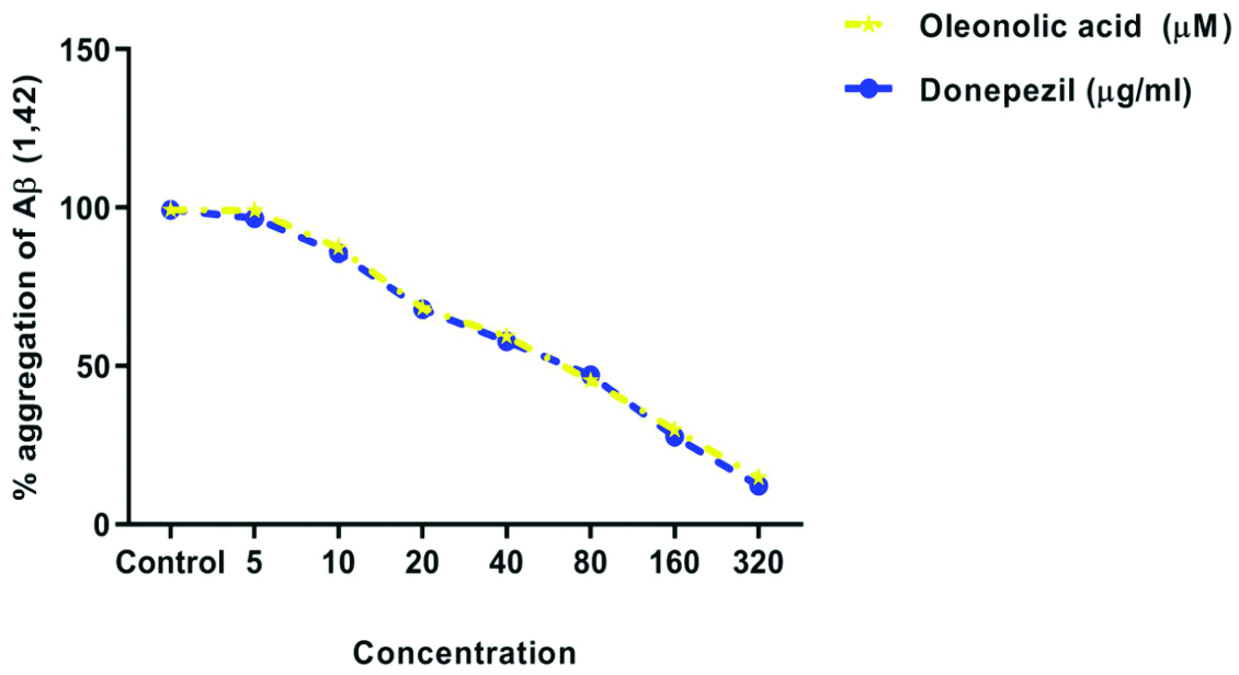
Amyloid-beta (Aβ) aggregation inhibition assay: From this graph, the authors can observe that OA and donepezil exhibit very similar effects regarding the inhibition of Aβ proteins. It is crucial to assess the inhibitory effect of the drug on Aβ protein aggregation, as this is one of the most significant features observed in a brain affected by AD. The percentage inhibition with OA and donepezil at 20 μM was 68.36±0.02% and 67.89±0.29%, respectively and at 320 μM, it was 14.63±0.04% and 12.33±0.24%, respectively [Table/Fig-5,7].
Graph showing effect of Oleanolic Acid (OA) Aβ proteins aggregation.
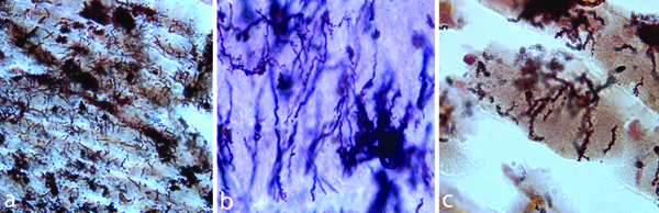
The following images [Table/Fig-8], magnified at 100,000x, show the effect of OA in the control group, indicating the amount of Aβ protein aggregation seen in AD. Donepezil, in conjunction with thioflavin T and amyloid-β, demonstrated a significant decrease in Aβ fibrillation. Similar results were observed with OA in combination with Thioflavin T and amyloid-β, indicating that OA is on par with standard donepezil.
Image represent the Aβ aggregation in: a) AD control; b) After treating with Oleanolic Acid (OA); c) After treating with donepezil.
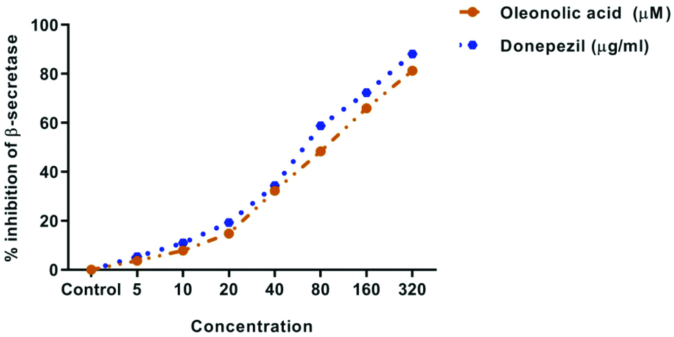
β-secretase inhibition assay: OA displayed a potent inhibitory effect on β-secretase. The percentage inhibition with oleanolic acid and donepezil at 20 μM was 14.77±0.09% and 19.28±0.08%, respectively and at 320 μM, it was 81.28±0.06% and 88.09±0.08%, respectively [Table/Fig-5,9].
Graph showing inhibition of β-secretase by Oleanolic Acid (OA).
Discussion
Alzheimer’s Disease (AD) is a degenerative neurological condition marked by worsening cognitive abilities, memory loss and behavioural issues. The key pathological processes involved include the accumulation of amyloid-beta (Aβ) plaques, hyperphosphorylation of tau proteins, oxidative stress and inflammation in the brain. Despite advances in understanding these mechanisms, effective treatments for AD are still lacking. Recent research suggests that OA, a naturally occurring triterpenoid present in various plants, shows potential neuroprotective properties that may be beneficial in addressing AD [14].
Ayeleso TB et al., 2017 have reported that OA exhibits anticancer effects, antidiabetic effects, antimicrobial effects on Listeria and Enterococcus bacteria, hepatoprotective activity, antihypertensive properties, antioxidant activity, anti-inflammatory potential and antiparasitic effects against Leishmania (L.) infantum [23]. Liu J et al., 2019 also demonstrated that OA is hepatoprotective at lower doses but can be hepatotoxic at higher doses [5]. The antioxidant and anti-Alzheimer’s effects of OA were observed using various assays and it was found to have significant antioxidant and anti-Alzheimer’s effects. From the results of the DPPH assay, ABTS assay and LPO assay, it is clear that OA has significant antioxidant effects when compared with ascorbic acid, a potent antioxidant. In both the DPPH and ABTS assays, it showed similar antioxidant effects to ascorbic acid at concentrations of 20 μM and 320 μM. Since OA displayed potent antioxidant effects in the DPPH, ABTS and LPO assays, we can confirm that it may be used to reduce oxidative stress in people suffering from neurodegenerative diseases.
OA also showed potent effects on inhibiting important enzymes involved in the mechanisms causing AD, such as ACh, β-Secretase and Aβ aggregation, almost on par with donepezil. Upon comparing the amyloid fibers observed in the control group with those treated with donepezil and OA, it was found that there was a reduction in the aggregation of amyloid beta protein, although this reduction was less substantial compared to that of donepezil. Upon analysing the results from the various assays, it can be confirmed that OA has a dose-dependent anti-Alzheimer’s effect, showing potent effects at higher concentrations, comparable to the standard drug. With all these inferences, it is clear that OA has potent antioxidant and anti-Alzheimer’s effects, as it was able to inhibit the major enzymes involved in the mechanisms causing AD [24].
Previous studies have also proven that OA has the effect of reducing oxidative stress, which is the main aetiology of AD [5]. The neuroprotective potential of OA was further demonstrated by research conducted by Gudoityte E et al., 2021 [25]. Zhang L et al., 2018 also proved that OA had an inhibitory effect on astrocytes, which are the predominant immunoregulatory cells in AD, thereby helping to reduce the neuroinflammation that causes neurotoxic injury in AD [26]. Terpenoids, such as ginsenosides extracted from Panax ginseng, were also found to promote the effect on Aβ proteins. Ginsenoside Rg3 specifically reduced Aβ production in CHO2B7 cells by 87% and by 31% in Tg2576 transgenic mice [27]. Δ9-tetrahydrocannabinol (THC), the major cannabinoid in Cannabis sativa, was found to have AChE-inhibiting activity and prevents AChE-induced Aβ aggregation, which is the main aetiology of AD [28,29]. Curcumin, found in turmeric, has demonstrated anti-inflammatory potential in various diseases, including arthritis, AD and other inflammatory conditions. It was also found to cause a significant reduction in Aβ aggregation [30]. Ocimum sanctum was found to have potent AChE-inhibiting properties and has the potential to be used as an anticholinergic agent [31].
In the present study, OA was compared with donepezil as a reference standard because donepezil is a well-established and widely used drug for the treatment of AD, recognised for its potent neuroprotective and antioxidative effects. Given its proven efficacy, donepezil serves as an ideal benchmark to evaluate the therapeutic potential of OA in both antioxidant and neuroprotective activities. Furthermore, focusing on a single, well-validated standard allows for a clearer and more precise assessment of OA’s comparative efficacy in these specific activities [32].
Limitation(s)
Due to the absence of elements such as the blood-brain barrier and systemic immunological responses, the present in-vitro study has intrinsic limitations that prevent it from fully replicating the complexity of real organisms. The doses of OA employed may not reflect realistic therapeutic levels in humans and potential long-term safety or cytotoxic consequences were not considered. Furthermore, focusing solely on oxidative stress ignores other critical pathways of Alzheimer’s pathogenesis, such as tau protein aggregation and neuroinflammation. The short duration of observation does not capture long-term or cumulative impacts. These limitations highlight the necessity for more in-vivo investigations and clinical trials to evaluate the therapeutic potential of OA in Alzheimer’s disease.
Conclusion(s)
The present study showed that OA possesses notable antioxidant and anti-Alzheimer’s properties. By reducing oxidative stress, a major contributor to neurodegeneration and targeting critical Alzheimer’s disease features such as amyloid-beta plaque buildup and tau protein hyperphosphorylation, OA demonstrates significant therapeutic potential. These results suggest that it may serve as a promising option for developing treatments that not only alleviate symptoms but also address the root causes of Alzheimer’s disease. Further investigation and clinical trials are needed to confirm its effectiveness and safety in humans.
Results are presented as mean±SEM (n=3); Differences between means were evaluated using two-way ANOVA, followed by Holm-Sidak post-hoc analysis. Statistical significance was indicated as ***p-value <0.001, **p-value <0.01 and *p-value <0.05, compared to the standard (ascorbic acid) group
Results are presented as mean±SEM (n=3); Differences between means were evaluated using two-way ANOVA, followed by Holm-Sidak post-hoc analysis. Statistical significance was indicated as ***p-value <0.001, **p-value <0.01 and *p-value <0.05, compared to the standard (Donepezil) group