Gender-based Morphometric Analysis of the Human Orbital Aperture Profile using 3D Reconstruction Computed Tomography: A Cross-sectional Retrospective Study
Muhammad Omer Afzal Bhatti1, Mujeeb UR Rehman Parrey2, Muhammad Ikram Ali3, Allah Rakhyo Shoro4, Abdulkrem Ahmed Alenazi5, Rayan Alhumaidi Alruwaili6
1 Lecturer, Department of Radiology, Northern Border University, Arar, Northern Border Region, Saudi Arabia.
2 Associate Professor, Department of Ophthalmology, Northern Border University, Arar, Northern Border Region, Saudi Arabia.
3 Associate Professor, Department of Forensic Medicine and Toxicology, Ziauddin University, Karachi, Pakistan.
4 Consultant, Department of Diagnostic Radiology, Tower Hospital, Arar, Northern Border Region, Saudi Arabia.
5 Medical Student, College of Medicine, Northern Border University, Arar, Northern Border Region, Saudi Arabia.
6 Medical Student, College of Medicine, Northern Border University, Arar, Northern Border Region, Saudi Arabia.
NAME, ADDRESS, E-MAIL ID OF THE CORRESPONDING AUTHOR: Dr. Mujeeb Ur Rehman Parrey, 7196 Ghazi Ash Shafi Al-Musaidiyah Unit No. 1, 73311-2931, Arar, Northern Border Region, Saudi Arabia.
E-mail: drparrey@gmail.com
Introduction
Sex estimation is a crucial aspect of forensic medicine and requires primary consideration when examining skeletal remains. Among the various anatomical features, the orbital region holds significant importance due to its distinct morphological variations. These variations provide valuable insights for sex determination, making orbital anatomy an indispensable tool in forensic investigations and analysis.
Aim
To measure and compare orbital apertures between males and females using 3D Computed Tomography (CT) in a sample from the Saudi population.
Materials and Methods
A cross-sectional retrospective study was conducted from January to June 2024 in the Radiology Department of Tower Hospital, Arar, Kingdom of Saudi Arabia, following the acquisition of ethical approval from the Local Bioethics Committee at Northern Border University. A total of 100 CT scans were randomly selected from the available dataset. The study encompassed CT scans of subjects from both genders, aged 18 years and above. However, CT scans of patients who had sustained head trauma or orbital injuries were excluded from the study. The data retrieved were analysed using Statistical Package for the Social Sciences (SPSS) version 23.0. All continuous data were tested for normality and expressed as Mean±Standard Deviation (SD). The significant differences in orbital measurements between males and females were assessed using the Independent t-test. A p-value of <0.05 was considered statistically significant.
Results
A total of 100 participants were included in the study, with the majority being females (52%). The mean age of male and female subjects was 40.5±13.19 years and 39.13±13.10 years, respectively. A statistically significant difference was found in right orbital width (p-value=0.011), left orbital area (p-value=0.04), and Interzygomatic Distance (IZD), with these measurements being higher in males compared to females (p-value=0.009).
Conclusion
The study revealed that males have greater orbital width, orbital area and IZD compared to females. These gender-specific variations in orbital and facial dimensions may play a crucial role in gender determination within forensic medicine.
Forensic anthropology, Interzygomatic distance, Orbital measurements
Introduction
Forensic anthropology is essential in addressing the challenges associated with identifying unknown human remains. Among the various skeletal structures, the orbital region stands out due to its morphological diversity and its potential role in sex differentiation. Accurate identification of human skeletal remains is vital in forensic investigations, with sex determination being a crucial initial step that influences subsequent analysis of age and stature [1]. Orbital anatomy and its morphological variations have far-reaching applications, particularly in forensic medicine, where they inform sex estimation and identification [2]. Understanding the complex morphology of the orbital region is essential for human identification [3].
Based on the skeleton or portions of the skeleton, several techniques are employed to estimate sex. Sex estimation is an integral and primary step in developing a reliable biological profile of skeletal remains. Accurate estimation of sex is vital for estimating age, ancestry and stature, as there are observable differences in ageing and growth patterns between sexes, along with variations in morphological traits related to ancestry [4]. Radiographs have been used to identify unidentified human remains since the early 1900s, with metric analysis on radiographs proving superior in terms of objectivity, accuracy and reproducibility [5]. The development of computer software has progressed significantly, as multiple segments of imaging of the bone can be obtained automatically; late-generation CT scanners can view even minor differences in contrast. Thus, it can be used as an effective tool for the anatomical measurement of a skeleton [6].
It is crucial to be aware of demographic variations in the skeleton while attempting to determine the sex of skeletal remains. Applying developed sex-estimating formulas to a sample from a different demographic does not yield the same level of accuracy. In addition to variations in environment, diet and culture, these distinctions are influenced by hormonal state [7].
The novelty of this study lies in its detailed exploration of orbital parameters using CT imaging to establish their utility in sex determination. By utilising cutting-edge imaging technology, the study will contribute to the growing body of forensic literature and provide a reliable methodology for human identification. Therefore, the present study aimed to measure and compare orbital apertures between genders in a Saudi sample to evaluate the potential of these measures for sex determination.
Materials and Methods
A cross-sectional retrospective study was conducted from January 2024 to June 2024 in the Radiology Department of Tower Hospital, Arar, Kingdom of Saudi Arabia. The study commenced following the acquisition of ethical approval (118/23/H) from the Local Bioethics Committee at Northern Border University. A total of 100 CT scans were randomly selected from the available dataset.
Inclusion criteria: CT scans of subjects from both genders, aged 18 years and above were included in the study.
Exclusion criteria: CT scans of patients who had sustained head trauma or orbital injuries were excluded from the study.
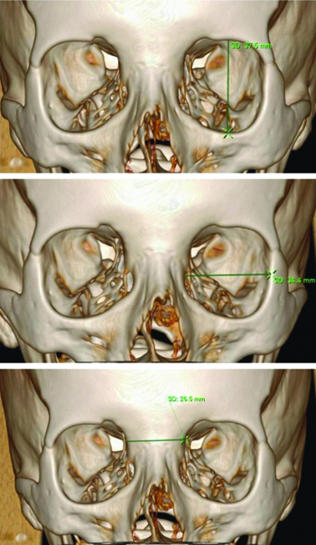
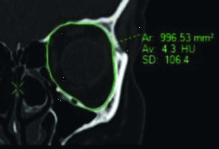
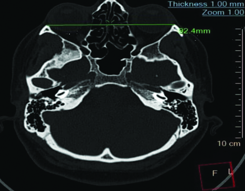
Measurement of orbital parameters: The measurements of orbital parameters, including orbital height, orbital width and interorbital distance (the distance between the medial walls of the orbits), in millimeters (mm) are illustrated in [Table/Fig-1]. The area of the orbit is given in mm2 [Table/Fig-2], and the IZD measured in millimeters, represents the maximum distance between the most prominent points on the right and left zygomatic arches [Table/Fig-3].
Top; (Orbital Height measured in millimeters). Middle; (Orbital Width measured in millimeters). Bottom; (Interorbital Distance, measured in millimeters).
Left orbital area (represented as mm2).
Interzygomatic Distance (measured in millimetres, representing the maximum distance between the most prominent points on the right and left zygomatic arches).
The orbital index (right and left) was calculated as the ratio of the orbit’s height to its width, multiplied by 100.
Statistical Analysis
The data retrieved were analysed using SPSS version 23.0. All continuous data were tested for normality and expressed as mean±SD. The significant differences in orbital measurements between males and females were assessed using the independent t-test. A p-value of <0.05 was considered statistically significant.
Results
A total of 100 participants were included in the study, comprising 52% females and 48% males. The mean age of male and female subjects was 40.5±13.19 years and 39.13±13.10 years, respectively. A statistically significant difference was found in right orbital width (p-value=0.011) between males and females [Table/Fig-4]. The comparison of coronal CT measurements between males and females revealed a significant difference in the left orbital area, with higher values in males than in their female counterparts (p-value=0.04) [Table/Fig-5]. The IZD was significantly higher in males than in females in the studied Saudi population, with a p-value of 0.009 [Table/Fig-6].
Comparison of 3D CT volume rendering orbital measurements between male and female subjects.
| Variable | Male | Female | p-value |
|---|
| Mean±SD |
|---|
| Age (years) | 40.5±13.19 | 39.13±13.10 | 0.997 |
| Right orbital height (mm) | 36.98±0.438 | 35.59±0.416 | 0.725 |
| Left orbital height (mm) | 36.93±0.438 | 35.6±0.446 | 0.834 |
| Right orbital width (mm) | 35.675±0.530 | 34.145±0.705 | 0.011* |
| Left orbital width (mm) | 35.67±0.567 | 34.210±0.68 | 0.072 |
| Right orbital index | 103.69±1.66 | 104.24±1.867 | 0.580 |
| Left orbital index | 103.55±1.65 | 104.09±1.797 | 0.485 |
| Interorbital distance (mm) | 25.938±0.479 | 24.522±0.421 | 0.462 |
Independent t-test; p-value <0.05*
Comparison of coronal CT measurements between male and female subjects.
| Variable | Male | Female | p-value |
|---|
| Mean±SD |
|---|
| Right orbital area (mm2) | 103.77±1.935 | 99.760±1.449 | 0.06 |
| Left orbital area (mm2) | 103.884±2.142 | 99.889±1.549 | 0.04* |
Independent t-test; p-value <0.05*
Comparison of axial CT measurements between male and female subjects.
| Variable | Male | Female | p-value |
|---|
| Mean±SD |
|---|
| Interzygomatic distance (mm) | 94.84±1.945 | 90.077±2.970 | 0.009* |
Independent t-test; p-value <0.05*
Discussion
The findings from the comparison of 3D CT volume rendering measurements reveal some noteworthy differences between male and female subjects in the studied Saudi population. The analysis of orbital dimensions shows that males have slightly higher mean values for both right and left orbital heights compared to females, but these differences are not statistically significant (p-values 0.725 and 0.834, respectively). Likewise, the findings of previous studies align with this study, showing no difference in the length of the orbit between males and females [8].
Contrary to the findings of the present study, previous studies have shown that the difference in orbital lengths was significantly different based on the gender of an individual. However, the indices were similar for the right and left orbits [9,10].
The findings of the study show that there was a significant difference in the right orbital width between males and females, with males having a wider mean right orbital width compared to females, as indicated by a p-value of 0.011. This suggests that male subjects tend to have a broader right orbital width. Consistent findings were observed in previous studies that reported that males had wider orbits than females [11,12].
A significant difference was observed in the left orbital area between males and females, with males showing a larger left orbital area compared to females, with a p-value of 0.04. The right orbital area was also larger in males, but this difference was not statistically significant (p-value=0.06). Similar findings were observed in studies conducted by El-Farouny RH et al., and Patra A et al., [6,13]. These findings suggest that males tend to have larger orbital areas, which might be attributed to the overall differences in craniofacial structures between genders.
The orbital index, which indicates the ratio between height and width, did not show any significant differences between males and females for either side. Similar findings were observed in a previous study on the Omani population, in which no significant difference was found between males and females regarding the orbital index [14].
The interorbital distance was found to be slightly greater in males compared to females, but this difference was not statistically significant (p-value=0.462). Similarly, IZD a crucial measurement indicating facial width, was significantly higher in males than in females (p-value=0.009). A study conducted on the Indian population also showed significantly higher values of IZD in males compared to females [15]. Moreover, these findings align with those of Kasaab N, who reported that the IZD in males is greater than in females [16].
This study provides important data on sex determination using digital CT in the Saudi Arabian population, which is a crucial aspect of forensic medicine and requires primary consideration when examining skeletal remains.
Limitation(s)
The study has limitations, particularly the single-centred approach, which limits the generalisability of the findings. Future studies should consider a multicentered approach to enhance the representativeness and applicability of the results across different populations.
Conclusion(s)
The study provides insightful evidence of distinct gender differences in both orbital and facial dimensions. These variations are significant and could have important implications for gender identification practices in the field of forensic medicine. The marked differences observed in the parameters of the orbits suggest that there are specific anatomical features that vary according to gender, particularly within the Saudi population. This indicates that understanding these anatomical differences is crucial for accurate gender estimation during forensic examinations, which can ultimately aid in criminal investigations and the resolution of unidentified remains.
Independent t-test; p-value <0.05*
Independent t-test; p-value <0.05*
Independent t-test; p-value <0.05*
Author Declaration:
Financial or Other Competing Interests: Funded by the Deanship of Scientific Research at Northern Border University, Arar, Saudi Arabia (Project number “NBU-FFR-2024-1531-01”).
Was Ethics Committee Approval obtained for this study? Yes
Was informed consent obtained from the subjects involved in the study? Yes
For any images presented appropriate consent has been obtained from the subjects. Yes
Plagiarism Checking Methods: [Jain H et al.]
Plagiarism X-checker: Oct 09, 2024
Manual Googling: Dec 12, 2024
iThenticate Software: Dec 14, 2024 (18%)
[1]. Ajanović Z, Ajanović U, Dervišević L, Hot H, Voljevica A, Talović E, A geometric morphometrics approach for sex estimation based on the orbital region of human skulls from Bosnian populationScanning 2023 2023(1):222313810.1155/2023/222313837089258PMC10121348 [Google Scholar] [CrossRef] [PubMed]
[2]. Krešić E, Bašić Ž, Jerković I, Kružić I, Čavka M, Erjavec I, Sex estimation using orbital measurements in the Croatian populationForensic Sci Med Pathol 2023 19(3):303-09.10.1007/s12024-022-00528-836151406 [Google Scholar] [CrossRef] [PubMed]
[3]. Diaconu SC, Dreizin D, Uluer M, Mossop C, Grant MP, Nam AJ, The validity and reliability of computed tomography orbital volume measurementsJ Craniomaxillofac Surg 2017 45(9):1552-57.10.1016/j.jcms.2017.06.02428747263 [Google Scholar] [CrossRef] [PubMed]
[4]. Krishan K, Chatterjee PM, Kanchan T, Kaur S, Baryah N, Singh RK, A review of sex estimation techniques during examination of skeletal remains in forensic anthropology caseworkForensic Sci Int 2016 261:165-e1.10.1016/j.forsciint.2016.02.00726926105 [Google Scholar] [CrossRef] [PubMed]
[5]. DiMichele DL, Spradley MK, Sex estimation in a modern American osteological sample using a discriminant function analysis from the calcaneusForensic Sci Int 2012 221(1-3):152-e1.10.1016/j.forsciint.2012.03.02622512944 [Google Scholar] [CrossRef] [PubMed]
[6]. El-Farouny RH, Hassanien SA, Azab RM, Morphometric evaluation of piriform and orbital aperture in sex discrimination by using computed tomography in Egyptian populationThe Egyptian J Forensic Sci App Toxicol 2021 21(1):01-02.10.21608/ejfsat.2021.54250.1182 [Google Scholar] [CrossRef]
[7]. Del Bove A, Menéndez L, Manzi G, Moggi-Cecchi J, Lorenzo C, Profico A, Mapping sexual dimorphism signal in the human craniumScientific Reports 2023 13(1):1684710.1038/s41598-023-43007-y37803023PMC10558540 [Google Scholar] [CrossRef] [PubMed]
[8]. Packirisamy V, Aljarrah K, Nayak SB, Morphometric evaluation of the orbital region for sex determination in a Saudi Arabian population using 3DCT imagesAnatomical Sci Int 2024 99(1):118-26.10.1007/s12565-023-00742-637721654 [Google Scholar] [CrossRef] [PubMed]
[9]. Botwe BO, Sule DS, Ismael AM, Radiologic evaluation of orbital index among Ghanaians using CT scanJ Physiol Anthropol 2017 36:01-06.10.1186/s40101-017-0145-728697737PMC5505022 [Google Scholar] [CrossRef] [PubMed]
[10]. Singh J, Ab Rahman R, Rajion ZA, Abdullah J, Mohamad I, Orbital morphometry: A computed tomography analysisJ Craniofacial Surg 2017 28(1):e64-70.10.1097/SCS.000000000000321827922969 [Google Scholar] [CrossRef] [PubMed]
[11]. Attia AM, Ghoneim M, Elkhamary SM, Sex discrimination from orbital aperture dimensions using computed tomography: Sample of Egyptian populationJ Forensic Radiology and Imaging 2018 14:32-38.10.1016/j.jofri.2018.08.007 [Google Scholar] [CrossRef]
[12]. Ghorai L, Asha ML, Lekshmy J, Rajarathnam BN, Kumar HM, Orbital aperture morphometry in Indian population: A digital radiographic studyJ Forensic Dent Sci 2017 9(2):61-64.10.4103/jfo.jfds_65_1629263609PMC5717774 [Google Scholar] [CrossRef] [PubMed]
[13]. Patra A, Singla RK, Mathur M, Chaudhary P, Singal A, Asghar A, Morphological and morphometric analysis of the orbital aperture and their correlation with age and gender: A retrospective digital radiographic studyCureus 2021 13(9):e1773910.7759/cureus.17739 [Google Scholar] [CrossRef]
[14]. Al Ajmi E, Al Subhi M, Al Maamari M, Al Dhuhli H, Sirasanagandla SR, Radiologic assessment of orbital dimensions among omani subjects: A computed tomography imaging-based study at a single tertiary centreSultan Qaboos University Medical Journal 2023 23(1):5510.18295/squmj.3.2022.02336865432PMC9974019 [Google Scholar] [CrossRef] [PubMed]
[15]. Singh A, Sreedhar G, George J, Shukla A, Vashishta V, Negi MP, Anthropometric study using craniofacial features to determine gender in Lucknow populationJ Forensic Dent Sci 2017 9(3):120-24. [Google Scholar]
[16]. Kassab N, Estimation of different facial landmarks as a guide in intercanine distance determination (Clinical Study)Al-Rafidain Dental Journal 2013 13(2):305-08.10.33899/rden.2020.165185 [Google Scholar] [CrossRef]