Images of Common Foreign Body at an Uncommon Site
Parindita Sarmah1, Sagar Gaurkar2, Prasad Deshmukh3
1 Junior Resident, Department of ENT, Jawaharlal Nehru Medical College, Sawangi (Meghe), Wardha, Maharashtra, India.
2 Professor, Department of ENT, Jawaharlal Nehru Medical College, Sawangi (Meghe), Wardha, Maharashtra, India.
3 Professor, Department of ENT, Jawaharlal Nehru Medical College, Sawangi (Meghe), Wardha, Maharashtra, India.
NAME, ADDRESS, E-MAIL ID OF THE CORRESPONDING AUTHOR: Dr. Parindita Sarmah, Junior Resident, Department of ENT, Jawaharlal Nehru Medical College, Sawangi (Meghe), Wardha, Maharashtra, India.
E-mail: parinditasharma@gmail.com
Dysphagia, Fish bone, Throat
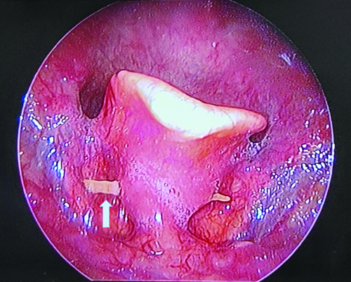
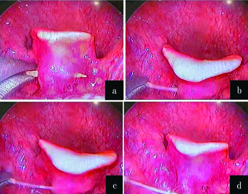
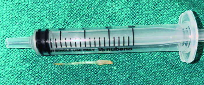
A 73-year-old male resident of Wardha presented to the Ear, Nose and Throat (ENT) Outpatient Department (OPD) with complaints of a foreign body sensation in his throat for the past eight days. He described ingesting a fish bone a week earlier and experiencing mild throat soreness. Subsequently, it developed into a persistent foreign body sensation in the throat and difficulty in deglutition, which did not subside with the intake of antacids and other throat lubricants. There was no history of fever following this incident. The posterior pharyngeal wall was found to be clear during the throat examination. Indirect laryngoscopy revealed that all hypopharyngeal structures were normal. However, during video [Video-1] direct laryngoscopy, a fish bone was discovered embedded in the lingual surface of the epiglottis [Table/Fig-1]. Upon receiving written consent, the embedded fish bone was removed with the help of long artery forceps under the guidance of video direct laryngoscopy [Table/Fig-2]. There was no evidence of bleeding, oedema, or congestion after the procedure. The foreign body measured approximately 1.5 cm and was causing the patient immense discomfort [Table/Fig-3]. The patient was prescribed a five-day course of antibiotics after the procedure, which included Tablet Amoxyclav 625 mg BD (Amoxicillin plus Clavulanic acid), Tablet Zerodol SP BD (Aceclofenac/Paracetamol/Serratiopeptidase), Tablet Pantop 40 mg OD (Pantoprazole), Tablet Levocet 10 mg HS (Levocetirizine), Syrup Mucaine gel 2 tsp BD (Aluminium hydroxide, magnesium hydroxide, and oxetacaine), and Betadine gargle TDS. He was advised to follow-up after five days to check for any delayed onset of postprocedure complications.
Embedded fish bone over the lingual surface of epiglottis.
a) Fish bone is held with a long artery forceps; b) Fish bone is pulled towards one side by a long artery forceps; c) Fish bone is subsequently pulled; d) Fish bone is removed from the lingual surface of epiglottis.
Post-removal of impacted fish bone measuring approx. 1.5 cm.
Foreign body impaction in the throat, particularly with fish bones, presents a common challenge in ENT clinics and emergency departments, especially in regions where fish consumption is prevalent. The case described highlights a typical scenario of a patient presenting with a foreign body sensation after ingesting a fish bone [1]. An impacted fish bone in the lingual surface of the epiglottis is indeed an uncommon and potentially serious situation. The epiglottis is a flap of tissue at the base of the tongue that prevents food and liquids from entering the airway during swallowing [2]. When a fish bone becomes lodged in the lingual surface of the epiglottis, it can cause significant discomfort, difficulty swallowing, and potentially compromise the airway if it obstructs breathing or triggers inflammation [2].
Symptoms of foreign body impaction in the throat typically include odynophagia (painful swallowing) and sharp pain upon swallowing. However, it’s essential to conduct a thorough evaluation, as symptoms may vary depending on the location and nature of the foreign body [3].
The diagnosis and management of such cases require careful history-taking, clinical examination, and the appropriate use of diagnostic tools such as laryngoscopy [4]. In this case, the patient’s symptoms persisted despite conservative measures, prompting further evaluation with video direct laryngoscopy, which revealed the embedded fish bone in the lingual surface of the epiglottis.
Alreefi M et al., reported a case of a migrating fish bone that got lodged in the right thyroid gland, causing a neck abscess. Incision and drainage of the abscess, along with the removal of the foreign body, and possibly a right hemithyroidectomy or total thyroidectomy, had to be performed [5].
Amirian A et al., presented a case of internal jugular vein injury after an episode of fish bone ingestion by a 65-year-old female, which required meticulous removal of the foreign body. This foreign body had caused a laceration in the medial aspect of the internal jugular vein, necessitating ligation distal to the site of injury [6].
Overall, prompt recognition and appropriate management of foreign body impaction in the throat are crucial to prevent potential complications such as perforation, infection, abscess formation, or airway compromise. A thorough clinical examination, often supplemented by endoscopic visualisation, is crucial in identifying the exact location of the impaction. Close follow-up after the removal of the foreign body is essential to monitor for any delayed onset of postprocedural complications. Raising awareness among clinicians regarding the presentation and management of such cases can improve patient outcomes and avoid unnecessary procedures.
Author Declaration:
Financial or Other Competing Interests: None
Was informed consent obtained from the subjects involved in the study? Yes
For any images presented appropriate consent has been obtained from the subjects. Yes
Plagiarism Checking Methods: [Jain H et al.]
Plagiarism X-checker: Jul 15, 2024
Manual Googling: Sep 14, 2024
iThenticate Software: Sep 16, 2024 (07%)
[1]. Klein A, Ovnat-Tamir S, Marom T, Gluck O, Rabinovics N, Shemesh S, Fish bone foreign body: The role of imagingInt Arch Otorhinolaryngol 2019 23(1):110-15.10.1055/s-0038-167363130647794 [Google Scholar] [CrossRef] [PubMed]
[2]. Scrase CD, Symonds P, Sino-nasal, oral, larynx and pharynx cancersWalter and Miller’s Textbook of Radiotherapy E-book: Radiation Physics, Therapy and Oncology 2012 :357 [Google Scholar]
[3]. Schaefer TJ, Trocinski D, Esophageal Foreign Body. [Updated 2023 Jan 30]In: StatPearls [Internet] 2024 Jan- Treasure Island (FL)StatPearls PublishingAvailable from: https://www.ncbi.nlm.nih.gov/books/NBK482131/ [Google Scholar]
[4]. Swain SK, Pani SK, Sahu MC, Management of fish bone impaction in throat. Our experiences in a tertiary care hospital of eastern IndiaEgyptian Journal of Ear, Nose, Throat and Allied Sciences 2017 18(1):27-30.10.1016/j.ejenta.2016.12.001 [Google Scholar] [CrossRef]
[5]. Alreefi M, Althawadi N, Patel A, Dwivedi R, A rare case of a migrating fishbone lodged in the larynx for 6 months in a patient with delayed presentation due to COVID-19 pandemicJ Surg Case Rep 2021 2021(5):rjab13110.1093/jscr/rjab13134025966 [Google Scholar] [CrossRef] [PubMed]
[6]. Amirian A, Ghoddusi Johari H, Karoobi M, Shahriarirad R, Ranjbar K, Internal jugular vein injury by fishbone ingestionCase Rep Med 2020 2020:918237910.1155/2020/918237932636881 [Google Scholar] [CrossRef] [PubMed]