Studies on the first rib have been conducted through inspection of dry bones as well as radiological examinations. The first rib is the shortest, most curved and atypical of all ribs. It consists of a head, neck, tubercle and shaft. The head of the first rib is small and round, featuring a circular articular facet. The neck is rounded and the tubercle has a medial oval articular facet. The angle and tubercle of the first rib coincide when it bends at the tubercle. The superior surface of the shaft is flat and crossed obliquely by two shallow grooves, which are separated by a slight ridge that ends at the internal border, where a small projection called the scalene tubercle is located. The anterior groove is associated with the subclavian vein, while the posterior groove is related to the subclavian artery and the lower trunk of the brachial plexus [1]. An angle visible between the head and the tubercle at the inferior portion of the neck, in a non anatomical position [2], can indicate the sex of the individual [3].
Congenital rib abnormalities are found in approximately 2% of the general population [4,5]. Pseudarthrosis of the first rib is a common finding in patients with severe calcifications. Radiologists should be aware of this frequent incidental finding, which should not be mistaken for pathology in Computed Tomography (CT) imaging following trauma [6]. The prevalence of these abnormalities can vary among different ethnic groups [7,8] or age categories [9,10]. Human first ribs demonstrate predictable, sequential changes in shape, size and texture with increasing age, thus serving as an indicator of age at death [11]. Rib measurements have also been utilised in biomechanical formulas for assessing respiration, truncal structure and identifying lateral asymmetry in the diagnosis of scoliosis [12]. There are no studies in the literature comparing the angle of the first rib between the right and left-sides in the Rajasthan population. With this background, the present study was conducted to observe and provide information about the morphometric and morphological variations of the first rib and to understand its clinical significance.
Materials and Methods
A cross-sectional study was conducted at SMS Medical College, Jaipur, in the Department of Anatomy, Rajasthan, India, from January 2023 to April 2024.
Inclusion criteria: Intact right and left first ribs were included in the present study.
Exclusion criteria: Damaged or broken first ribs and ribs with ossified first costal cartilage were excluded.
Study Procedure
A total of 90 adult human dry first ribs (45 right and 45 left) of unknown age and sex were studied. The following measurements were taken using a Vernier calliper and thread: length, height, angle, external length, internal length, shortest external length and shortest internal length of the first rib. Morphological variations, such as the scalene tubercle and vascular groove, were also observed. The angle of the rib was determined using the inverse sine function. This sine function was calculated in Microsoft Excel, using the formula Asin= Height/Length.
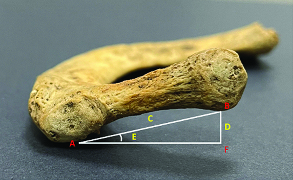
For the first rib, the following points were marked as illustrated in [Table/Fig-1]:
A- The point where the tubercle contacts the surface, B- The lowest point on the head of first rib, C=AB= Length from head to tubercle, D=BF= Height between head and surface, E= Angle of first rib, sin E=D/C= BF/BA.
The lowest point on the head.
The point where the tubercle contacts the surface.
Subsequent measurements were obtained in a non anatomical position:
Length: The distance between the head and the tubercle [Table/Fig-1].
Height: The distance between the head and the surface [Table/Fig-1].
Angle: The angle of the first rib was computed in Microsoft Excel using the inverse sine function [Table/Fig-1].
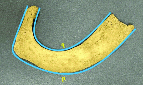
External length: Measured from the anterior sternal end to the lateral side of the head along the outer curvature of the first rib [Table/Fig-2].
Internal length: Measured from the posterior sternal end to the medial side of the head along the inner curvature of the first rib [Table/Fig-2].
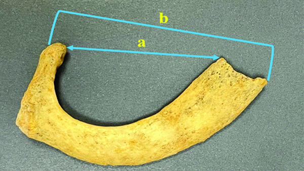
Shortest external length: The distance measured from the anterior sternal end to the lateral side of the first rib head [Table/Fig-3].
Shortest internal length: The distance measured from the posterior sternal end to the medial side of the first rib head [Table/Fig-3].
p-External length, q-Internal length of the first rib.
a- Shortest internal length of the first rib, b- Shortest external length of the first rib.
Variations regarding the scalene tubercle and vascular grooves were observed and recorded.
Statistical Analysis
The statistical analysis was performed using Microsoft Excel 2020 and IBM Statistical Package of Social Sciences (SPSS) version 23.0. It involved descriptive statistics to summarise the dataset’s characteristics and independent sample t-test were used to compare means between two groups (right and left-sides). Correlation analysis was conducted to assess the relationships between angular orientation and anatomical dimensions on both the right and left-sides, revealing patterns of association between variables.
Results
In the present study, the mean and Standard Deviation (SD) of the height and length on the right-side were 6.99±3.45 cm and 26.69±2.4 cm, respectively. The angle was greater on the left-side (16.59±6.18°). Statistical analysis indicated a non significant difference between the groups [Table/Fig-4].
Mean and standard deviation of various morphometric parameters of first rib.
| Parameters | Side | Mean±SD | t-test | p-value |
|---|
| Height (cm) | Right | 6.99±3.45 | -0.551 | 0.583 |
| Left | 7.35±2.55 |
| Length (cm) | Right | 26.69±2.41 | 1.070 | 0.287 |
| Left | 26.10±2.80 |
| Angle (degree) | Right | 15.14±7.27 | -1.016 | 0.313 |
| Left | 16.59±6.18 |
| External length (cm) | Right | 13.15±1.31 | 0.973 | 0.333 |
| Left | 12.88±1.34 |
| Internal length (cm) | Right | 8.85±1.00 | 1.642 | 0.104 |
| Left | 8.51±0.97 |
| Shortest external length (cm) | Right | 7.54±0.80 | 1.290 | 0.201 |
| Left | 7.34±0.68 |
| Shortest internal length (cm) | Right | 4.99±0.58 | 1.534 | 0.129 |
| Left | 4.80±0.63 |
p<0.05* statistically significant
Both sides exhibited strong positive correlations between angle and height (Right: r-value=0.986, p-value=0.001; Left: r-value=0.952, p-value=0.001) [Table/Fig-5,6].
Correlation of angle with various parameters of first rib of right-side.
| Parameters | r | p-value |
|---|
| Height | 0.986 | 0.001 |
| Length | 0.317 | 0.034 |
| External length | 0.352 | 0.018 |
| Internal length | 0.330 | 0.027 |
| Shortest external length | 0.356 | 0.016 |
| Shortest internal length | 0.254 | 0.092 |
p<0.05* statistically significant; Coefficient of correlation was used
Correlation of angle with various parameters of first rib of left-side.
| Parameters | r | p-value |
|---|
| Height | 0.952 | 0.001 |
| Length | -0.212 | 0.162 |
| External length | -0.349 | 0.019 |
| Internal length | -0.231 | 0.127 |
| Shortest external length | -0.198 | 0.192 |
| Shortest internal length | -0.153 | 0.314 |
p<0.05* statistically significant; Coefficient of correlation was used
Morphologically, 66.67% of the right ribs and 64.44% of the left ribs showed a normal scalene tubercle. The scalene tubercle was absent in 11.11% of the ribs on the right-side and in 22.22% of the ribs on the left-side [Table/Fig-7,8].
Variations of the scalene tubercle.
| Scalenetubercle | Right-side | Left-side |
|---|
| Frequency | Percentage | Frequency | Percentage |
|---|
| Present | 30 | 66.67% | 29 | 64.44% |
| Rudimentary | 10 | 22.22% | 6 | 13.33% |
| Absent | 5 | 11.11% | 10 | 22.22% |
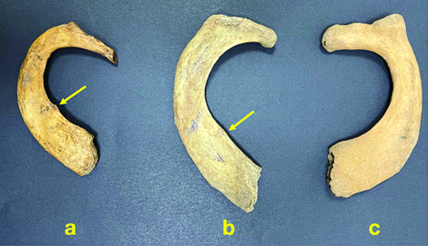
Variations of scalene tubercle: a- Present, b- Rudimentary, c- Absent.
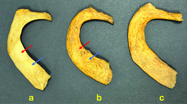
The present study also found that 71.11% of the right ribs and 75.56% of the left ribs displayed a normal vascular groove. The vascular groove was absent in 6.67% of ribs on the right-side and in 13.33% on the left-side [Table/Fig-9,10].
Variations of the vascular grooves.
| Vascular grooves | Right-side | Left-side |
|---|
| Frequency | Percentage | Frequency | Percentage |
|---|
| Present | 32 | 71.11% | 34 | 75.56% |
| Insignificant | 10 | 22.22% | 5 | 11.11% |
| Absent | 3 | 6.67% | 6 | 13.33% |
Variations of vascular groove: a- Present, b- Insignificant, c- Absent (red arrow- groove for subclavian artery, blue arrow- groove for subclavian vein).
Discussion
There have been very few studies focusing on the morphometric and morphological parameters of the first rib. In the present study, the mean angle for the right-side was found to be approximately 15.14°, while for the left-side, it was about 16.59°. D Souza A et al., found the mean angles of the rib on the right and left-sides to be 13.5° and 15.1°, respectively [3].
Elrod PW’s study indicated that there was a 60.2% chance that an individual’s sex could be ascertained solely by examining the angle of their first rib. If both the angle of the first rib and its entire length were considered, the chance increased to 70.5% [13].
In the current study, when analysing height, length, angle, external length, internal length, shortest external length and shortest internal length, the statistical analysis indicated no significant differences between the right and left-sides of the first rib (p-value> 0.05). This suggests that for these parameters, both sides exhibit similar characteristics. Compared to Elrod PW’s study, the values were lower, which might be attributable to racial differences. Elrod PW’s study showed that for females, the mean height of the first rib was 8.8 mm, the length was 23.3 mm and the angle was 22.5°, whereas for males, the mean height was 11.2 mm, the length was 25.4 mm and the angle was 26.4° [13].
Kubicka AM and Piontek J conducted a study for the estimation of sex in the Polish population. The exterior edge, anteroposterior diameter of the sternal end and depth of the arc were the most reliable parameters. Their results suggested that the first rib is dimorphic and that the described method can be used for sex assessment [14].
Lynch JJ et al., examined the use of the first rib for determining sex and reported that combining metric and geometric morphometric variables could achieve correct sex classification in European Americans with an accuracy of up to 88.05% and in African Americans with up to 70.86%. This underscores the influence of ancestry and race on rib characteristics [15].
Krogman W reported that the pelvic girdle, long bones and skull were the main areas of focus for sex assessment through the analysis of sexually dimorphic characteristics. However, in situations where the skull and pelvis are too damaged to analyse or are not available for investigation, the ribs may offer an alternative method for estimating an individual’s sex [16].
In the present study, the authors observed morphological variations such as the scalene tubercle and vascular grooves. The authors found that 11.11% of the ribs lacked a scalene tubercle on the right-side and 22.22% on the left-side. A rudimentary tubercle was reported in 22.22% of the right ribs and 13.33% of the left ribs, whereas vascular grooves were absent in 6.67% of the right ribs and 13.33% of the left ribs. Pillai NR et al., observed that nearly 50% of the ribs had either absent or insignificant vascular grooves. Moreover, rudimentary heads or tubercles were absent in 97.1% of right ribs and 91.4% of left ribs, with rudimentary or absent scalene tubercles found in approximately 50% of the ribs [17].
Rashia S and Zaidi SHH examined 50 first ribs and found that 46% of the ribs lacked scalene tubercles, 28% were missing vascular grooves and rudimentary tubercles and heads were present in 12% and 5.7% of the ribs, respectively [18].
Keerthi S et al., observed that scalene tubercles were absent in 13.75% of the cases, rudimentary in 23.33% and hypertrophied in 1.67% of the cases. The vascular groove was absent in 11.25% of the cases and the oblique ridge was absent in 5.83%. Additionally, the head and tubercle were observed to be rudimentary in 16.67% and 12.5% of the cases, respectively [19].
The first rib is less fragile than other skeletal elements, such as the pubic symphysis and is therefore more likely to survive in archaeological and forensic contexts [20]. Structural malformations of the first rib are common and when present, they may help in understanding the clinical significance of compression of the neurovascular bundle at the root of the neck. Awareness of the morphology of the first rib is important for anatomists, radiologists and thoracic surgeons dealing with this region.
Limitation(s)
The main limitation of the study was the relatively small sample size. The results would be more meaningful if the first ribs belonged to individuals of known age and sex.
Conclusion(s)
The morphometric parameters indicate non significant differences between the right and left-sides. Both sides exhibit strong positive correlations between angle and height, suggesting that as the angle increases, so does the height. This indicates a consistent relationship between angular orientation and vertical dimension across both sides of the observed entities. Further research is required on the first rib to supplement this information and assist anthropologists in identifying the age and sex of an individual.
p<0.05* statistically significant
p<0.05* statistically significant; Coefficient of correlation was used
p<0.05* statistically significant; Coefficient of correlation was used