Exencephaly: A Visual Insight into Cranial Anomaly
Mohini Gokuldas1, Dipak Kolate2, Meenal Patvekar3
1 Resident, Department of Obstetrics and Gynaecology, Dr. D. Y. Patil Medical College, Hospital and Research Centre, Pimpri, Pune, Maharashtra, India.
2 Associate Professor, Department of Obstetrics and Gynaecology, Dr. D. Y. Patil Medical College, Hospital and Research Centre, Pimpri, Pune, Maharashtra, India.
3 Head, Department of Obstetrics and Gynaecology, Dr. D. Y. Patil Medical College, Hospital and Research Centre, Pimpri, Pune, Maharashtra, India.
NAME, ADDRESS, E-MAIL ID OF THE CORRESPONDING AUTHOR: Dipak Kolate, Associate Professor, Department of Obstetrics and Gynaecology, Dr. D. Y. Patil Medical College, Sant Tukaram Nagar, Pimpri, Pune-411018, Maharashtra, India.
E-mail: dr.dipakkolate@gmail.com
Acrania, Neural tube defect, Ultrasonography
A 30-year-old, Gravida 3, parity 1, living 1, abortion 1 with previous lower segment Caesarean section came to the labour room with complaints of pain in the abdomen for one day. She has been married for seven years, with her first child being six years of age and one abortion was done three-year-ago. She has no co-morbidities and was vitally stable. On examination of per abdomen, the uterus was palpable and per vagina examination revealed os was closed.
Ultrasonography suggested a single live intrauterine gestation of 15 weeks four days, cranial vault is absent [Table/Fig-1], cephalad to the orbits with brain tissue herniating into the amniotic fluid suggestive of Exencephaly. After inducing labour, an abortus was delivered weighing 62 g with exencephaly as shown in [Table/Fig-1,2].
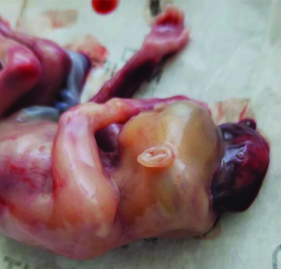
An abortus with exencephaly.
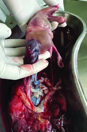
Exencephaly, a rare and devastating malformation of the neural tube, is characterised by the absence of the cranial vault, resulting in a large protrusion of brain tissue beyond the protective barrier of the skull [1]. Exencephaly is incompatible with extrauterine life and often precedes anencephaly, a severe neural tube defect where the base of the brain and facial structures are present, unlike in exencephaly. Typically associated with stillbirth, exencephaly can rarely be seen in live full-term foetuses, but their survival rate is extremely low [1].
The exposed brain tissue in exencephaly, with its shallow, disorganised gyri and sulci, is highly vascular and lacks proper neuronal differentiation, contributing to the poor prognosis of affected individuals [2].
Treatment options for exencephaly are limited due to the severe nature of this condition. The absence of the calvarium, which is a defining feature of exencephaly, complicates treatment options further, as it exposes the protruding brain tissue to external elements and increases the risk of infections and damage [1]. Exencephaly involves the formation of a brain without a protective skull or meninges, exposed to amniotic fluid. This exposure leads to the brain’s disintegration and prevents the development of the skull and cerebral hemispheres, resulting in anencephaly [3]. Effective preventive measures, particularly folic acid supplementation, play a vital role in reducing the risk of such congenital anomalies [3].
Comprehensive counselling and emotional support for affected families are essential components of managing this condition, helping them navigate the complex and challenging decisions they face. Continued research and public health efforts remain critical in preventing and understanding neural tube defects like exencephaly.
Author Declaration:
Financial or Other Competing Interests: None
Was informed consent obtained from the subjects involved in the study? Yes
For any images presented appropriate consent has been obtained from the subjects. Yes
Plagiarism Checking Methods: [Jain H et al.]
Plagiarism X-checker: Jul 09, 2024
Manual Googling: Jul 27, 2024
iThenticate Software: Aug 08, 2024 (6%)
[1]. Renuka IV, Sasank R, Devi SI, Vasundhara M, Exencephaly in a live, full term fetusJ Pediatr Neurosci 2009 4(2):134-36.Available from: https://www.ncbi.nlm.nih.gov/pmc/articles/PMC3162784/ [Google Scholar]
[2]. Oria M, Duru S, Figueira RL, Scorletti F, Turner LE, Fernandez-Alonso I, Cell necrosis, intrinsic apoptosis and senescence contribute to the progression of exencephaly to anencephaly in a mice model of congenital chranioschisisCell Death Dis 2019 10(10):01-11.[cited 2022 Dec 6]. Available from: https://www.nature.com/articles/s41419-019-1913-6/ [Google Scholar]
[3]. Bhusal A, Shrestha M, Rayamajhi A, Bista M, Das A, Discordant exencephaly in case of a twin delivery: A case reportAnn Med Surg (Lond) 2023 86(1):598-601.PMCID: PMC1078340410.1097/MS9.000000000000158938222692 [Google Scholar] [CrossRef] [PubMed]