Clinical Image of Tuberculosis Verrucosa Cutis: A Tropical Trouble
C Balakumaran1, NR Vignesh2, D Manoharan3, M Athira Deepthi4
1 Assistant Professor, Department of Dermatology, Venereology and Leprosy, Sree Balaji Medical College and Hospital, Chennai, Tamil Nadu, India.
2 Associate Professor, Department of Dermatology, Venereology and Leprosy, Sree Balaji Medical College and Hospital, Chennai, Tamil Nadu, India.
3 Professor, Department of Dermatology, Venereology and Leprosy, Sree Balaji Medical College and Hospital, Chennai, Tamil Nadu, India.
4 Junior Resident, Department of Dermatology, Venereology and Leprosy, Sree Balaji Medical College and Hospital, Chennai, Tamil Nadu, India.
NAME, ADDRESS, E-MAIL ID OF THE CORRESPONDING AUTHOR: Dr. D Manoharan, 7, CLC Works Road, Chromepet, Chennai-600044, Tamil Nadu, India.
E-mail: drmanomd@yahoo.co.in
Cutaneous tuberculosis, Directly observed therapy shortcourse, Prosector’s wart, Verrucous plaque
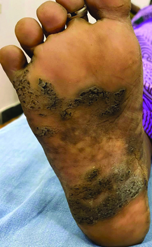
A 40-year-old female house help residing in Chennai, Tamil Nadu, presented with a rough, elevated lesion on her right sole that had persisted for 10 years, following a penetrating thorn prick injury. The lesion slowly evolved from a small, asymptomatic papule on the insole into a well-defined plaque extending across the instep of the right sole. Cutaneous examination revealed a well-defined, solitary, hyperpigmented, verrucous plaque with peripheral crypts and central clearing over the right plantar arch, approximately measuring 5×10 cm [Table/Fig-1]. Additionally, palpable, firm, non matted, and tender right superficial inguinal nodes were noted.
Image of right foot showing a solitary, hyperpigmented, verrucous plaque with peripheral crypts and central clearing over the plantar arch.
While psoriasis vulgaris was considered due to the involvement of the instep, the unilateral presentation and resistance to topical treatment argued against this diagnosis. Superficial fungal infections were ruled out by a negative potassium hydroxide mount. The chronicity, history of trauma, lymphadenopathy, and clinical features pointed towards a diagnosis of Tuberculosis Verrucosa Cutis (TBVC), which was confirmed by a positive Mantoux test and histopathological examination showing epithelioid granulomas with Langhans giant cells. The patient was planned to be started on antitubercular therapy but was lost to follow-up.
The diagnosis of a verrucous plaque on the foot can be challenging due to the similar presentations of various conditions, such as cutaneous tuberculosis, chromoblastomycosis, verruca vulgaris, psoriasis vulgaris, and tinea pedis. However, maintaining a high suspicion index for cutaneous tuberculosis is essential in endemic and tropical regions with a high prevalence of tuberculosis, as approximately one-third of cases are associated with systemic involvement [1].
TBVC, or warty tuberculosis, caused by Mycobacterium tuberculosis, is a chronic exogenous, paucibacillary form of cutaneous tuberculosis [2]. It usually occurs in previously sensitised immunocompetent individuals, often following trauma, and typically presents as painless papules or plaques on the feet and lower extremities, especially in tropical and developing countries. In developed countries, it is commonly seen on the hands of anatomists, butchers, or prosectors [2]. The plaque commonly exhibits slow peripheral extension and is susceptible to secondary infections, while central involution and scarring may occur rarely. Antitubercular therapy with isoniazid, rifampicin, ethambutol, and pyrazinamide is administered daily for two months, followed by isoniazid and rifampicin for four months, with expected evident resolution in 4 to 8 weeks. Multidrug resistant tuberculosis should be considered in cases where there is little resolution after four weeks of therapy.
Although cutaneous tuberculosis is rarer now, the incidence remains significant in endemic tropical regions, particularly among individuals engaged in practices that predispose them to trauma [3]. There is a difficulty in differentiating TBVC from other entities due to the low sensitivity and specificity of confirmatory investigations [4]. Nevertheless, it is imperative to initiate treatment, as the response to treatment may serve as a diagnostic criterion. This case highlights the need for every physician to consider cutaneous tuberculosis as a differential diagnosis in patients presenting with a chronic or recalcitrant verrucous plaque, especially in tropical countries, where early recognition is crucial to prevent further transmission and progression of the lesion.
Author Declaration:
Financial or Other Competing Interests: None
Was informed consent obtained from the subjects involved in the study? Yes
For any images presented appropriate consent has been obtained from the subjects. Yes
Plagiarism Checking Methods: [Jain H et al.]
Plagiarism X-checker: Jun 17, 2024
Manual Googling: Jul 20, 2024
iThenticate Software: Aug 10, 2024 (4%)
[1]. Kaul S, Kaur I, Mehta S, Singal A, Cutaneous tuberculosis. Part I: Pathogenesis, classification, and clinical featuresJ Am Acad Dermatol 2023 89(6):1091-103. [Google Scholar]
[2]. Sari TI, Rahardja JI, Utami AP, Pasaribu SD, Tuberculosis verrucosa cutis in immunocompetent patient: A case report: Tuberculosis verrucosa cutis: A case reportJ Pak Assoc Dermatol 2024 34(1):311-13. [Google Scholar]
[3]. Angreni AR, Adriani A, Amin S, Wahab S, Budhiani S, Massi MN, The many faces of tuberculosis cutis in Makassar, Indonesia-A case seriesEnferm Clin 2020 30:250-54. [Google Scholar]
[4]. Septiafni K, Pamudji R, Rusmawardiana R, Argentina F, Tuberculosis verrucous cutis mimicking chromoblastomycosis: A case report and diagnostic challengesInt J Mycobacteriol 2022 11(2):208-10. [Google Scholar]