Roth Spots in Sickle Cell Trait: Association or Chance Spot
K Harshitha Reddy1, Manjeet Kothari2, Vijaya Mallareddy3, Sunil Kumar4
1 Postgraduate Resident, Department of Medicine, Jawaharlal Nehru Medical College, Datta Meghe Institute of Higher Education and Research, Wardha, Maharashtra, India.
2 Postgraduate Resident, Department of Medicine, Jawaharlal Nehru Medical College, Datta Meghe Institute of Higher Education and Research, Wardha, Maharashtra, India.
3 Postgraduate Resident, Department of Ophthalmology, Jawaharlal Nehru Medical College, Datta Meghe Institute of Higher Education and Research, Wardha, Maharashtra, India.
4 Professor, Department of Medicine, Jawaharlal Nehru Medical College, Datta Meghe Institute of Higher Education and Research, Wardha, Maharashtra, India.
NAME, ADDRESS, E-MAIL ID OF THE CORRESPONDING AUTHOR: Dr. K Harshitha Reddy, Postgraduate Resident, Department of Medicine, Jawaharlal Nehru Medical College, Datta Meghe Institute of Higher Education and Research, Wardha-442004, Maharashtra, India.
E-mail: harshureddy95@gmail.com
Haemorrhages, Retinopathy, Sickle cell anaemia
Roth spots are white-centred retinal haemorrhages that are a hallmark of infective endocarditis caused by septic emboli inside the retina [1-3]. Several theories have been postulated on the pathophysiology, but the most widely recognised is that endothelial cell dysfunction causes retinal capillary rupture and intraretinal haemorrhage due to platelet-fibrin plugs. Visual disruption caused by Roth spots is uncommon but can occur when the macula is involved. Treatment is frequently supportive and directed at the underlying reason. Sickle cell trait (AS pattern) is a benign disorder characterised by only one defective haemoglobin beta gene allele in the affected individual. These individuals do not have sickling symptoms and, as a result, have a higher quality of life than patients with sickle cell disease [2,3].
A 20-year-old girl presented with complaints of floaters and blurring of vision for one month. She had been diagnosed with sickle cell trait (AS pattern) since childhood. On general examination, pallor, icterus, cyanosis, clubbing, lymphadenopathy, and pedal oedema were absent. The patient had no other co-morbidities. On physical examination, the patient was afebrile, with a blood pressure of 120/80 mmHg, a pulse rate of 78/min, and a respiratory rate of 16/minute. Her cardiac system S1 and S2 were heard. Her respiratory, gastrointestinal, and neurological systems were normal.
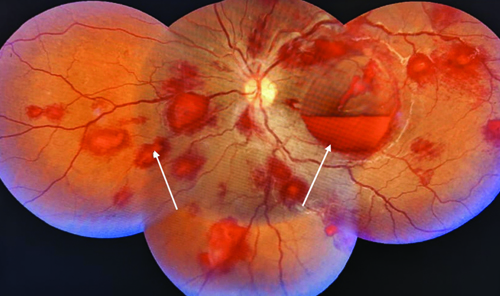
Due to her visual complaints, an ophthalmology referral was made, and a fundus examination revealed multiple haemorrhages with white centres in the retinas of both eyes, indicating Roth spots [Table/Fig-1].
Multiple haemorrhages with white centres in the retinas of both eyes, indicating Roth spots with white arrows (Montage fundus).
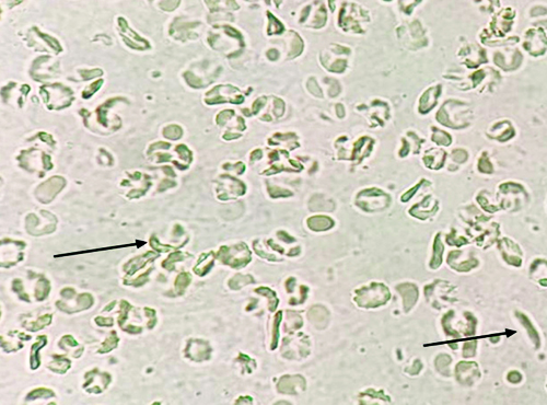
On investigations, haemoglobin was 8 g/dL, white blood count was 4200 cumm, and thrombocytopenia was 70,000 cumm. Peripheral blood smears showed microcytic hypochromic Red Blood Cells (RBCs) and sickle-shaped RBCs [Table/Fig-2]. Serum lactate dehydrogenase was elevated (386 U/L, normal <250 U/L). Other laboratory parameters like liver and renal function tests were within the reference range. A 2D echocardiography did not reveal any vegetation. The patient was asked to follow-up in the Ophthalmology Department every three months or if the patient had any complaints of loss of vision, but the patient was lost to follow-up.
RBC sickling (AS pattern) pointed with a black arrow.
Roth spots are white-centred retinal haemorrhages that are a hallmark of infective endocarditis caused by septic emboli inside the retina [4,5]. Infective endocarditis, severe anaemia, white blood and platelet disorders, collagen vascular disease, hypertensive and diabetic retinopathy, and hypoxia are all causative factors of Roth spots [5,6].
A known complication of sickle cell disease is papillary cell necrosis due to increased concentration in the medulla as it can reach 1200 mOsm/kg from the counter-current multiplier mechanism, and also the total blood flow to the medulla is less than ten percent, causing hypoxia. This causes water to pull out of cells, leading to increased red cell polymerisation, acidity, and pH [5].
The exact pathogenesis of Roth spot in sickle cell trait is unknown; however, the proposed hypothesis may be RBC polymerisation due to hypoxia, increased viscosity, and dehydration [5,6]. The retina has an end artery supply by the central retinal artery, so in sickle cell trait, it may have low oxygen tension significantly increased with dehydration, leading to hypoxia, ischaemia, and haemorrhage, as happened in the present case.
Author Declaration:
Financial or Other Competing Interests: None
Was informed consent obtained from the subjects involved in the study? Yes
For any images presented appropriate consent has been obtained from the subjects. Yes
Plagiarism Checking Methods: [Jain H et al.]
Plagiarism X-checker: Aug 11, 2023
Manual Googling: Oct 26, 2023
iThenticate Software: Dec 04, 2023 (8%)
[1]. Chandra A, Chakraborty U, Ganai S, Ray AK, Roth spots in acute myeloid leukaemiaBMJ Case Rep 2020 13(9):e238133 [Google Scholar]
[2]. Li EJ, Carroll VG, Sickle cell trait and renal papillary necrosisClin Pediatr (Phila) 2014 53(10):1013-15.Epub 2014 May 710.1177/000992281453341824807983 [Google Scholar] [CrossRef] [PubMed]
[3]. Ruddy SM, Bergstrom R, Tivakaran VS, Roth spotsIn StatPearls [Internet] 2022 StatPearls Publishing [Google Scholar]
[4]. Zehetner C, Bechrakis NE, White centered retinal hemorrhages in vitamin B12 deficiency anaemiaCase Rep Ophthalmol 2011 2(2):140-44. [Google Scholar]
[5]. Mahajan J, Talwar D, Kumar S, Acharya S, Kakade Y, ECAST syndrome (Exercise Collapse Associated with Sickle Cell Trait): First knock of sickle cell in a young bodybuilderJ Pharm Res Int 2021 33(Issue 56A):74-48.10.9734/JPRI/2021/v33i56A33887 [Google Scholar] [CrossRef]
[6]. Sundd P, Gladwin MT, Novelli EM, Pathophysiology of sickle cell diseaseAnnu Rev Pathol 2019 14:263-92. [Google Scholar]