The reason for restoring teeth that have undergone endodontic treatment is based on the fact that these teeth are more prone to damage due to their reduced moisture content and increased brittleness. When a tooth undergoes endodontic treatment, it loses both internal and external structure, making it less resistant to stress and a poor candidate for supporting dental work. Additionally, these treated teeth are more susceptible to developing additional decay due to the loss of neural stimuli [1].
Historically, numerous methods for restoring endodontically treated teeth have been employed. It is always advisable to reinforce endodontically treated teeth before placing crown or bridge or using them as abutments [2]. The primary purpose of utilising a post is to provide support for a core that replaces the lost coronal tooth structure and ultimately retains the permanent coronal restoration [3]. Variables such as post length, diameter, design and surface configuration, as well as, type of luting cement used influence the post’s ability to perform optimum function [1,3,4].
The evolution from the Richmond crown led to the development of cast post and cores, which have been found to be highly effective for treating endodontically treated teeth with moderate-to-severe coronal damage [4,5]. Historically, metallic posts have been more widely used for restoring endodontically treated teeth. They were used more commonly because of their favourable physical properties. One factor that has, over the years, reduced their use is ‘Aesthetics’. Metal posts may be visible through translucent ceramic restorations, which can cause the zenith line to appear dark. To address this, white and/or translucent posts have been developed [6].
Currently, aesthetic tooth-coloured prefabricated fibre-reinforced posts are perceived as promising alternatives to metal post especially with the increasing use of aesthetic restorations [7]. Researchers claim several advantages of tooth-coloured fibre post that include improved aesthetics; through increasing the transmission of light within the root and overlying gingival tissues, thereby eliminating the dark appearance that is often associated with the use of a metal post [8]. Moreover, the modulus of elasticity of fibre post is close to that of dentine, which reduces the risk root fracture of endodontically treated teeth as a result of even distribution of forces in the root. In addition, aesthetic fibre posts have shown the capability of bonding to dentine as well as, to resin composite core material with the use of adhesive systems and resin cements [7].
The literature reports that fibre post may have the potential of reinforcing endodontically treated teeth [8]. The flexible nature of these materials is believed to enable teeth to bend under pressure, which helps distribute stress more evenly between the dental post and the surrounding dentin. This can decrease the likelihood of the root fracturing. However, it’s important to note that stress could still build up between the cement and the endodontic post, potentially raising the risk of adhesive failure. Some experts also argue that using flexible materials might lead to the development of additional decay around the edges of the final restorations, especially on the palatal area of the front teeth [9]. However, very few clinical studies focused on the survival of aesthetic posts in the maxillary anterior region [10]. Additionally, none of the studies have compared the clinical performance of Zircon-enriched silicon fibre and glass fibre-reinforced posts.
This prospective clinical study aimed to evaluate the survival rate of endodontically treated teeth restored using two different aesthetic post systems over a 12-month period in the maxillary anterior region. The null hypotheses for the study were that there would be no difference in the clinical service provided by the two types of aesthetic posts.
Materials and Methods
The prospective intervention study was conducted in the Department of Prosthodontics, Manav Rachna Dental College (Faridabad) for a period of 12 months from “Nov 2022- Oct 2023”. During this study the standard guidelines by world Medical Association Declaration of Helsinki regarding ethical conduct of research involving human subjects and/or animals were followed appropriately. Institutional ethical clearance was obtained before initiating the study, Ref No. (MRDC/IEC/2022/533). All the patients who fullfilled the eligibility criteria were informed about the study in their native language and only the willing patients were asked to sign the Participant Informed Consent Form (PICF).
Inclusion criteria: For cases where a post-retained crown was needed in single-rooted maxillary anterior teeth, adequately obturated root canal with absence of any periapical pathology, perforation and root fracture. Healthy and stable periodontium around the tooth, with no bleeding on probing and No mobility of the endodontically treated tooth were selected.
Exclusion criteria: Endodontically treated teeth with occlusal problems, periapical/periodontal pathology and fixed dental prosthesis opposite to the tooth to be restored.
Sample size estimation: Most studies have shown that there is no difference in survival rates between post types [8,9,11,12]. Therefore, the sample size calculation was performed based on the assumption that the standard and experimental treatments are equivalent. This calculation determined that at least 40 participants (teeth) were required to ensure a 90% confidence interval.
A total of 40 patients with endodontically treated maxillary teeth were enroled for this clinical study. The selected patients were split into two groups of 20 each. They were mostly from the local population in the age group of 15-55 years.
Group-I consisted of 20 clinical cases restored with zircon-enriched silicon fibre posts (EASYPOST: DENTSPLY INDIA) which has cylindrical-conical form and a longitudinal modulus of elasticity and shear strength close to that of dentine. This was followed by a core build-up with composite resin and a porcelain fused-to-metal crown.
Group-II consisted of 20 clinical cases restored with glass fibre-reinforced posts (Tenax Fibre Trans: Coltene/Whaledent) which has a cylindrical-conical form with 4% taper in the bottom third and modulus of elasticity similar to dentine. This was followed by a core build-up with composite resin and a porcelain fused-to-metal crown.
Single-rooted maxillary anterior teeth that were obturated and showed no evidence of pathology, perforation, root fracture with a minimum apical seal of 4 mm were selected for the study. Teeth were not taken into consideration if there was any obvious occlusal interference or presence of mobility in the tooth to be restored.
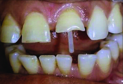
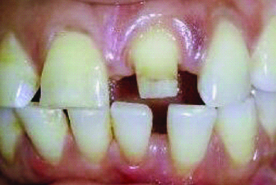
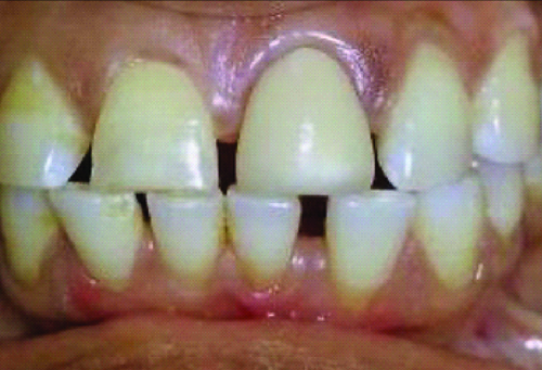
Clinical procedure: The suitability of each tooth for post placement was determined by conducting clinical and radiographic examinations before canal preparation. Based on the preoperative radiograph, an appropriate size of glass fibre post was selected for each canal. Isolation of the tooth was done using a rubber dam and post space preparation was initiated at least two days after obturation, using a piezo reamer drill at a speed of 5000 rpm to a depth that leaves a minimum of 4 mm apical seal. The post length was assessed and noted, then the adjusted post was positioned in the canal to validate its length. The root dentin was then etched with a 37% phosphoric acid gel. A primer (Rely a bond, Reliance Products USA) was applied with a micro brush and the adhesive was light-cured. Adhesive resin cement (RelyX ARC, 3M ESPE, USA) was dispensed into the root canal, the aesthetic endodontic post was placed appropriately and the cement was light-cured [Table/Fig-1]. The alignment of the post was confirmed with the help of a radiograph. Thereafter, core was built up by adding composite resin in bulk increments of no more than 2 mm2, and then shaped to accommodate a metal coping [Table/Fig-2]. All teeth were prepared for full coverage crown restorations by creating a ferrule of 2 mm. Gingival retraction was done using gingival retraction cord (ROEKO Stay-put No. 0) and impressions were made of each tooth using Polyvinyl Siloxane (PVS) impression material. (Affinis, Coltene-Whaledent India). Lab fabrication of the crown was initiated and till that time a temporary crown of polycarbonate was cemented with the temporary cement (TempBond Kerr, Italy). The final prepared and glazed crowns were cemented using zinc phosphate cement [Table/Fig-3]. A radiograph was obtained after each crown was cemented. Oral hygiene instructions and compliance with the follow-up appointments were reinforced to all the patients.
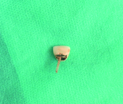
Seating of endodontic post in the post space.
Postoperative Evaluation
All patients were evaluated after 1, 6 and 12 months’ interval post after cementation. If patients showed up in between or after regular intervals of recall observation were performed. During each follow-up visit, a periapical radiograph was taken and the clinical performance was noted. These findings were compared with the radiographs taken immediately after the treatment. The success or failure of the restoration, both clinically and radiographically was assessed. Restored tooth was considered futile, if any of the following criteria were met: mobility of the crown under finger pressure, secondary caries at the crown margin, fracture of the restoration or root, periapical or periodontal pathology requiring crown removal.
Statistical Analysis
The data obtained was arranged systematically and the information was transferred onto a master chart which was prepared in Microsoft Excel (2007) with the values obtained for: (i) movement of crown margin under finger pressure; (ii) recurrent caries at crown margin; (iii) fracture of the restoration; (iv) fracture of the post; and (v) periapical or periodontal pathology. Statistical Package for Social Sciences (SPSS) version 20.0 was used for analysis. To find the significance of the study parameters, the Fisher’s exact test was used and a p-value of <0.05 was considered to be statistically significant.
Results
During the observation period of 12 months, two failures occurred (one absolute and one relative) at six months [Table/Fig-4,5]. Based on the success parameters, the overall survival rate of both the aesthetic post systems were calculated to be 95%.
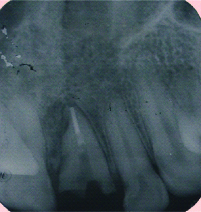
Immediate post-op of the fractured restoration case.
Fractured restoration after six months.
Movement of the Crown Margin under Finger Pressure
Neither of the post and core restorations showed movement of the crown margin with finger pressure at 1-year follow-up; both the glass fibre- and zircon-enriched silicon fibre posts showed success rate of 100%.
Recurrent caries detected at crown margin:
There were no restorations of zircon-enriched silicon fibre posts or glass fibre-reinforced posts that presented any secondary caries at various time intervals; hence, giving the success rate of 100%.
Fracture of the Restoration
One case of post-fracture occurred with glass fibre-reinforced posts during the evaluation period of 12 months; it was the same case that showed failure at six months. Thus, the success rate was 95% [Table/Fig-6,7].
Showing results based on 5 criteria considered for clinical survival.
| Criteria | Timeline | Group-I | Group-II | p-value |
|---|
| Fracture of the restoration | 1 month | 0 | 0 | 0.02 |
| 6 months | 0 | 1 |
| 12 months | 0 | 1 |
| Periapical or periodontal pathology requiring crown removal. | 1 month | 0 | 0 | 0.02 |
| 6 months | 0 | 1 |
| 12 months | 0 | 1 |
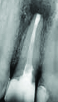
Twelve months follow-up of fractured post case.
No post fracture was observed in the zircon-enriched silicon fibre-reinforced posts on clinical and radiographic examinations at different follow-up hence, the success rate was 100%.
Fracture of the Root
There were no restorations of zircon-enriched silicon fibre posts or glass fibre-reinforced posts that presented any fracture of the root on clinical and radiographic examinations at various time intervals, hence; the success rate was 100%.
Periapical or Periodontal Pathology Requiring Crown Removal
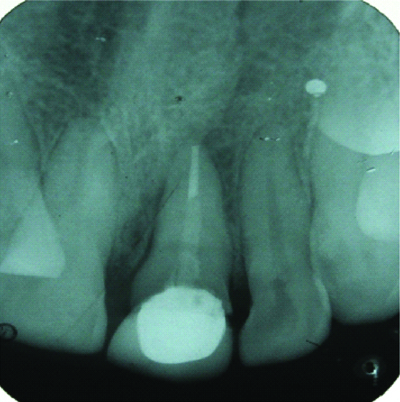
A single case of periapical radiolucency was noticed in the present study, six months after the completion of the treatment in glass fibre-reinforced posts; thus, the success rate was 95%. No case of periapical radiolucency was seen with the zircon-enriched silicon fibre posts; thus, the success rate was 100% [Table/Fig-8].
Tooth restored with the zircon-enriched silicon fibre posts showing periapical radiolucency.
Discussion
There was no significant difference in the clinical outcome provided by the two types of posts, indicating that the use of different fibre post materials does not remarkably contribute to the clinical performance of the restoration. Hence, the null hypothesis tested for the study was rejected. However, significant difference was seen between the two-post system for Fracture of the restoration and periapical pathology. Published retrospective studies on the clinical performance of fibre posts have highlighted that there is insufficient control of all the variables that may arise under clinical conditions [10,11]. Various clinical studies have investigated restorations involving endodontic posts and have recorded the important reasons of failure. The main reported causes include: caries, loss of retention of the post, loss of retention of the crown, root fracture, post distortion, and post fracture [Table/Fig-9] [10-15].
Comparative analysis of various studies done on endodontically treated teeth restored with post and core.
| Author and year | Place of study | Type of post | Type of testing | Type of failure observed |
|---|
| Ribeiro MTH et al., 2023 [10] | Brazil | Fibre Glass Post (FGP) | 15000 cycles of loading | Fatigue failure |
| Penteado MM et al., 2023 [11] | Brazil | custom Glass Fibre Post (CTM), and universal 2-piece glass Fibre-Reinforced Composite (FRC) resin post (UNI) | 10000 cycles of loading | Fatigue failure |
| Asmussen E et al., 1999 [12] | Denmark | posts of zirconia (Biopost, Cerapost), titanium (PCR), and carbon fibre (Composipost) | 450 angle load in the Instron machine | Composipost had the lowest values for stiffness, elastic limit, and strength |
| Souza JCM et al., 2022 [13] | Portugal | Glass Fibre-Reinfored Resin Composite (GFRC) with surface treatment | Push-out/shear bond strength | Grit-blasting or etching that promoted a mechanical interlocking of the adhesive and resin-matrix cements decrease the risk of clinical failures by fracture and detachment of endodontic posts |
| Volom A et al., 2023 [14] | Budapest, Hungary | Fibre-Reinforced Composite (FRC) systems | 40000 cycles of loading | Fatigue failures |
| Molnár J 2022 [15] | Szeged, Hungary | Flowable SFRCs | 40000 cycles of loading | Fatigue failures |
| Present study | Faridabad, Haryana, India | Zircon-enriched silicon fibre posts and glass fibre posts | Clinical and radiographical analysis | Fracture of restoration and periapical pathology with glass fibre posts |
In a prospective study, many of the possible influencing factors are controlled at the stage of case selection, making the experimental groups more similar except for the specific variable under study. In present study, the experimental groups were controlled for the type of tooth (maxillary anteriors), a single operator, and the type of endodontic treatment, which was performed using the same technique for all the teeth. Therefore, the only significant factor under study, the use of different aesthetic post systems for post endo restoration, was the main factor responsible for the variability in the clinical performance of the teeth over time.
Because the moduli of elasticity of fiber-reinforced composite posts are closer to that of dentine, many studies have demonstrated that they offer advantages over metal posts [12,14,16]. This phenomenon of ‘modulus compensation of stress-induced root fractures’ results in a positive effect on the longevity of post-core restorations. However, it should be recognised that the material’s modulus is just one of the parameters that can influence stress. Hence, each parameter of the present study will be discussed with relevance to the published literature of the present study findings.
In the present study, no failures were seen with respect to the movement of crown margin under finger pressure in both the glass fibre post systems during the recall period of 12 months. The results were therefore in agreement with a previous study which concluded 10% failure in relation to the mobility of the crown margin under finger pressure for cast posts and carbon fibre posts but a 100% success rate for glass fibre posts over a recall period of 12 months [13]. In another study, mobility of the single PFM crown restored with glass fibre post at one month follow-up was noticed, possibly due to the inaccuracy in marginal fit of the crown because of error in fabrication at lab [14].
No case of recurrent caries at crown margin was detected in either of the aesthetic fibre post systems during the recall period of 12 months. The durability of restorations against recurrent caries is influenced by three main factors: patients’ caries activity, the quality of treatment provided, and the cariostatic effects of the restorative material. In the current clinical study, patients received oral hygiene instructions, which could have impacted their compliance and motivation levels, thus potentially affecting the absence of recurrent caries in the two groups. Also, another important factor to be mentioned is the short duration of the present study, which could be a reason for the absence of recurrent caries.
In the present study, one case of post fracture occurred with glass fibre reinforced post during the evaluation period of 12 months whereas no post fracture was observed in the other system. The difference was, however, statistically significant. The concerned tooth was extracted and a three-unit porcelain-fused to metal bridge was given to the patient. The overall failure rate was in accordance with the results of the previous retrospective [10,16,17] and prospective studies [15,18,19].
In the present study, no failures occurred due to fracture of the root in either of the post systems over the evaluation period of 12 months. The results were therefore, in agreement with the results of the previous studies [11,13-15]. Dean JP et al., carried out an in-vitro comparison was conducted between carbon fibre and conventional cast posts [20]. The study revealed that there were no root fractures associated with carbon fibre posts whereas 50% of the teeth with cast posts experienced root fractures.
The failure of fibre posts is never due to a root fracture [11,21], unlike cast posts [10,21], where root fracture poses a huge risk. A potential reason for the frequent occurrence of root fractures with cast posts may be due to the friction generated along the walls and the rigidity of the metallic materials compared to dentin [22]. On the other hand, the modulus of elasticity of fibre posts is quite close to dentin which gives it increased resistance to root fractures. Glass fibre posts have demonstrated the capacity to fracture at the coronal section of a tooth restoration under extreme forces, without causing damage to the root. This is a significant reason for their use and documented success [14].
A single case of periapical radiolucency was noticed in the present study, six months after the completion of the treatment in one of the post systems, i.e., glass fibre reinforced post system. No case of periapical radiolucency was seen with the other post system. The difference was statistically significant. The result was slightly higher than the results obtained in some of the previous studies where no periapical lesion was observed during the evaluation period that indicated reasonable healing [13,14]. The reinfection was treated with appropriate antibiotics and resolved within a few months, not necessitating the removal of the fibre post.
The current study compared the performance of two types of translucent fibre posts that were used with the same adhesive and restorative materials under similar clinical scenarios. The results showed no significant difference in the performance of the two classes of posts. The frequency and patterns of failure were similar to studies which were commenced and reported in literature [15,23]. Long follow-up data on translucent-fibre posts and resin composites used for core build ups are expected from a prospective study currently in progress.
Limitation(s)
The follow-up period was 12 months, so this observation period may not be considered as an optimum time for evaluating the clinical success. Endo-crowns are upcoming these days and can be a viable treatment option with advances in dental material and adhesion sciences. Future assessment of glass fibre posts and CAD-CAM dentin posts with endo-crowns shall assist in establishing better clinical recommendations. A study may be conducted in the future to evaluate the post’s versatility based on patient preference, dentist convenience, and cost analysis.
Conclusion(s)
Within the limitation of the present clinical study, the following conclusions could be drawn that the clinical survival rate of both the aesthetic post systems was acceptable over a period of 12 months. The Zircon-enriched silicon fibre did not show any signs of failure in the above-mentioned criteria over the course of the study; however, the glass fibre-reinforced posts showed two cases of failure. The failures seen were with respect to the fracture of the post and periapical radiolucency seen after post cementation.
Fibre posts, used in combination with adhesive techniques, enable the creation of a homogeneous and integrated unit, demonstrating a positive impact on the mechanical properties of the tooth and potentially ensuring excellent clinical outcomes and survival.