Recurrent Deep Vein Thrombosis Shrouding a Sinister Colorectal Carcinoma in a Young Adult
Nikhil Pantbalekundri1, Shilpa Gaidhane2
1 Resident, Department of General Medicine, Jawaharlal Nehru Medical College, Datta Meghe Institute of Higher Education and Research, Wardha, Maharashtra, India.
2 Professor, Department of General Medicine, Jawaharlal Nehru Medical College, Datta Meghe Institute of Higher Education and Research, Wardha, Maharashtra, India.
NAME, ADDRESS, E-MAIL ID OF THE CORRESPONDING AUTHOR: Dr. Nikhil Pantbalekundri, Resident, Department of General Medicine, Jawaharlal Nehru Medical College, Datta Meghe Institute of Higher Education and Research, Wardha-442107, Maharashtra, India.
E-mail: nikhil6996pant@gmail.com
Adenocarcinoma, Khorana, Protecht, Venous thromboembolism, Vienna
Dear Editor,
Venous Thromboembolism (VTE) is an essential concern for cancer patient, as they have a notably higher risk than non cancer patients, which can result in significant morbidity and mortality. Approximately 15% of cancer patients suffer from VTE, with risk persisting throughout the illness [1]. Colorectal cancer, with genetics as a primary risk determinant, exhibits a pronounced association with VTE. However, comprehensive guidelines for managing VTE in colorectal cancer patients remain elusive due to limited research.
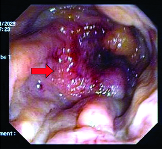
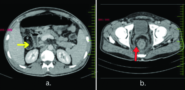
A 38-year-old tailor presented to the Outpatient Department (OPD) with complaints of left upper limb pain persisting for seven days. It significantly impaired his daily activities. His medical history revealed a recent diagnosis of Deep Vein Thrombosis (DVT) in the right leg, for which he was currently on warfarin therapy with no notable history of travel, trauma, or surgeries. Clinical examination indicated tachycardia and tachypnea; laboratory investigations indicated elevated D-dimer levels of 1240 ng/dL (normal: less than 300 ng/dL). Subsequent imaging studies, including colour Doppler imaging and pulmonary angiography, confirmed extensive DVT involving the left subclavian and ulnar veins, along with bilateral pulmonary emboli. Concurrently, the patient experienced distressing symptoms related to defecation and occasional rectal bleeding. With the suspicion of malignancy, explaining multiple episodes of venous thrombosis, a colonoscopy was done, revealing a friable mass in the rectum. On gross examination, it was reported as single, significant, ulcero-infiltrative and circumferential growth. Histopathology analysis suggested an ill-differentiated adenocarcinoma of the rectum [Table/Fig-1]. Contrast-enhanced Computed Tomography (CECT) unveiled widespread metastatic disease involving the colon and para-aortic lymph nodes [Table/Fig-2]. Consequently, the patient was referred to the Department of Surgical Oncology for palliative management, ultimately undergoing colonic bypass surgery.
Colonoscopy showing large friable mass partially obstructing descending colon and rectum (red arrows).
CECT of abdomen and pelvis showing: a) Mass with ill-defined margins extending to hepatic flexure transverse colon (yellow arrow); b) A large 6×3 cm mass with an ill-defined margin in the rectum (red arrow).
A well-researched cancer consequence, DVT is thought to occur in 4-17% of individuals who present with underlying malignancies. Patients with cancer have a 7% chance of developing VTE episodes, which is partially because they are more likely to be exposed to different incidental risk factors such as medication, immobility, and surgery throughout their illness. Furthermore, prothrombotic states are frequently linked to malignancy and can result in VTE that is resistant to anticoagulants or be clinically asymptomatic [2]. Patients who have both cancer and thrombotic events have a worse survival rate when compared to cancer patients who do not have thrombosis.
Cancer patients are more susceptible to VTE due to multiple underlying pathophysiological comorbid causes. All three of the Virchow’s triad predisposing factors to thrombus formation are abnormal in cancer patients: increased viscosity and stasis, which slows blood flow; increased platelet aggregation and activation, which increases procoagulant factors along with decreased fibrinolytic and anticoagulant factors; endothelial cells that become prothrombotic due to inflammatory cytokines, which leads to a dysfunctional endothelium. A hypercoagulable state appears to be the result of an imbalance between the fibrinolytic and coagulation systems brought on by cancer. According to a study, patients with colorectal cancer as well as those with pancreatic, haematological, lung, and brain tumours had an increased incidence of VTE [3]. GPIIIA PlA2 polymorphism, 4G/4G genotype, and MTHFR 677T variant allele are the most researched risk factors linked to VTE and cancer. Additionally, obesity poses a serious risk for both arterial thrombosis and VTE [4].
Clinical thromboembolism can be the earliest sign of a malignancy, arising before the cancer has developed symptoms. Thrombotic events typically present as pulmonary embolism and classical DVT of the limbs in cancer patients. On the other hand, Disseminated Intravascular Coagulation (DIC), thrombotic microangiopathy, and thrombosis of the vena cava and visceral circulation can also occur. The fact that there is a significantly greater body of information about VTE than arterial thrombosis in cancer indicates that the former occurs more frequently than the latter. Although the link between arterial thrombosis and cancer has been established over time, the underlying processes remain unknown. Clinical signs of arterial thrombosis can include DIC, thrombotic thrombocytopenic purpura, and localised arterial occlusion [5]. It is advised that the risk of VTE be evaluated prior to starting chemotherapy. Several risk models have been developed to categorise lung cancer patients based on their likelihood of experiencing VTE. One widely recognised tool is the Khorana Risk Score (KRS), which considers factors such as the location of the cancer, platelet and leucocyte counts, haemoglobin levels, and Body Mass Index (BMI) to predict VTE risk. A higher KRS indicates a higher risk of VTE and is also associated with poorer prognosis. An enhanced risk assessment tool called the PROTECHT score builds upon the Khorana score by incorporating additional factors, such as specific chemotherapy types like gemcitabine or platinum-based treatments. The PROTECHT score has demonstrated superior ability in identifying cancer patients at high-risk for VTE compared to the Khorana score alone [6]. This indicates ongoing efforts to refine risk prediction in cancer-associated thrombosis, aiming to improve patient outcomes through better targeted preventive measures. P-selectin and D-dimer are indicators of platelet activation and clotting cascade activation, respectively. They were incorporated into the Vienna Prediction Model, an enhanced version based on the original Khorana score, significantly improving the prediction accuracy for VTE in cancer patients. In clinical practice, assessing VTE risk in these patients involves using scores like Khorana, PROTECHT, and Vienna [7]. Factors such as advanced cancer stage, recent surgery, immobilisation, and chemotherapy are key predictors of VTE episodes. These scoring systems help clinicians identify patients at higher risk of thrombotic complications, guiding preventive measures and optimising patient care.
In young people, the identification of recurrent DVT may be suggestive of colorectal cancer. Therefore, we propose that in addition to coagulation disorders, malignancies should be considered as a differential diagnosis in patients who experience recurrent venous thrombotic events. We also emphasise the need for a multifactorial perspective on thrombotic disease in order to effectively assess risk, customise anticoagulant prophylaxis, and target therapies.
Author Declaration:
Financial or Other Competing Interests: None
Was informed consent obtained from the subjects involved in the study? Yes
For any images presented appropriate consent has been obtained from the subjects. Yes
Plagiarism Checking Methods: [Jain H et al.]
Plagiarism X-checker: Apr 12, 2024
Manual Googling: May 17, 2024
iThenticate Software: Aug 09, 2024 (6%)
[1]. Mulder FI, Horváth-Puhó E, van Es N, van Laarhoven HWM, Pedersen L, Moik F, Venous thromboembolism in cancer patients: A population-based cohort studyBlood 2021 137(14):1959-69.Available from: https://doi.org/10.1182/blood.2020007338 [Google Scholar]
[2]. Qdaisat A, Wechsler AH, Cruz Carreras MT, Menendez JR, Lipe D, Highsmith EA, Concomitant deep vein thrombosis in cancer patients with unsuspected pulmonary embolismCancers 2022 14(18):4510Availble from: https://doi.org/10.3390/cancers14184510 [Google Scholar]
[3]. Girardi L, Wang T, Ageno W, Carrier M, Updates in the incidence, pathogenesis, and management of cancer and venous thromboembolismArterioscler Thromb Vasc Biol 2023 43(6):824-31.Available from: https://doi.org/10.1161/ATVBAHA.123.318779 [Google Scholar]
[4]. Peng J, Wu Z, MTHFR act as a potential cancer biomarker in immune checkpoints blockades, heterogeneity, tumour microenvironment and immune infiltrationDiscov Onc 2023 14:112Available from: https://doi.org/10.1007/s12672-023-00716-0 [Google Scholar]
[5]. Ades S, Pulluri B, Holmes CE, Lal I, Kumar S, Littenberg B, Risk factors for venous thromboembolism in metastatic colorectal cancer with contemporary treatment: A SEER-Medicare analysisCancer Med 2022 11(8):1817-26.10.1002/cam4.4581 [Google Scholar] [CrossRef]
[6]. Van Es N, Di Nisio M, Cesarman G, Kleinjan A, Otten HM, Mahé I, Comparison of risk prediction scores for venous thromboembolism in cancer patients: A prospective cohort studyHaematologica 2017 102(9):1494-501.10.3324/haematol.2017.169060 [Google Scholar] [CrossRef]
[7]. Donnellan E, Khorana AA, Cancer and venous thromboembolic disease: A reviewOncologist 2017 22(2):199-207.10.1634/theoncologist.2016-0214 [Google Scholar] [CrossRef]