The mandible (from Latin “mandibula,” meaning “jawbone”) is the largest and strongest bone of the face, developing from the first pharyngeal arch. It has a horseshoe-shaped body that houses the teeth and a pair of rami that project upwards from the posterior ends of the body [1-3].
The mandible, comparable to a long bone, is movable under the control of masticatory, facial expression, and some suprahyoid muscles, which are antagonistic to the maxilla. The mandibular foramen lies a little above the center of the ramus of the mandible and runs obliquely downward and forward within the ramus, leading into the MC [3].
The MC descends and runs horizontally forward into the body of the mandible at the level of the occlusal surfaces of the teeth, opening into the mental foramen, where it is positioned beneath the alveoli and communicates with them through small openings, along with the inferior alveolar artery, vein, and nerve. Anatomically, the MC is known to be a single bilateral structure, but variations of the MC have also been observed [4,5].
Morphological variations of the MC occur due to abnormal interactions of tissues during dental embryonic development. These variations can be based on shape (oval, round, or pear-shaped) or can include various accessory canals, such as bifid, trifid, or temporal crest canals. The terms bifid and trifid originate from Latin, meaning “split into two” and “split into three,” respectively. It has been described that during embryonic development, three inferior dental nerves fuse during maturation to form a single nerve [6]. Thus, bifid and trifid MCs may result from the incomplete fusion of these three nerves. The temporal crest canal, is a rare anatomical variation of the MC. It is an anomalous canal whose posterior opening is located posterior to the temporal crest, while its anterior opening is located anterior to the temporal crest (i.e., in the retromolar fossa) [4,7-9].
A detailed understanding of the MC’s anatomy, including its contents, position, course, and morphology, is important for managing various surgical procedures, including dental implant placement, third molar surgery, dental anaesthesia, mandibular osteotomy, bone harvesting from the ramus and body of the mandible, bone plating in the angle and body regions of the mandible, and any other surgical procedure involving the mandible [4,10].
Failure of local anaesthesia in the buccal and inferior alveolar nerve blocks can be attributed to variations in anatomy. When these variations are overlooked, they can lead to damage to these nerves during dental procedures, such as lesion excision, difficulties in implant placement, and osteotomy procedures [4,6-8].
These anatomical variations in the mandibular region can be evaluated using 2D imaging modalities such as panoramic radiographs, as well as 3D imaging modalities like Computed Tomography (CT) and CBCT. Among all these imaging modalities, CBCT offers a 3D view with high image resolution and lower radiation exposure compared to CT. It is an effective imaging modality for assessing the course, location, configuration, and variations of Mandibular Canals (MCs). Therefore, this study was conducted to determine the prevalence of MC variations among the Indian population. The objectives were to measure the diameter, length, and location of the MCs.
Materials and Methods
A retrospective observational study was conducted in the Department of Oral Medicine and Radiology at Rajarajeswari Dental College and Hospital, Ramohalli Cross, Mysuru Road, Bengaluru, Karnataka, India over a peroid of six months from October 2023 to April 2024. Institutional Ethical Committee approval was obtained (IEC NUMBER: RRDCH/IEC/2022/044). In present study, 190 images were randomly retrieved from the archives of CBCT in the Department of Oral Medicine and Radiology.
Inclusion criteria: CBCT images of the mandible in the cross-sectional view of subjects aged 16 to 75 years, irrespective of gender, ethnicity, and the presence or absence of teeth, were selected.
Exclusion criteria: CBCT images of fractures of the mandible, congenital or dental anomalies of the maxillofacial complex, and images showing errors or artifacts that obscured the visibility of the structure of the mandibular canal were excluded.
Study Procedure
All images were assessed and measured using the On Demand 3D and Scanora software of the CBCT machine. The cross-sectional view was used to locate the mandibular canal, starting from the mandibular foramen and extending to the mental foramen.
The following measurements were taken using the measuring tool in ONDEMAND 3D software, following the methodology of the authors given (2009) with modifications provided by Rashsuren O et al., (2014) [11]. The classifications by Naitoh M et al., (2009) and Rashsuren O et al., (2014) for bifid and trifid canals were utilised solely to assess their prevalence in the Indian population [11]. Bifid canals were classified as Type 3, and trifid canals as Type 5, without further categorisation. Similarly, author applied the classification by Han SS et al., to evaluate the general prevalence of temporal crest canals, rather than examining each specific type [12]. The classifications are as follows:
Type I (The retromolar canal): A bifid canal that reaches the retromolar region.
Type II (The dental canal): A bifid canal that extends to the root apex of the second or third molar.
Type III (The forward canal):
(A) Forward canal without confluence: A bifid canal that arises from the superior wall of the MC and courses forward toward the second molar region.
(B) Forward canal with confluence: A bifid canal that arises from the superior wall of the MC, courses anteriorly, and then joins the main MC.
Type IV (The buccolingual canal): A bifid canal that arises from the buccal or lingual wall of the MC.
Type V (The trifid canal):
(A) Two accessory canals of the retromolar canal type.
(B) Two accessory canals, one of the retromolar and one of the dental canal type.
(C) Two accessory canals of the dental canal type.
(D) Two accessory canals, one of the dental and one of the forward canal type.
(E) Two accessory canals of the retromolar canal type with two mandibular foramina.
The Temporal Crest Canal is classified into two types based on the study by Han SS et al., [12]:
(A) Type 1 TCC: Increasingly narrow and curved.
(B) Type 2 TCC: Uniformly wide and slightly curved.
Morphometric analysis: The variables that were measured included the length and diameter of the MC. The length of the MC was measured in the reformatted panoramic view using a scale in the CBCT software. The diameter of the MC was measured in the coronal view at the distal part of teeth 38 and 48, also using a scale.
Statistical Analysis
The Statistical Package for Social Sciences (SPSS) for Windows Version 22.0, released in 2013, was used to perform statistical analyses. Descriptive analysis of all explanatory and outcome parameters was conducted using the mean and Standard Deviation (SD) for continuous variables, and frequency and proportions for categorical variables. The Chi-square test was used to compare the anatomical variations of the MC based on age and gender. A paired Student’s t-test was employed to compare the mean lengths of the MC (in mm) as well as the mean diameter width and length between the right and left-sides. An Independent Student’s t-test was used to compare the mean length, mean diameter length, and mean diameter width based on the age group of the study subjects and gender.
Results
The study sample comprised 380 sides of CBCT images, which included patients aged 16-75 years. The mean age of the study population was 37.96±14.63 years. Among the 190 images, 105 (55.3%) belonged to males, and 85 (44.7%) belonged to females.
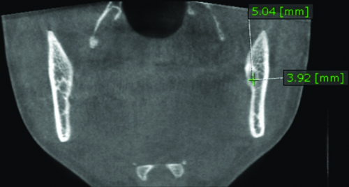
The mean vertical and horizontal diameters of the MC were 4.98±1.27 mm and 2.95±0.95 mm, respectively [Table/Fig-1,2]. When comparing the right and left-sides, the horizontal diameter was greater on the left-side, and this difference was statistically significant (p-value ≤0.001*) [Table/Fig-1]. There was no statistically significant difference in the diameter of the MC when comparing samples below 35 years with those above 35 years for both the right and left-sides [Table/Fig-3]. Among the two genders, the vertical diameter was significantly smaller in females on both the right and left-sides of the mandible. The difference in horizontal diameter was not significant between genders [Table/Fig-4].
Mean diameter of Mandibular Canal (MC) using student’s paired t-test.
| Parameters | Sides | N | Mean±SD | Mean diameter | Mean diff. | p-value |
|---|
| Vertical | Right | 190 | 5.03±1.08 | 4.98±1.27 | 0.10 | 0.26 |
| Left | 190 | 4.93±1.46 |
| Horizontal | Right | 190 | 2.87±0.76 | 2.95±0.95 | -0.17 | 0.001* |
| Left | 190 | 3.04±0.74 |
Measurement of diameter of MC.
Mean diameter of Mandibular Canal (MC) based on the age group using independent student’s t-test.
| Side | Age | N | Vertical diameter | Horizontal diameter |
|---|
| Mean±SD | Mean diff. | p-value | Mean±SD | Mean diff. | p-value |
|---|
| Right | ≤35 y | 96 | 5.12±1.11 | 0.19 | 0.24 | 2.97±0.80 | 0.19 | 0.08 |
| >35 y | 94 | 4.93±1.05 | 2.77±0.70 |
| Left | ≤35 y | 96 | 4.99±1.26 | 0.11 | 0.60 | 3.10±0.72 | 0.11 | 0.32 |
| >35 y | 94 | 4.87±1.64 | 2.99±0.77 |
Mean diameter of Mandibular Canal (MC) based on the gender using independent student’s t-test.
| Side | Gender | n | Vertical diameter | Horizontal diameter |
|---|
| Mean±SD | Mean diff. | p-value | Mean±SD | Mean diff. | p-value |
|---|
| Right | Male | 105 | 5.12±1.12 | 0.40 | 0.01 | 2.96±0.73 | 0.20 | 0.08 |
| Female | 85 | 4.81±0.99 | 2.76±0.78 |
| Left | Male | 105 | 5.10±1.57 | 0.37 | 0.04 | 3.12±0.75 | 0.18 | 0.10 |
| Female | 85 | 4.73±1.29 | 2.94±0.73 |
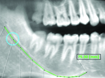
The mean length of the MC was 67.15±1.56 mm, and it was significantly longer on the left-side [Table/Fig-5,6]. There was no statistically significant difference in the lengths of the MC among different age groups [Table/Fig-7]. The MC was significantly longer in males on both the right and left-sides, with values of 67.64±5.23 mm and 69.36±5.35 mm, respectively [Table/Fig-8].
Mean length of Mandibular Canal (MC) using student’s paired t-test.
| Parameter | Sides | N | Mean±SD | Mean length | Mean diff. | p-value |
|---|
| Length | Right | 190 | 66.16±5.77 | 67.15±1.56 | -1.99 | <0.001* |
| Left | 190 | 68.14±5.64 |
Measurement of mean length of MC.
Comparison of mean length of Mandibular Canal (MC) based on age group using independent student’s t-test.
| Side | Age | n | Mean±SD | Mean diff. | p-value |
|---|
| Right | ≤35 y | 96 | 66.44±5.68 | 0.58 | 0.49 |
| >35 y | 94 | 65.86±5.87 |
| Left | ≤35 y | 96 | 68.70±5.59 | 1.14 | 0.17 |
| >35 y | 94 | 67.57±5.66 |
Comparison of mean length of Mandibular Canal (MC) based on age group using independent student’s t-test.
| Side | Gender | n | Mean±SD | Mean diff. | p-value |
|---|
| Right | Male | 105 | 67.64±5.23 | 3.32 | <0.001* |
| Female | 85 | 64.32±5.90 |
| Left | Male | 105 | 69.36±5.35 | 2.73 | 0.001* |
| Female | 85 | 66.63±5.65 |
The MC was located below the apices of the molars before opening into the mental foramen in the premolar region [Table/Fig-9].
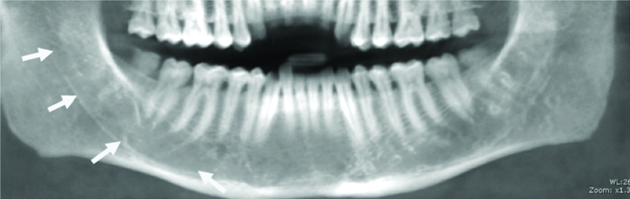
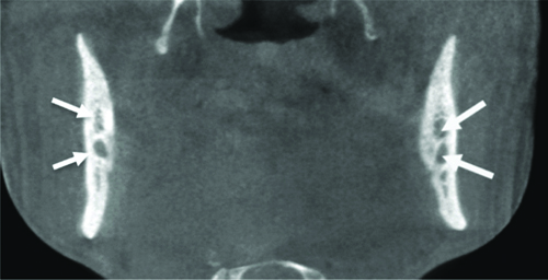
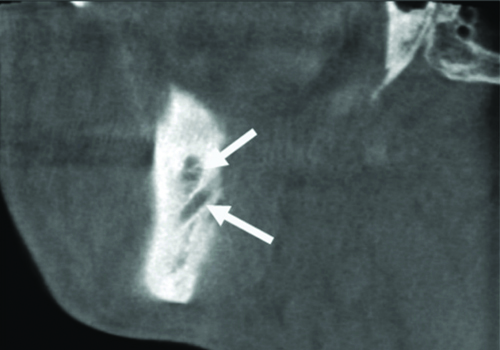
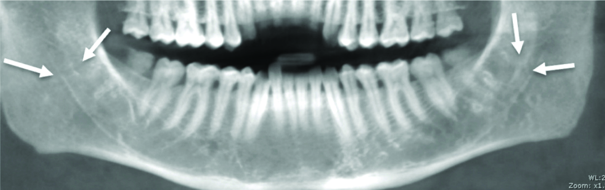
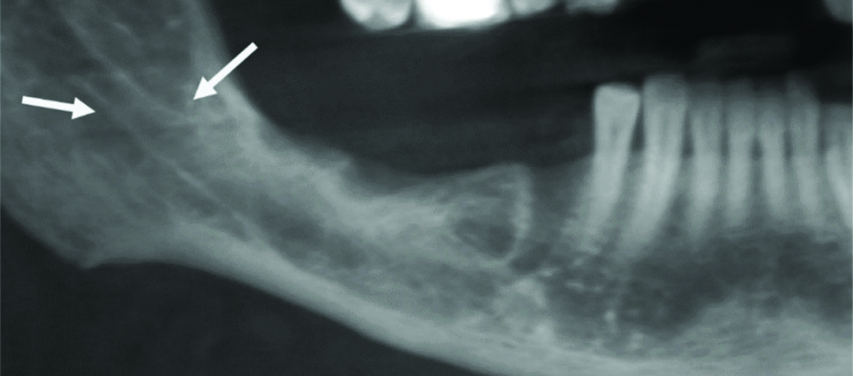
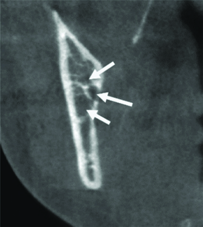
Among the three variations included in this study, the bifid canal was the most common variation (14.7%), followed by the temporal crest canal (5.3%) and the trifid canal (3.7%) [Table/Fig-10]. The bifid canal was more common on the left-side (5.7%) [Table/Fig-11,12], while the temporal crest canal was more common on the right-side (3.2%) [Table/Fig-13,14]. The trifid canal was observed in 3.7% of the population [Table/Fig-15].
Prevalence of anatomical variation in Mandibular Canal (MC).
| Variable | Total present | % | Right | Left | Bilaterally |
|---|
| n (%) | n (%) | n | % |
|---|
| Bifid | 28 | 14.7% | 8 (4.2) | 11 (5.7) | 9 | 4.7 |
| Trifid | 7 | 3.7% | 2 (1.1) | 1 (0.5) | 4 | 2.1 |
| Temporal crest canal | 10 | 5.3% | 6 (3.2) | 3 (1.6) | 1 | 0.5 |
Coronal section of CBCT image showing bifid canal bilaterally.
Left cross-sectional CBCT image showing bifid canal.
Bilateral reformatted panoramic view of temporal crest canal.
Right reformatted panoramic view of temporal crest canal.
Left cross-section of CBCT image showing trifid canal.
All the anatomic variations were more common in images belonging to patients below 35 years, but this difference was not statistically significant [Table/Fig-16]. None of the anatomic variations showed a statistically significant difference regarding gender predilection [Table/Fig-17].
Comparison of anatomical variation in the Mandibular Canal (MC) based on the age group using Chi-square test.
| Variables | Category | ≤35 y | >35 y | p-value |
|---|
| n (%) | n (%) |
| Bifid | Present | 18 (18.8) | 10 (10.6) | 0.12 |
| Absent | 78 (81.3) | 84 (89.4) |
| Trifid | Present | 5 (5.2) | 2 (2.1) | 0.26 |
| Absent | 91 (94.8) | 92 (97.9) |
| Temporal crest canal | Present | 6 (6.3) | 4 (4.3) | 0.54 |
| Absent | 90 (93.8) | 90 (95.7) |
Comparison of anatomical variation in Mandibular Canal (MC) based on gender using Chi-square test.
| Variables | Category | Males | Females | p-value |
|---|
| n (%) | n (%) |
|---|
| Bifid | Present | 16 (15.2) | 12 (14.1) | 0.83 |
| Absent | 89 (84.8) | 73 (85.9) |
| Trifid | Present | 3 (2.9) | 4 (4.7) | 0.50 |
| Absent | 102 (97.1) | 81 (95.3) |
| Temporal crest canal | Present | 3 (2.9) | 7 (8.2) | 0.10 |
| Absent | 102 (97.1) | 78 (91.8) |
Discussion
The mean vertical and horizontal diameters of the mandible in present study were greater than those reported in previous studies conducted on Indian, Japanese, and Chinese populations, which were performed by Komal A et al., (2.36±2.131 mm), Kuribayashi A et al., (2.02-4.63 mm), and Saeed TA et al., (vertical diameter 2.93 mm and horizontal diameter 2.19 mm), respectively [8,13,14]. Three other studies conducted previously by Rashsuren O et al., (2.85±3.28 mm), Zhou X et al., (4.54±6.02 mm), and Elnadoury EA et al., (3.98±1.31 mm) measured only the vertical diameter, with values that were nearly similar to those obtained in our study [15-17].
In present study, the vertical and horizontal diameters were measured posterior to the third molar region, while the other studies measured the diameter at anterior locations. Additionally, Komal A et al., observed that the diameter of the mandible progressively decreased from the third molar region to the first premolar region [8]. Therefore, the larger diameters observed in present study compared to previous studies may be attributed to this difference in measurement location.
The present findings indicated that the horizontal diameter of the mandible was significantly higher on the left-side, which contrasts with the study conducted by Komal A et al., who found no significant difference in the mean horizontal diameter between the right-side (2.131 mm) and the left-side (2.120 mm) [8]. Similarly, a study by Elnadoury EA et al., reported that the diameter was significantly larger on the right-side (3.98 mm, p-value=0.019) compared to the left-side (3.80 mm, p-value=0.056), which again contrasts with our findings [17]. These discrepancies may be attributed to variations in the ethnicities of the populations studied.
In present study, there was no statistically significant difference in the diameter of the MC with respect to age. These results are similar to those obtained by Zhou X et al., and Safi Y et al., [16,18]. The present study compared the diameter of the MC by gender and found that the vertical diameter was significantly smaller in females bilaterally, while the horizontal diameter showed no sexual dimorphism. Similarly, a study conducted by Elnadoury EA et al., showed that the diameter of the MC was significantly smaller in females, but only on the right-side [17]. Another study by Safi Y et al., also indicated that the mean diameter of the MC was significantly greater in males than in females [18].
The mean length of the MC was measured to be 66.16±5.77 mm and 68.14±5.64 mm on the right and left-sides, respectively, in the present study. These values were similar to those found in the studies conducted by Komal A et al., (mean value on the right-side: 65.175±5.578 mm; on the left-side: 66.039±5.753 mm) but were lower than those found in the study by Muñoz G et al., (mean length for the MC: 70.8±5.3 mm on the right-side and 71±5.8 mm on the left-side) [8,19]. All these studies found that the MC was longer on the left-side than on the right-side.
In present study, with respect to age, the length of the MC showed no statistically significant difference across different age groups. However, the MC was significantly longer in males than in females. Similarly, Saeed TA et al., also found that the MC was significantly longer in males, with no difference when compared by age [14].
The location of the MC was noted to be below the apices of the molars, being closest to the roots of the third molars, although precise measurements were not made in present study. In present study, the MC was found to be in closest proximity to the roots of the third molars, and the diameter of the MC was measured distal to the third molar. A study conducted by Komal A et al., assessed the location of the MC by measuring the distance from the outermost margin of the MC to the buccal cortical plate and the lingual cortical plate. They found that the canal was located at a mean distance of 4.331 mm from the lateral aspect of the buccal cortical plate and 1.819 mm from the medial aspect of the lingual cortical plate in the third molar region [8]. The clinical implication of the MC’s location is to raise awareness of the orientation of the neurovascular bundle, which should be assessed preoperatively to avoid inadvertent injury in cases of anatomical variation.
In present study, bifid canals were detected in 14.7% of present cases. This finding is in accordance with the studies conducted by Chanda S et al., Kuribayashi A et al., Kang JH et al., and Yoon TYH et al., who reported prevalence rates of 12%, 15.6%, 10.2%, and 13.4%, respectively [11,13,20,21]. Most other studies indicated a higher prevalence rate, with a maximum prevalence of 40% reported in a study by Okumuş Ö et al., within the Turkish population [22]. Conversely, a study conducted by Afsa M and Rahmati H, in the Iranian population showed a lower prevalence of 6.9% [23].
Our study found that bifid canals were more common on the left-side, whereas studies by Chanda S et al., and Okumuş Ö et al., indicated a higher prevalence on the right-side [11,22]. The study by Yoon TYH et al., also reported a greater presence of bifid canals on the left-side, aligning with present results [21]. Bilateral bifid canals were found to be rare, occurring in 4.7% of present cases. The prevalence of bilateral bifid canals was observed to be higher in a study by Muinelo-Lorenzo J et al., (8.89%) [24] and lower in a study by Yoon TYH et al., (2.1%) [21].
When comparing bifid canals concerning age, no significant differences were found in our study. This result is consistent with studies conducted by Rashsuren O et al., (where bifid and trifid MCs were found in 22.6% of 500 patients and 16.2% of 755 sides, with no significant differences between genders and among age groups) and Kang JH et al., (who observed bifid MCs in 198 (10.2%) of 1933 patients) [15,20]. The most frequently observed type of bifid MC in present study was the retromolar canal 104 (52.5%), with no significant differences in the incidence of each canal based on age or gender [15,20].
In present study, bifid canals were more common in males, though this finding was not statistically significant. This result is similar to that of Muinelo-Lorenzo J et al., who also found a male predilection for bifid canals [24]. Conversely, studies conducted by Chanda S et al., Yoon TYH et al., Correr GM et al., and Orhan K et al., reported a higher prevalence of bifid canals in females [11,21,25,26]. Additionally, two other studies by Rashsuren O et al., and Kang JH et al., found no gender predilection [15,20]. These variations may be attributed to differences in the ethnicity of the populations studied.
The next common variant of the MC is the trifid canal, which showed a prevalence of 3.7% in present study. The prevalence of trifid canals was higher in studies conducted by Rashsuren O et al., and Elnadoury EA et al., which reported values of 5.8% and 8.7%, respectively [15,17]. Other studies by Chanda S et al., Okumuş Ö et al., and Afsa M et al., showed prevalences of 1%, 2.4%, and 1.7%, respectively, all of which are lower than those found in our study [11,22,23].
Regarding laterality, trifid canals were more commonly found on both sides of the mandible, with a prevalence of 2.1%. These results are similar to those of the study conducted by de Castro MAA et al., although the prevalence of trifid MC in their study was 4.9% [27]. There was no significant correlation between trifid canals and age, which is supported by previous studies conducted by Rashsuren O et al., and Kang JH et al., [15,20].
Multiple MCs, including trifid canals, were more common in females, as indicated by previous studies conducted by Chanda S et al., Correr GM et al., and Orhan K et al., which align with present findings regarding trifid canals [11,25,26]. Other studies by Rashsuren O et al., Kang JH et al., and de Castro MAA et al., reported no gender predilection [15,20,27].
In present study, the Trifid Canal Complex (TCC) was found in 5.3% of cases, predominantly on the right-side, with no age predilection and a higher occurrence in females. A study conducted by Yalcin ED and Akyol S in a Turkish population reported a prevalence of TCC at 0.97%, with a preference for the left-side of the mandible and a higher occurrence in males [12]. Another study by Hasani M et al., in an Iranian population showed an equal distribution of TCC on the right and left-sides of the mandible [28]. These results differ from those obtained in our study and may be attributed to ethnic differences among the populations studied.
Inspite of TCC being a rare entity, we included it in present study as it may lead to failure in obtaining adequate anaesthesia during dental and surgical procedures and may be damaged during jaw surgeries. CBCT delivers a higher radiation dose than panoramic radiographs but is more beneficial in detecting precise details in three dimensions. Muinelo-Lorenzo J et al., mentioned that while panoramic radiographs can detect bifid canals in the range of 0.82% to 8.3%, CBCT provides a higher detection rate of 10% to 66% [24]. Similar findings were reported by Rashsuren O et al., [15]. Due to the limited ability of 2D imaging, panoramic radiographs have relatively low specificity and sensitivity in measuring anatomical landmarks and detecting variations. All these factors make CBCT a superior imaging modality for radiographic visualisation and morphometric analysis of jaw bones.
Limitation(s)
The large sample size should be taken into consideration. The MC variations are not subclassified, which, if they were, could have provided a clearer picture.
Conclusion(s)
The evaluation of MC variations is of paramount importance due to their potential implications for surgical procedures in the maxillofacial region and the associated risk of complications. In particular, a deeper understanding of the temporal crest canal variations is crucial to equip oral health professionals with accurate information, thereby mitigating unwarranted complications and reducing their recurrence rates.