Thyroid hormones, through their action on almost all nucleated cells, are central to growth, neuronal development, reproduction, regulation of energy metabolism, and the proliferation of blood corpuscles. Thyroid dysfunction, though common, readily identifiable, and easily treatable, can have profound adverse consequences if left untreated or undiagnosed [1]. The diagnosis of thyroid dysfunction is primarily based on biochemical parameters, with TSH levels being the most sensitive indicator [2]. Micronutrients, in the form of iodine, iron, zinc, and selenium, are essential prerequisites for the synthesis and metabolism of thyroid hormones. Deficiencies in iodine and autoimmunity stand as the leading causes of primary hypothyroidism [3]. A large multicentre study has reported remarkably high rates of hypothyroidism in India, nearly 11%, compared to only 2% in the United Kingdom (UK) and 4-6% in the United States (US) [4,5].
Hypothyroidism is characterised by a significant metabolic slowdown, with manifestations varying, depending on the age of onset and the deficiency or inefficacy of thyroid hormones. Haematopoietic tissue is one of the primary organs affected, with anaemia being the most common and frequently underestimated clinical presentation [6]. Research indicates that mice deficient in thyroid hormone receptors have been found to have reduced PCV values [7]. Conversely, Beard J et al., reported diminished thyroxine production in age-matched male rats depleted of iron through dietary means [8]. Consequently, it appears that hypothyroidism and anaemia can influence one another, leading to non specific symptoms of ill health and a further reduced quality of life. The Lancet has published that the age-standardised incidence of hypothyroidism globally is three times higher in females as compared to males, whereas another paper reports that the total prevalence of anaemia in the world population is twice as common in females compared to males [9,10]. The elevated co-occurrence of these two conditions points towards a pivotal connection between anaemia and hypothyroidism [7]. Studies have shown a comparatively higher prevalence of anaemia in hypothyroid patients compared to the euthyroid population [11,12]. However, a limited number of studies have addressed the gender-specific association of thyroid hormones with haematological parameters [13].
In light of the aforementioned considerations, the present study was designed to evaluate and compare the gender-based association of TSH with haematological parameters within a population-based hypothyroid cohort.
Materials and Methods
This was a cross-sectional observational study conducted at the Department of Biochemistry in a tertiary care teaching hospital in Hyderabad, Telangana, India, from October 2022 to February 2023. The study was initiated after obtaining ethical clearance from the Institutional Ethics Committee (Ref IEC/MAMS/2022/037). Patients were enrolled after providing informed written consent, and their complete anonymity was maintained.
Inclusion criteria: All newly diagnosed, biochemically proven primary hypothyroid patients aged 18 to 50 years with TSH levels greater than 4.5 μIU/L [14] who attended the Biochemistry Laboratory were included in the study.
Exclusion criteria: All known hypothyroid patients currently undergoing treatment, hypothyroid patients with chronic diseases that may affect blood parameters, those with known intrinsic erythrocyte abnormalities, pregnant females, individuals with menstrual cycle abnormalities, those with evident nutritional deficiencies, and patients who declined to give consent were excluded from the study.
Sample size: To determine the sample size, the Cochran’s formula: n=Z2 PQ/d2 was used, where, Z=1.96, P for prevalence of anaemia in overt hypothyroidism was taken from a previous study [15], i.e., 0.11; Q=1-0.11, and d=allowable error i.e., 5% or 0.05. Thus, the calculated sample size was n=151.
Study Procedure
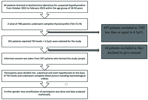
Maintaining aseptic precautions, approximately 5 mL of fasting venous blood was withdrawn from all patients referred to the Biochemistry Laboratory for screening thyroid disorders. The collected blood samples underwent a complete thyroid profile test, which included assessing thyroxine, triiodothyronine, and TSH levels using a fully automated chemiluminescence analyser with commercially available kits (Mindray CL1000i). Patients with TSH levels less than or equal to 4.5 μIU/L were excluded from the study. All patients with TSH levels greater than 4.5 μIU/L were included in the study and subsequently underwent a complete blood count test on a cell counter (Benesphera H33S) for all the haematological parameters, including erythrocyte count, Hb, PCV, MCV, MCH, MCHC, and Red cell Distribution Width (RDW).
Study participants were divided into two groups based on their TSH levels: subclinical and overt hypothyroidism. Patients with TSH levels higher than 4.5 μIU/L and up to 10 μIU/L, with normal thyroxine and triiodothyronine levels, were categorised as having subclinical hypothyroidism. Overt hypothyroidism was defined as TSH levels greater than 10 μIU/L, accompanied by lower-than-normal thyroxine and triiodothyronine levels, in agreement with the National Academy of Clinical Biochemistry (NACB) guidelines [14]. Anaemia was defined according to World Health Organisation (WHO) criteria (Hb <13 g/dL in males and <12 g/dL in females) [16]. The quality of results throughout the study period was validated by participation in routine internal quality control procedures and external quality assessment schemes [Table/Fig-1].
Flowchart depicting the study’s sequence.
Statistical Analysis
The data was collated and expressed in the form of frequencies, percentages, means, and standard deviations. Statistical differences were calculated among subclinical and overt hypothyroid patients for all blood parameters, separately for males and females, using Student’s unpaired t-test. Categorical data underwent the Chi-square test to identify statistical differences. Univariate linear regression analysis was conducted to find the potential association between haematological parameters and the category of hypothyroidism for males and females separately, with TSH values entered as independent variables and haematological parameters as dependent variables. All analyses were performed using IBM SPSS Statistics for Windows, Version 22.0, Armonk, NY: IBM Corp., and a p-value of <0.05 was considered statistically significant.
Results
A total of 347 biochemically proven primary hypothyroid patients, with a mean age of 37.12±12.12 years, formed the study sample. Among the 347 participants included in the study, 110 (31.7%) were males, while 237 (68.3%) were females. Within the total hypothyroid cohort, 184 (53.03%) experienced subclinical hypothyroidism, while 163 (46.97%) exhibited overt hypothyroidism [Table/Fig-2].
Demographic distribution of the study participants.
| Gender | SubclinicalHypothyroid n (%) | OvertHypothyroid n (%) | Total |
|---|
| Males | 58 (52.72) | 52 (47.27) | 110 (31.7) |
| Females | 126 (53.16) | 111 (46.83) | 237 (68.3) |
| Total | 184 (53.02) | 163 (46.98) | 347 (100) |
The subclinical and overt hypothyroid patients were age-matched. A significant difference in T3, T4, and TSH values was observed between subclinical and overt hypothyroid patients for both males and females [Table/Fig-3].
Gender-based distribution of baseline characteristics of the study participants.
| Variables | Gender | Subclinical hypothyroidMean±SD | Overt hypothyroidMean±SD | p-value |
|---|
| Age (years) | Males | 36.37±12.2 | 37.21±11.6 | 0.3501 |
| Females | 34.19±9.3 | 39.78±8.6 | 0.3322 |
| T3 (ng/dL) | Males | 114.2±19.1 | 97.3±57.6 | 0.0264* |
| Females | 112.5±30.3 | 99.7±33 | <0.001** |
| T4 (μg/dL) | Males | 9.3±10.4 | 5.9±2.2 | <0.001** |
| Females | 6.97±1.5 | 4.68±2.9 | 0.0002** |
| TSH (μIU/L) | Males | 7.18±1.4 | 63.2±40.4 | <0.001** |
| Females | 7.48±1.2 | 47.3±46.3 | <0.001** |
The prevalence of anaemia in the hypothyroid cohort was estimated to be 151 (43.5%), which was higher among patients with overt hypothyroidism (88, or 54%) compared to those with subclinical hypothyroidism (63, or 34.24%). There was a notable gender disparity in anaemia prevalence, with females exhibiting a higher prevalence than males. The majority of the patients suffered from microcytic anaemia. Even subgroup analysis revealed microcytic anaemia as the most commonly encountered variant among both males and females, though the difference was found to be statistically insignificant (p-value=0.9920) [Table/Fig-4].
Prevalence and type of anaemia among the study participants.
| Variables | Normocytic anaemia N (%) | Microcytic anaemia N (%) | Macrocytic anaemia N (%) | Total prevalence of anaemia N (%) |
|---|
| Male(110) | Subclinical hypothyroid (58) | 5 (8.62%) | 4 (6.90%) | 1 (1.72%) | 10 (17.2%) |
| Overt hypothyroid (52) | 5 (9.61%) | 6 (11.54%) | 1 (1.92%) | 12 (23.07%) |
| Female(237) | Subclinical Hypothyroid (126) | 17 (13.49%) | 30 (23.81%) | 6 (4.76%) | 53 (42.06%) |
| Overt hypothyroid (111) | 24 (21.62%) | 42 (37.84%) | 10 (9.01%) | 76 (68.47%) |
χ2 (6, N=151)=1.9616, p=0.9920
The mean haematological parameters, when compared between subclinical and overt hypothyroid patients, were found to differ. However, gender-based analysis highlighted that the differences were statistically significant only among females (except for Hb, which was significant for males) and not among males. Moreover, the difference for RDW was highly significant for both males and females (p-value <0.001) [Table/Fig-5].
Gender-based comparison of haematological parameters of the study participants.
| Variables | Gender | Subclinical hypothyroidMean±SD | Overt hypothyroidMean±SD | p-value |
|---|
| Hb (gm/dL) | Males | 13.38±1.9 | 12.5±1.5 | 0.0070** |
| Females | 11.69±1.3 | 10.82±1.7 | 0.0560 |
| MCV (fL) | Males | 88.17±5.5 | 87.9±4.4 | 0.1524 |
| Females | 87.96±5.2 | 85.5±4.5 | <0.001** |
| MCH (pg) | Males | 32.3±1.4 | 32.5±2.5 | 0.1761 |
| Females | 31.4±2.8 | 30.3±1.2 | <0.001** |
| MCHC (%) | Males | 33.3±1.4 | 32.98±1.4 | 0.0827 |
| Females | 32.3±2.4 | 32.1±1.7 | 0.0016* |
| PCV (%) | Males | 44.8±2.5 | 43.5±3.0 | 0.194 |
| Females | 43.7±2.9 | 42.1±3.1 | <0.001** |
| TRBC (million/mm3) | Males | 4.88±0.5 | 4.62±0.7 | 0.0189 |
| Females | 4.23±0.4 | 4.01±0.5 | 0.009* |
| RDW (%) | Males | 13.55±1.4 | 14.65±1.5 | <0.001** |
| Females | 13.7±1.2 | 14.6±1.8 | <0.001** |
*p<0.05 (Significant); **p<0.001 (Highly significant); p>0.05 (Insignificant); Hb: Haemoglobin; MCV: Mean corpuscular volume; MCH: Mean corpuscular haemoglobin; MCHC: Mean corpuscular haemoglobin concentration; PCV: Packed cell volume; TRBC: Total red blood cell; RDW: Red blood cell distribution width
The linear regression analysis between TSH (the independent variable) and haematological parameters (the dependent variable) revealed a highly significant correlation between the two variables. The correlation coefficient was higher for females compared to males for all parameters, as depicted in [Table/Fig-6].
Linear regression analysis showing the effect of TSH on other haematological parameters.
| Variables | Gender | df | SS | Co-efficient | F | r | p-value |
|---|
| Hb | Males | 1 | 40.9 | 9.85 | 19.5 | 0.27 | <0.001 |
| Females | 1 | 83.6 | 13.75 | 36.08 | 0.50 | <0.001 |
| MCV | Males | 1 | 196.6 | 78.7 | 5.77 | 0.22 | <0.001 |
| Females | 1 | 853.7 | 70.70 | 25.54 | 0.31 | <0.001 |
| MCH | Males | 1 | 159.6 | 28.08 | 4.57 | 0.20 | <0.001 |
| Females | 1 | 293.02 | 25.2 | 33.2 | 0.35 | <0.001 |
| MCHC | Males | 1 | 42.99 | 33.7 | 25.9 | 0.44 | <0.001 |
| Females | 1 | 454.38 | 30.73 | 58.4 | 0.44 | <0.001 |
| PCV | Males | 1 | 153.8 | 39.2 | 8.98 | 0.27 | <0.001 |
| Females | 1 | 116.2 | 32.0 | 12.66 | 0.59 | <0.001 |
| RBC | Males | 1 | 1.52 | 4.86 | 3.89 | 0.19 | <0.001 |
| Females | 1 | 0.144 | 4.14 | 0.41 | 0.05 | <0.001 |
| RDW | Males | 1 | 35.8 | 13.5 | 17.12 | 0.37 | <0.001 |
| Females | 1 | 207.5 | 13.5 | 129 | 0.59 | <0.001 |
*p<0.05 (Significant); **p<0.001 (Highly significant); p>0.05 (Insignificant); Hb: Haemoglobin; MCV: Mean corpuscular volume; MCH: Mean corpuscular haemoglobin; MCHC: Mean corpuscular haemoglobin concentration; PCV: Packed cell volume; TRBC: Total red blood cell; RDW: Red blood cell distribution width
Discussion
Thyroid disorders are the most prevalent endocrine abnormalities worldwide, with hypothyroidism reportedly affecting one in ten Indian adults [4]. Hypothyroidism has long been associated with abnormal haematological parameters, including anaemia [11,12]. In the current study, the gender-based association between haematological parameters and TSH levels among individuals with hypothyroidism was evaluated.
A total of 347 patients were included in the study, which had a female preponderance, with a sex ratio of M:F=1:2.1. The female predominance reported among the hypothyroid cohort in this study was consistent with findings by van Vliet NA et al., and Ahmed SS and Mohammed AA, [11,17]. The mean age of the hypothyroid cohort was 37.12±12.12 years, with an age range of 18 to 50 years, which was comparable to the study by Ahmed SS and Mohammed AA [17].
The prevalence of anaemia in the hypothyroid cohort was observed to be 43.52%, which was slightly lower than that reported by Omar S et al., (57.10%) [18]. Similarly, the UK Biobank cohort also reported higher odds (OR 1.12) of anaemia among hypothyroid patients compared to euthyroid individuals [11]. The prevalence of anaemia was especially higher among those with overt hypothyroidism (54%) compared to those with subclinical hypothyroidism (34.24%). This finding was consistent with the results of Mehmet E et al., who reported the prevalence of anaemia to be 43% in overt hypothyroidism and 39% in subclinical hypothyroidism [6]. In parallel, a meta-analysis by Wopereis DM et al., involving 42,162 individuals, revealed higher odds of anaemia among those with overt hypothyroidism (1.84) compared to those with subclinical hypothyroidism (1.21) [7]. These results highlight the diversity in findings regarding the association between hypothyroidism and anaemia across different studies, but it is clear that anaemia is more prominent in overt hypothyroidism compared to subclinical hypothyroidism.
Gender-stratified analysis revealed that the prevalence of anaemia in males was lower than in females among both subclinical as well as overt hypothyroid participants, consistent with the findings of Mehmet E et al., and Reddy M and Reddy D [6,19]. Anaemia observed in hypothyroidism can be multifactorial. Thyroid hormones typically stimulate the secretion of erythropoietin; therefore, hypothyroidism can lead to reduced levels of erythropoietin, resulting in a decreased erythrocyte count. Literature indicates the presence of TSH and thyroxine receptors on erythrocyte precursors, indicating a direct influence of thyroid hormones on erythropoiesis. Additionally, thyroxine has been observed to stimulate both the initiation and completion of Hb protein chains in-vitro, further implicating thyroid hormones in erythropoiesis. These factors, along with the chronic suppression of bone marrow in the context of hypothyroidism, may contribute to the overall decline in red blood cell production observed among hypothyroid patients [7]. Furthermore, hypothyroidism is associated with menorrhagia, which may contribute to the elevated prevalence of anaemia observed among the female hypothyroid cohort in the study.
Based on the MCV values, the study participants were classified into normocytic, microcytic, and macrocytic anaemia. The most prevalent form of anaemia observed in the study among both subclinical as well as overt hypothyroid patients was microcytic anaemia, constituting 54.6% of the anaemic cases. Normocytic (34%) and macrocytic (12%) variants of anaemia followed closely. These results are in concordance with those of Das C et al., who reported that the majority (43.3%) of adult primary hypothyroid patients suffered from microcytic anaemia, while Bilonia SK et al., described normocytic anaemia as the most common type reported [12,20]. The prevalence of microcytic anaemia was highest among overt hypothyroid female patients compared to other subgroups. Hypothyroidism-induced malabsorption can lead to deficiencies in micronutrients such as iron, vitamin B-12, and folate, which may manifest as either microcytosis or macrocytosis, depending on the specific deficiency. Thyroid hormones play a critical role in iron transport and utilisation, which are essential for erythropoiesis, helping to explain the microcytosis observed in the study. Additionally, the reduced activity of thyroid peroxidase associated with hypothyroidism can contribute to iron deficiency, further accounting for the presence of microcytosis [7].
The gender-stratified analysis of haematological parameters among subclinical and overt hypothyroid patients revealed reduced values of Hb, MCV, MCH, MCHC, PCV, and Total Red Blood Cell (TRBC) counts. However, the haematological parameters were severely affected in overt hypothyroid patients compared to those with subclinical hypothyroidism, consistent with the findings of Dorgalaleh A et al., [21]. The authors additionally noted that the difference between subclinical and overt hypothyroidism was particularly pronounced among females and depicted a high level of statistical significance when compared with males.
Interestingly, RDW, a crucial haematological parameter studied in this research, and its association with TSH has not been extensively reported. RDW exhibited a highly significant difference between subclinical and overt hypothyroid patients, both among males as well as females. This observation aligns with studies conducted by Ahmed SS and Mohammed AA and Montagnana M et al., [17,22]. The increased RDW, along with microcytosis observed in the hypothyroid cohort, could indicate iron deficiency anaemia in the study population.
The regression analysis of various haematological parameters, with TSH as an independent variable, concluded that all haematological parameters had a significant association with TSH, which was better correlated in females compared to males. The highest correlation, though weak, was found between TSH and RDW among hypothyroid females (r=0.59), consistent with the findings of Karkoutly S et al., [23].
Limitation(s)
This was a cross-sectional study; therefore, a cause-and-effect relationship was not established. Gender-based longitudinal studies are needed to determine the causality between hypothyroidism and anaemia, especially in females. Additionally, hidden nutritional deficiencies that could not be ruled out may have influenced the observed relationship between anaemia and hypothyroidism.
Conclusion(s)
The prevalence of anaemia in this hypothyroid cohort was 43.5%. There was a notable gender disparity in anaemia prevalence, with females exhibiting a higher prevalence compared to males. A significant association was found between TSH and all the haematological parameters in both males and females. This association was stronger in females compared to males in both subclinical and overt hypothyroidism. This study adds strength to the fact that hypothyroidism could be a major hidden cause of anaemia. Thus, in patients with anaemia of unknown origin without dietary deficiencies, hypothyroidism should be considered with high suspicion, especially in females. Collaborative, gender-specific efforts among healthcare professionals are essential for the comprehensive management of patients with concurrent hypothyroidism and anaemia, aiming to alleviate symptoms, improving quality of life and optimising health outcomes.
χ2 (6, N=151)=1.9616, p=0.9920
*p<0.05 (Significant); **p<0.001 (Highly significant); p>0.05 (Insignificant); Hb: Haemoglobin; MCV: Mean corpuscular volume; MCH: Mean corpuscular haemoglobin; MCHC: Mean corpuscular haemoglobin concentration; PCV: Packed cell volume; TRBC: Total red blood cell; RDW: Red blood cell distribution width
*p<0.05 (Significant); **p<0.001 (Highly significant); p>0.05 (Insignificant); Hb: Haemoglobin; MCV: Mean corpuscular volume; MCH: Mean corpuscular haemoglobin; MCHC: Mean corpuscular haemoglobin concentration; PCV: Packed cell volume; TRBC: Total red blood cell; RDW: Red blood cell distribution width