Crouzon Syndrome (CS) with Acanthosis Nigricans is a disorder characterised by the premature joining of certain bones of the skull {Craniosynostosis (CSO)} during development and a skin condition called Acanthosis Nigricans. Two cases of CSO with abnormal facies were referred to the present tertiary genetic centre. The first case was a one-year-three-month-old female with CSO, normal Intelligence Quotient (IQ) and normal limbs, clinically diagnosed as CS. The second case was a two-year-two-month-old male child with CSO, choanal atresia, hydrocephalus, normal limbs, post-surgical follow-up and suspected as a syndromic CSO. Both cases showed the presence of diffuse Acanthosis Nigricans and the final diagnosis was CS with Acanthosis Nigricans. This condition is distinct from classical CS due to its dermatological features, hydrocephalus, choanal atresia and impaired glucose values. CS with Acanthosis Nigricans (CAN) showed a specific mutation in the Fibroblast Growth Factor Receptor 3 (FGFR3) gene, while Classical CS, in contrast, has a mutation in the FGFR 2 gene. Both of these cases were confirmed by molecular testing as CAN. This is a very rare condition in our population because of its peculiar clinical associations.
Case Report
The authors described two cases of the rare {Craniosynostosis (CSO)} syndrome. Informed consent was obtained from the parents for publishing the case details and photographs.
Case 1
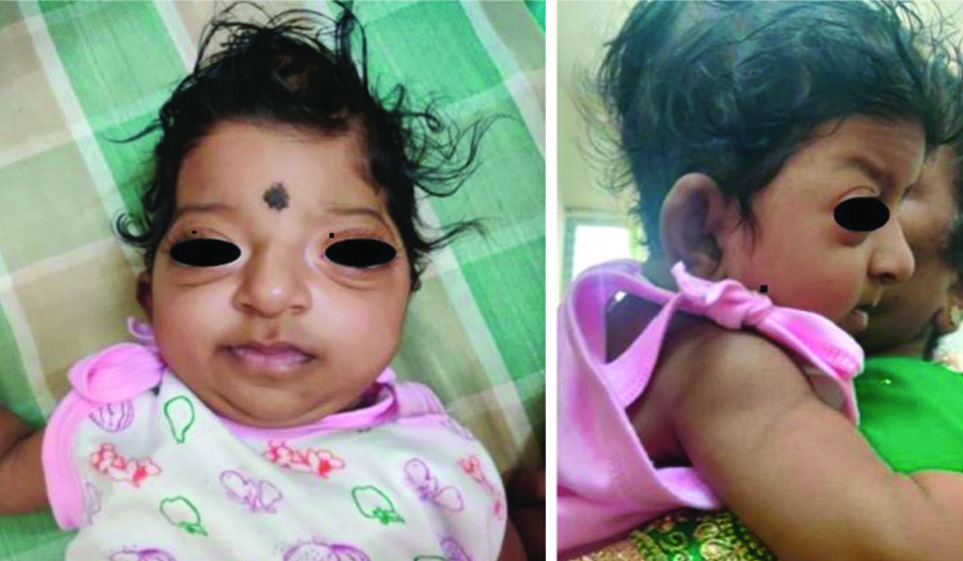
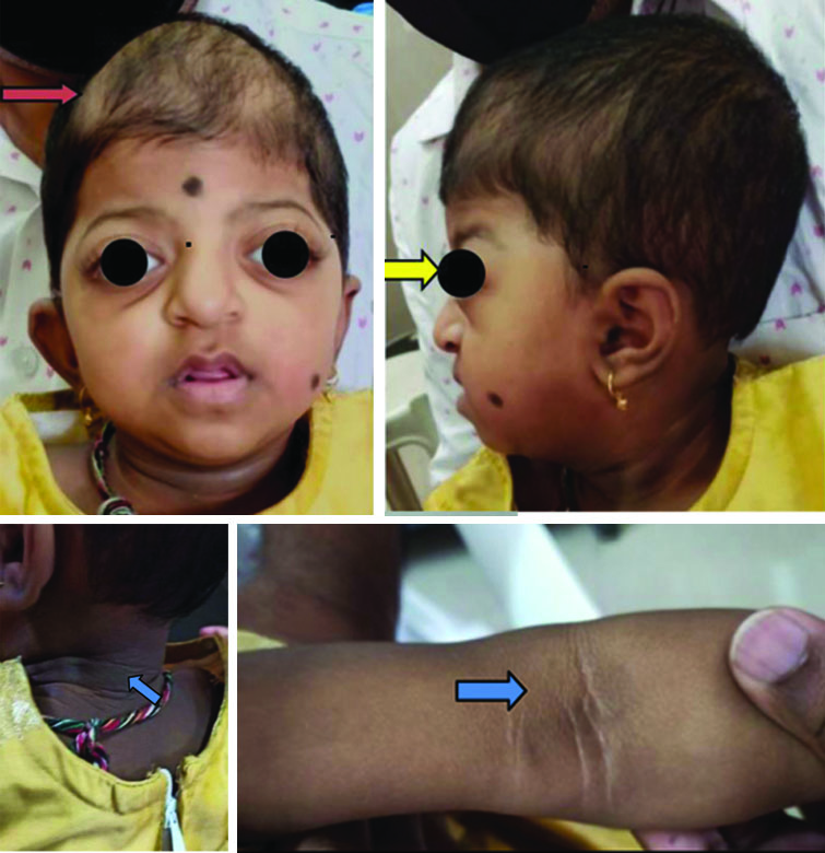
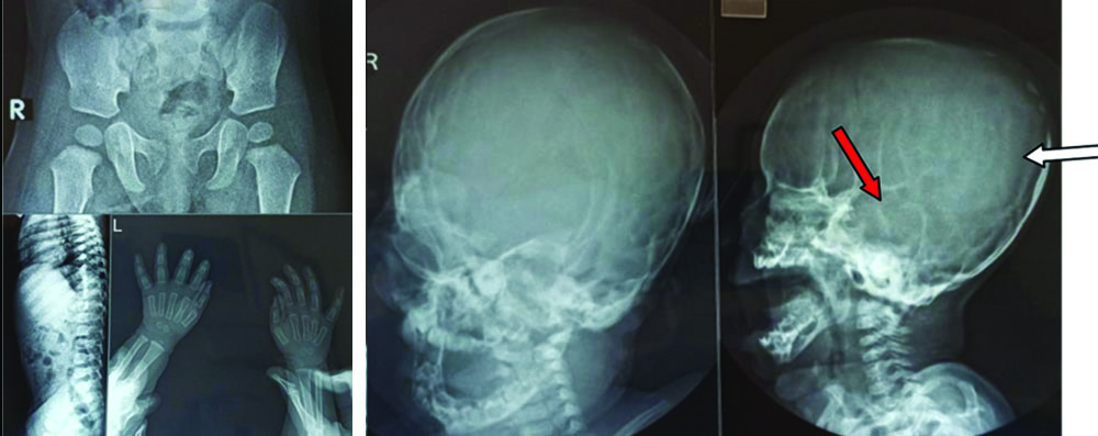
A one-year-three-month-old female child, firstborn to non consanguineous healthy Indian parents, with an uneventful antenatal course. The pregnancy was terminated at 36 weeks due to severe oligohydramnios detected late. The three-generation family tree and family history were normal. A Lower Segment Caesarean Section (LSCS) was performed and the baby cried immediately after birth. The birth weight was 2.2 kg and feeding was also started. After a few hours, the baby experienced some respiratory distress and was shifted to the Neonatal Intensive Care Unit (NICU). Intravenous medications and nasal prongs for oxygen support were initiated for three days, after which the baby was shifted out. Membranous choanal atresia was suspected and unusual facial and cranial features were described. The patient was discharged at two weeks with no residual feeding or breathing issues. At the genetic clinic evaluation at two months of age, the baby was suspected to have CSO and was clinically diagnosed with CS. Basic genetic workup, including ophthalmologic assessment, hearing evaluation, echocardiography, renal scan, Computed Tomography (CT) brain and Magnetic Resonance Imaging (MRI), was performed at four months of age, revealing no additional abnormalities. At 11 months of age, she was evaluated at the general Paediatric Department and typical Crouzon facial features with normal limbs were noted [Table/Fig-1]. However, due to the Coronavirus Disease 2019 (COVID-19) pandemic and related issues, the patient missed the follow-up. She was reviewed again at 15 months of age. This time, additional features such as motor milestone delay (evaluated at our developmental paediatric clinic using the Trivandrum Development Screening Chart) [1], disturbed sleep with snoring and incomplete closure of both eyes during sleep were documented. Anthropometry showed weight and head circumference below the 3rd percentile and length between the 15th and 50th percentile. In particular, she had diffuse skin pigmentation suggestive of Acanthosis Nigricans involving mainly the neck and flexural areas [Table/Fig-2]. Skeletal survey using a plain X-ray showed a copper beaten pattern skull (white arrow), depressed nasal bridge (red arrow), shallow orbit, normal spine and hip with normal hands (to rule out other skeletal dysplasia) [Table/Fig-3].
Case 1 at 11 months of age showing turribrachycephaly, proptosis, beaked nose and microcephaly.
At 15 months with crouzonoid facial features, turricephaly, microcephaly (red arrow) bulging eyes, prominent nose, midface retrusion (yellow arrow) Acanthosis nigricans on neck fold and antecubital fossa (blue arrow).
Case 1 skeletal survey with plain X-ray showing copper beaten pattern skull (white arrow), depressed nasal bridge (red arrow), shallow orbit, normal spine and hip with normal hands (to rule out other skeletal dysplasia).
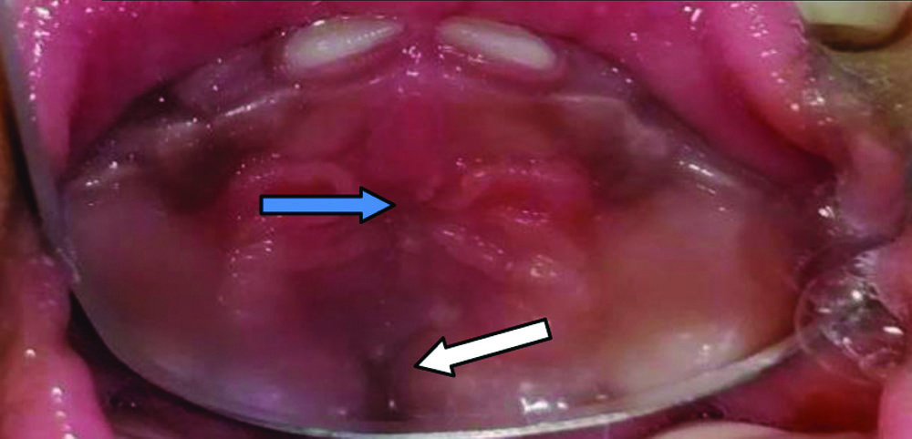
Clinically, the baby was suspected to have CAN and was advised molecular testing. Clinical exome sequencing showed a mutation in C.1178 C>A (p.Ala393Glu) in exon 9 of FGFR3 (+), confirming the diagnosis. Parental Sanger sequencing was done and the baby was screened for early-onset diabetes and hypothyroidism, but all reports came back normal. An endocrinology opinion was sought and advised follow-up at regular intervals, while ophthalmology consultation showed normal results. Dental evaluation revealed microstomia, maxillary hypoplasia, a high-arched palate, delayed teething and bilateral localised palatal swelling giving the appearance of “pseudoclefting” [Table/Fig-4]. No additional jaw lesions were detected and no treatment was provided. Yearly follow-up was recommended.
Dental evaluation showing high arch palate (blue arrow) and pseudoclefting (white arrow).
Post-genetic test counselling was conducted and the child is now under multidisciplinary management. Yearly follow-up was recommended at the respective departments. The recurrence risk and prenatal options were discussed with the parents.
Case 2
A two-year-two-month-old male, the second child born to non-consanguineous, normal Indian parents, was delivered by LSCS to a 37-year-old mother at term gestation. The birth weight was 3.2 kg and the baby cried after birth. The postnatal period was uneventful. Three-generation pedigree analysis, family history and antenatal history were unremarkable. Antenatal ultrasonographic evaluations were normal. No respiratory issues were noted, but feeding problems suggested choanal atresia. Parents noticed an abnormal head shape around five months of age and a CT evaluation at six months of age revealed obstructive hydrocephalus, which was treated with endoscopic third ventriculostomy at a different hospital. A follow-up MRI after four months showed low-lying cerebellar tonsils, crowding of the foramen magnum and supratentorial hydrocephalus.
The clinical examination revealed [Table/Fig-5] CSO causing turricephaly, frontal bossing, midface retrusion, maxillary hypoplasia, proptosis, blue sclera, posteriorly rotated ears and diffuse Acanthosis Nigricans mainly distributed over neck folds. The limbs were normal without polysyndactyly, which more strongly suggested CS over other CSO syndromes. Anthropometric evaluation fell within the normal range (3rd-97th percentile). The developmental history indicated delays in gross motor milestones and speech. Cognition appeared normal. The child was still unable to walk alone without experiencing frequent falls.
Case 2 showing tower shaped skull, broad forehead (red arrow), blue sclera, proptosis, low set posterior rotated ears (blue arrow), midface retrusion, normal limbs, post-surgical scar seen on right frontoparietal area. Acanthosis Nigricans can be seen over the neck from anterior and posterior view of neck (green arrow).
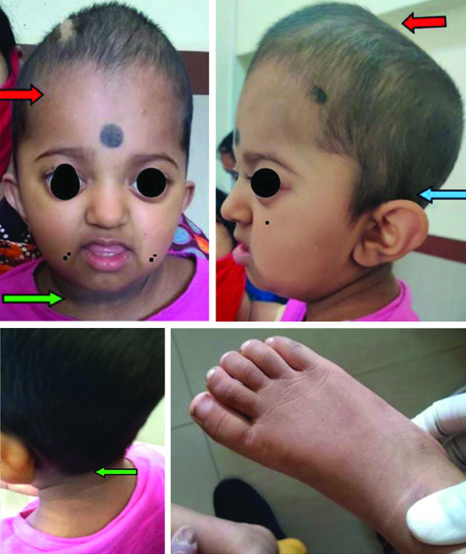
Dermatology and endocrinology opinions were sought and baseline evaluations were conducted for a general check-up, thyroid function, hyperlipidemia and glycaemic status, all of which were reported as normal. With the features mentioned above, the authors suspected CAN and conducted genetic testing- clinical exome sequencing. The report confirmed FGFR3 (+) c.1178 C>A in Exon 9 and diagnosed CAN. Post-test genetic counselling was provided and the child was followed-up in the Neurodevelopmental clinic.
Discussion
The CSO is defined as the premature fusion of one or more cranial sutures, leading to specific skull abnormalities. The morphology of the skull was first described by Hippocrates in literature and a detailed description of premature sutural fusion was delineated by Virchow in 1851 [2,3]. CSO can occur either in isolation or in association with other abnormalities, which constitute a specific syndrome. With a described frequency of 1 in 1400 to 2100 live births, CSO and associated syndromes require multidisciplinary management [4]. Common syndromes associated with CSO include Apert, Crouzon, Pfeiffer and Muenke, each with a specific underlying genetic mutation. CS features bicoronal CSO, proptosis, a beaked nose, normal IQ and normal limb findings. It has a frequency of 1 in 50,000 newborns [5].
A German dermatologist, Curth HO, in 1968 first described the association between CSO and Acanthosis Nigricans [6]. Reddy BS et al., in 1985 described a single case of Crouzon with acanthosis [7]. The mutation in CS related to the FGFR 2 gene was first described by Reardon W et al., in 1994 [8]. The present condition, which is distinct from classical CS, is CAN {Crouzono dermo skeletal syndrome (CDSS)} and Meyers GA et al., in 1995 described a specific transmembrane mutation in the Fibroblast Growth Factor Receptor (FGFR3) as the cause of CAN [9].
The CAN is a very rare condition (1 in 1 million) compared to classic CS (1 in 50,000) and constitutes around 1-2% of total syndromic CSO cases. A female preponderance of 2:1 is noted, in contrast to classic CS (1:1) [9]. All ethnic groups are equally affected with an autosomal dominant inheritance, with a suspected parental age effect yet to be confirmed. CAN includes a facial gestalt of “crouzonoid features”-bicoronal CSO, microcephaly, prominent bulging eyes due to shallow orbits (exorbitism), ocular hypertelorism, a “parrot-beaked nose,” and low-set ears [5]. In addition to this, CAN is also associated with dermatological and skeletal features such as Acanthosis Nigricans, jaw lesions, short stature and an increased association with choanal atresia and hydrocephalus [9].
The CAN and classical CS are both related to FGFR mutations. FGFRs are secreted protein ligands of transmembrane receptors that bind to the FGF class of proteins, which are involved in various cellular signaling processes, proliferation, differentiation, migration and selective apoptosis in embryogenesis. There are four members (FGFR 1/2/3/4) coded by different genes on different chromosomes, yet they share a high homology (56-71%) [10]. Structurally, they have a “canonical RTK architecture” consisting of an extracellular domain, transmembrane domain and an intracellular split tyrosine kinase domain. Mutations are mainly gain-of-function and are clinically associated with two groups of developmental disorders: CSO and the Achondroplasia family of skeletal dysplasias [6].
Molecular genetics have shown an association of CAN with FGFR3 instead of FGFR2 (Classical CS) [8]. This mutation (Ala391Glu) is found to be located near the locus for the mutation in Achondroplasia (Gly380Arg). This explains the association of mild Achondroplasia (ACH) skeletal features in CAN patients as reported in the literature (not found in the present cases). Cohen in 1995 proposed the term Crouzono Dermo Skeletal Syndrome (CDSS) instead of CAN to emphasise these associations with epidermal alterations and skeletal features [5].
In CAN, choanal atresia is more specific and also shows increased severity compared to other CSOs [9]. Severe upper airway obstruction is expected in postnatal life and may require intervention. Additionally, cleft palate, bifid uvula and a high-arched palate can also be present. Maxillary hypoplasia and mandibular prognathism can lead to dental malocclusion and an underbite. Jaw lesions may include cementomas and desmoosteoblastoma.
There is a frequent association of hydrocephalus with ventriculomegaly with or without signs of raised Intracranial Pressure (ICP). The distortion of the skull shape may mask the signs of hydrocephalus, so prompt neuroimaging and neurosurgical evaluation are necessary [5]. Chiari I malformation, posterior fossa hypoplasia and tonsillar herniation have also been reported.
Dermatological lesions in CAN include Acanthosis Nigricans, melanocytic nevus and hypopigmented post-surgical scars [9]. The occurrence of Acanthosis Nigricans is postulated to be due to the stimulation of FGFR3 seen in epidermal keratinocytes and dermal fibroblasts, leading to local epidermal hyperplasia. These may not be apparent in the early stages of life but can present early compared to other causes. Around 80% of cases will have acanthosis within the first decade of life and a 100% association is seen before puberty. The bodily distribution can also be atypical, with occurrences around the chest, abdomen, periorbital and perioral areas [9].
The FGFR3 proteins are negative regulators of bone and chondrocyte growth. An increase in tyrosine kinase signaling due to activating mutations results in skeletal changes. Skeletal features of CAN include short stature, scoliosis and broad, short fingers. Reports mention spinal canal stenosis with a narrowing of the Interpedicular (IP) distance, a finding similar to other FGFR3-related skeletal dysplasias {(ACH and Hypochondroplasia (HCH)}. Limbs are typically not involved [11,12]. Renal involvement is seen with a variable presentation of dysplastic kidney, Membranous Glomerulonephritis (MGN) and renal failure [13]. The renal system was not affected in either of the cases.
Conclusion(s)
The variation of CAN from classical CS can be summarised as a mutation in the FGFR3 gene instead of the FGFR2 gene. In CAN, the presence of diffuse and atypical Acanthosis Nigricans, an increased association of choanal stenosis, hydrocephalus with Chiari I malformation, jaw involvement and abnormal scar formation are observed. These features are not 100% specific but can be used as diagnostic clues for clinicians to differentiate CAN from classical Crouzon in clinical practice. The initiation of an early multidisciplinary approach is crucial for better survival and quality of life and individuals with CAN typically have a normal lifespan if properly intervened.
[1]. Nair MK, Nair GS, George B, Suma N, Neethu C, Leena ML, Development and validation of Trivandrum Development Screening Chart for children aged 0-6 years {TDSC (0-6)}Indian J Pediatr 2013 80(2):S248-55. [Google Scholar]
[2]. Dimopoulos VG, Kapsalakis IZ, Fountas KN, Skull morphology and its neurosurgical implications in the Hippocratic eraNeurosurg Focus 2007 23(1):E10 [Google Scholar]
[3]. Alden TD, Lin KY, Jane JA, Mechanisms of premature closure of cranial suturesChilds Nerv Syst 1999 15(11-12):670-75.Available from: https://dx.doi.org/10.1007/s003810050456 [Google Scholar]
[4]. Boulet SL, Rasmussen SA, Honein MA, A population-based study of craniosynostosis in metropolitan Atlanta, 1989-2003Am J Med Genet 2009 146A(8):984-91. [Google Scholar]
[5]. Cohen MM, Let’s call it “Crouzonodermoskeletal syndrome” so we won’t be prisoners of our own conventional terminologyAm J Med Genet 1999 84(1):74 [Google Scholar]
[6]. Curth HO, The necessity of distinguishing four types of acanthosis nigricans. In: Jadassohn W, Schirren CG, editorsCongressus internationalis dermatologiae. Munich vol1 1968 1BerlinSpringer:557-58. [Google Scholar]
[7]. Reddy BS, Garg BR, Padiyar NV, Krishnaram AS, An unusual association of acanthosis nigricans and crouzon’s disease: A case reportJ Dermatol 1985 12(1):85-90.Available from: https://dx.doi.org/10.1111/j.1346-8138.1985.tb01542.x [Google Scholar]
[8]. Reardon W, Winter RM, Rutland P, Pulleyn LJ, Jones BM, Malcolm S, Mutations in the fibroblast growth factor receptor 2 gene cause Crouzon syndromeNat Genet1994(8)(1):98-103.Available from: https://dx.doi.org/10.1038/ng0994-98 [Google Scholar]
[9]. Meyers GA, Orlow SJ, Munro IR, Przylepa KA, Jabs EW, Fibroblast growth factor receptor 3 (FGFR3) transmembrane mutation in Crouzon syndrome with acanthosis nigricansNat Genet 1995 11(4):462-64. [Google Scholar]
[10]. Goetz R, Mohammadi M, Exploring mechanisms of FGF signalling through the lens of structural biologyNat Rev Mol Cell Biol 2013 14(3):166-80.Available from: https://dx.doi.org/10.1038/nrm3528 [Google Scholar]
[11]. Robson CD, Mulliken JB, Robertson RL, Proctor MR, Steinberger D, Barnes PD, Prominent basal emissary foramina in syndromic craniosynostosis: Correlation with phenotypic and molecular diagnosesAJNR Am J Neuroradiol 2000 21(9):1707-17. [Google Scholar]
[12]. Schweitzer DN, Graham JM Jr, Lachman RS, Jabs EW, Okajima K, Przylepa KA, Subtle radiologic findings of achondroplasia in patients with Crouzon syndrome with acanthosis nigricans due to an Ala391Glu substitution in FGFR3Am J Med Genet 2001 98(1):75-91. [Google Scholar]
[13]. Vajo Z, Francomano CA, Wilkin DJ, The molecular and genetic basis of fibroblast growth factor receptor 3 disorders: The achondroplasia family of skeletal dysplasias, Muenke craniosynostosis, and Crouzon syndrome with acanthosis nigricansEndocr Rev 2000 21(1):23-39. [Google Scholar]