Hyperhaemolysis syndrome, often associated with sickle cell disease, can also occur in non sickle cell conditions following viral infections and autoimmune disorders. This case report presents a rare instance of hyperhaemolysis in the context of familial anaemia. A 41-year-old female with a history of unexplained anaemia presented with four episodes of haematemesis, abdominal pain, and headache. She had received a blood transfusion a month prior. Upon admission, she was diagnosed with pancytopenia and hypovolemic shock and was treated with packed cell volume transfusions. The patient had a significant family history of anaemia in her sisters, who also required intermittent blood transfusions. Following admission, she developed thrombocytopenia, a drop in Haemoglobin (Hb), petechiae, and subdural haemorrhage. Further investigations confirmed hyperhaemolysis syndrome. Treatment included blood transfusions, random donor platelet transfusions, and corticosteroids. The patient was discharged on prednisolone and cyclosporine. Over six months, her condition improved significantly, with no further bleeding episodes and stable Hb levels. Diagnosing hyperhaemolysis syndrome is challenging and requires a high index of suspicion. This case underscores the importance of considering hyperhaemolysis in patients with familial anaemia and unexplained haemolytic episodes. Early recognition and appropriate management are crucial for favourable outcomes.
Case Report
A 41-year-old female presented with four episodes of coffee-brown haematemesis (100-150 mL each) and black stools over two days, accompanied by mild intermittent lower abdominal pain and a dull, holocranial headache. She reported past episodes of melena over the last six months, with no prior evaluation or use of Non Steroidal Anti-Inflammatory Drugs (NSAIDs)/steroids. She had a family history of anaemia in her sisters, who also had intermittent blood transfusions. On examination, she exhibited pallor, bilateral sluggishly reactive pupils, an extensor left plantar reflex, normal deep tendon reflexes, and no neck stiffness. The abdominal examination revealed epigastric and periumbilical tenderness, as well as an enlarged spleen. She was in hypovolemic shock, with a BP of 70/50 mmHg and a pulse rate of 110 bpm. Immediate treatment included a pantoprazole infusion and transfusion of two units of packed red blood cells. Upon admission, pancytopenia was noted, and a subsequent day’s haemogram values were mentioned in [Table/Fig-1].
Blood parameters during hospital stay.
| Parameters | Day 1 | Day 4 | Day 6 | Day 7 | Day 10 |
|---|
| Haemoglobin (Hb) (gm%) | 7.1 | 6.6 | 6.4 | 6.5 | 6.9 |
| White blood cell counts (cells/μL) | 2600 | 2400 | 2100 | 3100 | 4800 |
| Platelet counts (μL) | 16000 | 6000 | 6100 | 7000 | 81000 |
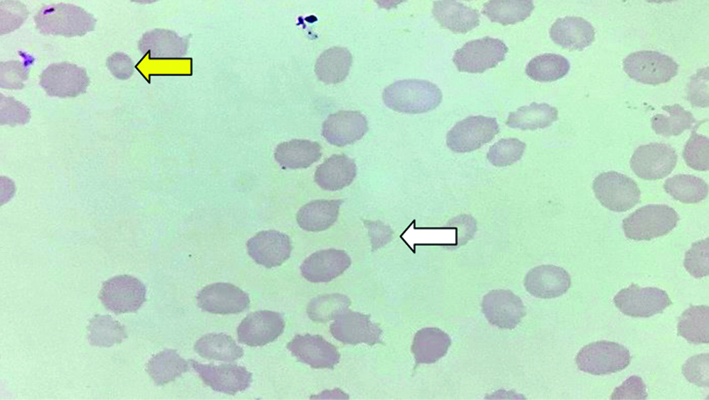
Pancytopenia was a concern. On day 1, a peripheral blood smear was sent for analysis. The peripheral blood smear [Table/Fig-2] revealed mild hypochromia, mild microcytosis, moderate anisocytosis with occasional tear-drop cells, and fragmented Red Blood Cells (RBCs). There was mild lymphopenia and marked thrombocytopenia present as well.
Mild hypochromia, mild microcytosis, moderate anisocytosis, microspherocyte (yellow arrow) with occasional tear-drop cells and fragmented RBCs (schistocyte) (white arrow) (H&E, 100x).
Liver Function Tests (LFT) and Renal Function Tests (RFT) were normal. Bleeding time was two minutes and 40 seconds, while clotting time was four minutes and 15 seconds. Prothrombin Time and International Normalised Ratio (PT-INR) as well as activated partial thromboplastin time were normal. Urine routine and microscopy showed trace proteinuria, 2-3 RBCs, and 1-2 pus cells. Iron studies and serum vitamin B12 were within normal limits. Stool occult blood was positive, and Human Immunodeficiency Virus (HIV) was negative.
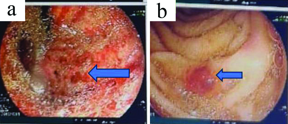
Endoscopy indicated erosive pangastritis with gastric and duodenal ulcers, classified as Forrest grade IIc [Table/Fig-3a,b], and the rapid urease test was negative. The patient was started on high-dose intravenous pantoprazole and oral liquid antacids.
a) Showing small oval shaped ulcers with regular margins in body and fundus with linear streaks of erythema suggesting pangastritis of the stomach; b) Showing small oval shaped ulcers in the 2nd part of duodenum.
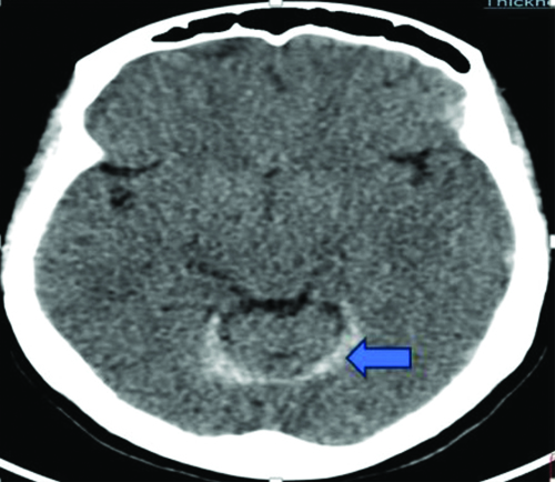
A Non Contrast Computed Tomography (NCCT) scan of the brain demonstrated a rim of acute subdural haemorrhage along the right leaf of the tentorium cerebelli [Table/Fig-4]. Subsequently, she was started on injection mannitol 100 mL intravenously three times a day.
Showing rim of acute subdural haemorrhage along the right leaf of tentorium cerebelli.
Ultrasonography of the abdomen and pelvis showed mild fatty liver with a mildly dilated portal vein. The spleen was enlarged, measuring 14.8 cm in size, but was normal in shape and echotexture, with no perisplenic collaterals noted. A small intramural uterine fibroid was also observed.
Four days after admission, the patient developed gross haematuria, with a urine routine showing 90-100 RBCs. Laboratory investigations were repeated, revealing a Hb level that dropped to 6.6 g/dL, Total Leukocyte Count (TLC) of 2400, and platelet count of 6000/cm3. Given the persistent low platelet count along with low Hb and significant haematuria, a haemolytic anaemia work-up was conducted, as depicted in [Table/Fig-5]. The differential diagnosis included paroxysmal nocturnal haemoglobinuria, paroxysmal cold haemoglobinuria, platelet dysfunction, sickle cell anaemia, thalassaemia, sideroblastic anaemia, and enzyme abnormalities.
Depicting the specific laboratory investigations regarding haemolytic anaemia.
| Tests | Values |
|---|
| Serum vitamin B12 (pg/dL); Normal: (187-883) | 780 |
| Reticulocyte count (%); Normal: 0.5-2.5 | 5 |
| Erythrocyte Sedimentation Rate (ESR) (mm/hour); Normal: upto 20 {age <50 years} | |
| Direct Coombs Test (DCT) | Positive |
| Indirect Coombs Test (ICT) | Negative |
| Serum D-dimer (ng/dL); Normal: 0-500 | 398 |
| Serum fibrinogen (mg/dL); Normal: 238-498 | 263 |
| Glucose-6 Phosphate Dehydrogenase (G6PD) | Negative |
| Sickling test | Negative |
| Hb electrophoresis | Negative |
| Serum Lactate Dehydrogenase (LDH) (U/L); Normal: 238-498 | 201 |
| Serum haptoglobin | 41 units |
| Urine haemoglobinuria | Negative |
| Urine myoglobinuria | Negative |
| Anti neutrophilic antibody by immunofluorescence | Negative |
| Anti-neutrophilic antibody by blot | Negative |
| Peri-nuclear antineutrophilic antibody | Negative |
| Cytoplasmic antineutrophilic antibody | Negative |
| Complement C3 (gm/L); Normal: (0.80-1.70) | 0.63 |
| Complement C4 (gm/L); Normal: 0.12-0.36 | 0.22 |
| Cold agglutinin test | Negative |
| Donath-landsteiner autoantibody | Negative |
| Serum ferritin (ng/mL); Normal: 4.63-204 | 516 |
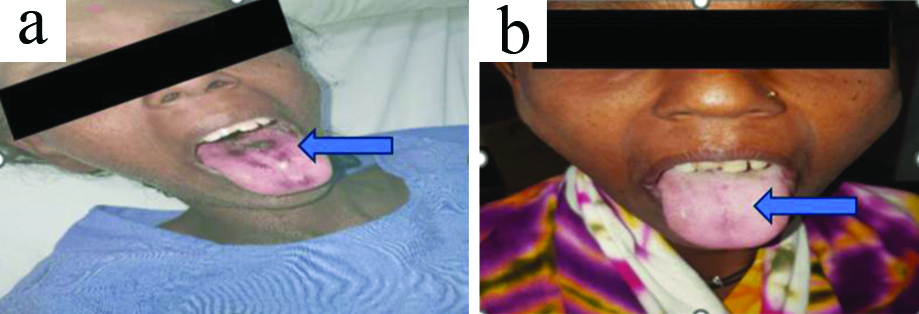
On day 6 of the hospital stay, the patient developed reddish spots on the tongue [Table/Fig-6a] and palate, which improved after treatment [Table/Fig-6b]. Thus far, 32 random donor platelets had been transfused to the patient to prevent further enlargement of the subdural haemorrhage.
a) Showing petechial haemorrhages over tongue; b) Showing oral petechial lesions which subsided post-treatment.
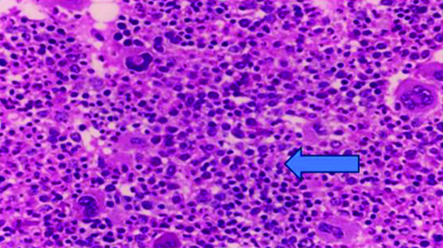
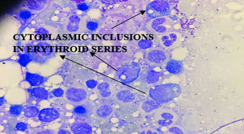
On day 7, a haematologist’s opinion was sought, and she was advised to undergo a bone marrow examination. The bone marrow examination suggested hypercellular marrow with dimorphic erythropoiesis and mild dysplastic changes in the erythroid and megakaryocytic lineages. Some cytoplasmic inclusions were also observed in the erythroid series, pointing towards haemolysis due to postviral infections or myelodysplastic syndrome [Table/Fig-7,8].
Bone marrow biopsy with hypercellular marrow spaces and erythroid hyperplasia (H&E, 40x).
Bone marrow imprint shows cytoplasmic inclusions in erythroid series (Leishman stain- 100x oil immersion field).
After review by the haematologist, the patient was started on intravenous dexamethasone 32 mg once daily for four days, followed by oral prednisolone at a dose of 1 mg/kg, tapered over two months, and cyclosporine 100 mg twice daily. She was advised to monitor her Complete Blood Count (CBC), LFT, RFT, and serum electrolytes weekly for the next three months. The viral panel for inclusion bodies and myelodysplastic panel results were as follows: Parvovirus B19 (ELISA), Epstein-Barr Virus (EBV), and cytomegalovirus PCR RNA were negative. The myelodysplastic panel was also negative, and karyotyping for both the patient and her siblings was normal. Therefore, the diagnosis was established as immune-mediated haemolytic anaemia.
A CT scan of the brain was repeated on the 10th day, which suggested a decrease in volume and attenuation of the acute component of the bleed, along with a decrease in mass effect [Table/Fig-9]. The oral lesions also subsided [Table/Fig-6b].
Showing rim of acute subdural haemorrhage has decreased in size.

After 10 days of therapy with prednisolone, a repeat investigation was conducted. After 20 days, patient’s CBC showed a Hb level of 7.20 g/dL, a TLC of 5200, and a platelet count of 117,000/cm3. The reticulocyte count was 2.5%. Urine Routine Microscopy (R/M) results indicated that protein, glucose, acetone, and bile pigments were all negative, and no RBCs were present. The patient is regularly attending follow-up appointments, and her haemograms are within normal limits. Currently, she is on a tapering dose of steroids and continues to take cyclosporine with regular monitoring.
Discussion
Hyperhaemolysis syndrome is a complication that primarily affects patients with underlying haemoglobinopathies, most commonly seen in those with sickle cell disease [1]. However, it can also occur in patients with other haemoglobinopathies, such as thalassaemia, Hb C, and Hb SC disease, following blood transfusions [2]. Additionally, hyperhaemolysis syndrome can be associated with other haematologic disorders, including myelofibrosis, anaemia of chronic disease, and lymphoma. The syndrome is diagnosed when severe anaemia occurs paradoxically after a blood transfusion, resulting in post-transfusion haemoglobin levels that are lower than pretransfusion levels [3,4].
In the present study, a 41-year-old female with a history of unexplained anaemia presented with four episodes of haematemesis, abdominal pain, and headache. She had received a blood transfusion one month prior. Upon admission, she was diagnosed with pancytopenia and hypovolemic shock and was treated with packed RBC transfusions. Shankar K et al., reported that hyperhaemolysis syndrome affects individuals with haemoglobinopathies needing frequent transfusions, causing a sudden drop in haemoglobin levels post-transfusion [5]. In this case, the reaction occurred 20 days after the patient’s last transfusion. Win N noted that hyperhaemolysis can be acute (within seven days, Direct Antiglobulin Test (DAT) negative, no alloantibodies) or delayed (beyond seven days, DAT positive, alloantibodies present) [6]. This case was delayed, occurring 20 days post-transfusion with a positive DAT. Management involves stopping further transfusions.
Paroxysmal nocturnal haemoglobinuria was considered due to microscopic haemoglobinuria but was excluded due to the lack of renal involvement. Paroxysmal cold haemoglobinuria was ruled out based on a negative Donath-Landsteiner autoantibody test. Platelet dysfunction was dismissed as bleeding and clotting times were normal. Sickle cell anaemia, thalassaemia, sideroblastic anaemia, and enzyme abnormalities were ruled out through sickling tests, haemoglobin electrophoresis, and family member screening. Bone marrow examination excluded malignancies, postviral infections, and other infections. Autoimmune connective tissue diseases and vasculitis were ruled out by a negative antinuclear antibody test and Antineutrophil Cytoplasmic Antibodies (ANCA) profile.
Hyperhaemolysis is an atypical transfusion reaction that can occur in any cytopenic condition requiring multiple transfusions. The mechanism involves “bystander haemolysis,” where both autologous and transfused RBCs are destroyed, even if autologous cells lack the target antigen [7]. It is important to distinguish hyperhaemolysis syndrome from delayed transfusion reactions, noting that hyperhaemolysis lacks alloantibodies. Diagnostic indicators include decreased reticulocyte counts and elevated ferritin and Lactate Dehydrogenase (LDH) levels, which normalise post-treatment. In present case, patient also had reduced reticulocyte counts and elevated LDH and ferritin levels [8]. According to Eberly LA et al., hyperhaemolysis syndrome can occur in patients without haemoglobinopathies, even with delayed haemolytic transfusion responses [9]. Although this syndrome is typically seen in sickle cell anaemia and thalassaemia major, in present case patient’s sickling test and Hb electrophoresis results were negative.
The treatment consisted of blood transfusions, random donor platelet transfusions, and corticosteroids. The patient was discharged with prednisolone and cyclosporine. Over six months, her condition improved significantly, with no further bleeding episodes and stable Hb levels. Win N et al., reported using intravenous methylprednisolone and Intravenous Immunoglobulins (IVIg) to treat hyperhaemolysis syndrome by suppressing macrophages that destroy RBCs, thereby avoiding further transfusions in conditions like sickle cell anaemia and myelofibrosis [10]. Although IVIg has antiviral and antibacterial effects, it carries risks such as renal damage, stroke, and thrombosis. Steroids, which affect CD4 T-cells and B-cells, have a delayed onset. In index patient, platelet levels significantly improved after the 7th to 8th doses of intravenous dexamethasone.
Conclusion(s)
This case highlights the importance of considering hyperhaemolysis in patients with familial anaemia and unexplained haemolytic episodes. Early recognition and appropriate management are essential for achieving favourable outcomes.
[1]. Danaee A, Inusa B, Howard J, Robinson S, Hyperhemolysis in patients with hemoglobinopathies: A single-center experience and review of the literatureTransfus Med Rev 2015 29(4):220-30.10.1016/j.tmrv.2015.06.001 [Google Scholar] [CrossRef]
[2]. Dasgupta A, Wahed A, Clinical Chemistry, Immunology and Laboratory Quality Control 2014 Elsevier [Google Scholar]
[3]. Banks M, Shikle J, Hyperhemolysis syndrome in patients with sickle cell diseaseArch Pathol Lab Med 2018 142(11):1425-27.10.5858/arpa.2017-0251-RS30407854 [Google Scholar] [CrossRef] [PubMed]
[4]. Stainsby D, Jones H, Wells AW, Gibson B, Cohen H, SHOT Steering GroupAdverse outcomes of blood transfusion in children: Analysis of UK reports to the serious hazards of transfusion scheme 1996-2005Br J Haematol 2008 141(1):73-79.10.1111/j.1365-2141.2008.07022.x [Google Scholar] [CrossRef]
[5]. Shankar K, Shah D, Huffman DL, Peterson C, Bhagavatula R, Hyperhemolysis syndrome in a patient with sickle cell disease and acute chest syndromeCureus 2021 13(1):e13017 [Google Scholar]
[6]. Win N, Hyperhemolysis syndrome in sickle cell diseaseExpert Rev of Hematol 2009 2(2):111-15. [Google Scholar]
[7]. El Alaoui K, Benghiat FS, Colard M, An unusual case of delayed hemolytic transfusion reaction with hyperhemolysis syndrome due to anti-Jkb and anti-Fya alloantibodiesJ Hematol 2022 11(2):66-70. [Google Scholar]
[8]. Menakuru SR, Priscu A, Dhillon V, Salih A, Acute hyperhemolysis syndrome in a patient with known sickle cell anaemia refractory to steroids and IVIG treated with tocilizumab and erythropoietin: A case report and review of literatureHematol Rep 2022 14(3):235-39. [Google Scholar]
[9]. Eberly LA, Osman D, Collins NP, Hyperhemolysis syndrome without underlying hematologic diseaseCase Rep Hematol 2015 2015(1):180526 [Google Scholar]
[10]. Win N, Yeghen T, Needs M, Chen FE, Okpala I, Use of intravenous immunoglobulin and intravenous methylprednisolone in hyperhaemolysis syndrome in sickle cell diseaseHematology 2004 9(5-6):433-36. [Google Scholar]