Worldwide, bladder cancer is the seventh most common cancer and the most prevalent malignant neoplasm of the urinary tract. It is three to five times more frequent in men than in women, due to factors like smoking, tobacco use, the protective effect of female hormones, and occupational exposure. The mean age of incidence is between 65 and 75 years [1,2]. Incidence, morbidity, recurrence, progression, and mortality rates vary among countries and ethnic groups. Due to the high rate of tumour recurrence and the need for frequent cystoscopy, bladder cancer is one of the cancers with the highest cost per patient in our healthcare system.
Bladder cancer is morphologically heterogeneous; more than 90% of bladder cancer cases are urothelial (transitional cell) carcinoma, whereas primary squamous cell carcinoma, adenocarcinoma, small cell carcinoma, and other tumours are less common [3]. Clinically, urothelial carcinoma of the bladder presents as two distinct phenotypes: the superficial (non muscle invasive) type, which is more common, and muscle-invasive urothelial carcinoma, which represents one-third of all cases. Approximately 50% of superficial tumours recur as non muscle invasive tumours, and 10-15% progress to muscle invasive disease. The mainstay of therapy for superficial tumours is Transurethral Resection (TURB), with or without intravesical chemotherapy and immunotherapy (BCG), while invasive tumours need aggressive combined treatment modalities, including cystectomy and chemotherapy.
According to the World Health Organisation (WHO) classification of tumours of the urinary tract, 5th edition, urothelial tumours are divided into invasive urothelial neoplasms (invasive urothelial carcinoma) and non invasive urothelial neoplasms. Non invasive urothelial neoplasms are classified into urothelial papilloma, inverted urothelial papilloma, Papillary Urothelial Neoplasm of Low Malignant Potential (PUNLMP), low-grade, high-grade non invasive papillary urothelial carcinoma, and urothelial carcinoma in situ. Histologic grading is one of the most important prognostic factors in bladder cancer, and this grading guides the choice of treatment options [1,3].
Several recent studies have revealed a significant correlation between the loss of p16 expression and progression in non invasive (pTa) and superficially invasive (pT1) urothelial carcinoma, highlighted the role of p16 as a prognostic biomarker. Loss of p16 has been found to be associated with high-grade and invasion in urothelial carcinoma [4-6]. p16 (CDKN2A), located at chromosome 9p21, is a tumour suppressor gene that inhibits Cyclin D-Dependent Protein Kinases (CDK), playing a vital role in the regulation of the G1-S transition and is proved to be commonly deleted or mutated in the oncogenesis of bladder cancer. Therefore, loss of its function results in abnormal cell proliferation. About 40-50% of urothelial carcinomas exhibit a heterozygous loss of p16, which is a common carcinogenic mechanism in urothelial carcinomas [7,8].
Understanding the expression of p16 can aid in predicting the grade and invasive nature of lesions. This is particularly useful when biopsy samples or in the absence of deep biopsy is not performed, as well as in cases where muscle tissue is not sampled in the deep biopsy specimen. Moreover, bladder biopsies and transurethral resections are prone to tissue artifacts from cautery and tangential histologic sectioning, further complicating the interpretation of tumour grade and invasion. Owing to the above reasons, accurate identification of lamina propria invasion by urothelial carcinoma might be difficult in histopathology sections [9,10].
In this background, this study was undertaken in South Indian population to estimate the frequency of p16 expression in urothelial carcinoma of the urinary bladder and to understand its utility in determining tumour invasiveness. This will help clinicians in prognostication and in determining treatment strategies. Treatment for non invasive urothelial carcinoma aims to reduce recurrence and prevent progression to more advanced disease, while in invasive carcinoma treatment strategy for invasive carcinoma includes more aggressive therapies, such as cystectomy and targeted therapies, to improve patient survival.
Materials and Methods
This was a cross-sectional study involving 37 cases of urothelial carcinoma, conducted in the Department of Pathology at Pushpagiri Institute of Medical Sciences and Research Centre, a tertiary care institution in South Kerala, India. The study was carried out over a period of one year and six months, from December 2019 to May 2021. Approval was obtained from the ethics committee, with the Institutional Ethical Committee (IEC) reference number PIMSRC/E1/388A/41/201.
Inclusion criteria: All TURBT specimens received in the Department of Pathology at Pushpagiri Institute of Medical Sciences and Research Centre that were diagnosed as urothelial carcinoma (both non invasive and invasive) on histopathology during the study period were included in the study.
Exclusion criteria: Patients with recurrence of urothelial carcinoma, absence of deep biopsy, squamous cell carcinoma, adenocarcinoma, papilloma, PUNLMP, and patients who had received prior treatment in the form of chemotherapy or radiotherapy were excluded from the study.
Study Procedure
IHC staining for p16 (PRM121 clone from PATH INSITU), with staining localisation in the nucleus and cytoplasm, was performed on 3 μm sections obtained from formalin-fixed paraffin-embedded blocks. The sections stained by IHC were examined alongside Haematoxylin and Eosin (H&E)-stained sections to determine the expression of p16. Tonsil tissue was used as the control for p16 staining.
p16 staining was classified as positive or negative depending on nuclear staining.
The samples were stratified into three groups according to the percentage of positive cell nuclei [4].
Less than 10% nuclear staining, or the absence of nuclear staining with only cytoplasmic staining, was considered negative.
Positive expression was divided into 10-50%, which corresponded to low expression and represented a normal nuclear staining pattern.
More than 50% nuclear staining was considered positive, staining with high expression of p16.
Patient demographics, including age and sex distribution, were analysed, as well as the association of the above parameters with the grade and invasiveness of the tumour. p16 expression was analysed in all cases, along with its association with the age and sex of the patient, and the tumour grade {according to WHO and International Society of Urological Pathology (ISUP) 2004} and its invasive nature were also sought for [1,3].
Statistical Analysis
The calculated data obtained was entered into a Microsoft Office Excel 2021 sheet. This data was then analysed using SPSS version 16.0. The frequency of p16 expression in urothelial carcinoma of the bladder and its association with grade and invasiveness were evaluated using Fisher’s-exact test. A p-value of <0.05 was considered statistically significant.
Results
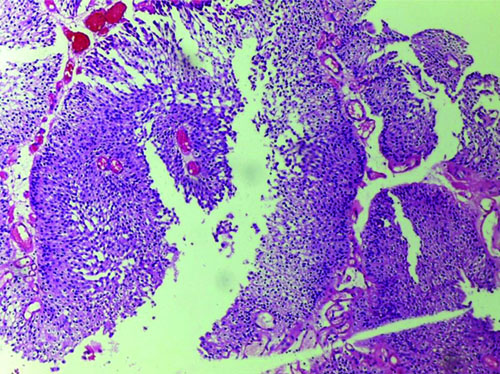
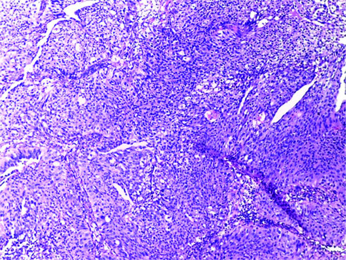
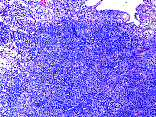
Out of 37 cases, there were 19 cases of low-grade non invasive papillary urothelial carcinoma [Table/Fig-1], four cases of high-grade non invasive papillary urothelial carcinoma [Table/Fig-2], and 14 cases of high-grade infiltrating urothelial carcinoma [Table/Fig-3].
Low-grade non invasive papillary urothelial carcinoma (H&E,10x).
High-grade non invasive papillary urothelial carcinoma (H&E,10x).
High-grade infiltrating urothelial carcinoma (H&E,10x).
In this study, the age of the patients with urothelial carcinoma of the bladder ranged from 40 years to 88 years, with a mean age of 69.81±10.59 years. There was a male predominance, with 32 out of 37 patients (86.5%) being male and only five (13.5%) female. Grading and invasiveness of the tumour had no statistically significant association among the various age groups and between genders.
The grading of the tumour exhibited an equal distribution among the various age groups, and hence the difference was found to not statistically significant (p-value=1.000). In this study, there was a male predominance, there was no significant association between gender and the grade of the tumour (p-value=0.660). Additionally, no significant differences were found between age groups (p-value=0.790) or gender (p-value=1.00) concerning the invasion status of the tumours.
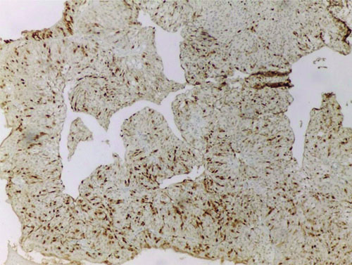
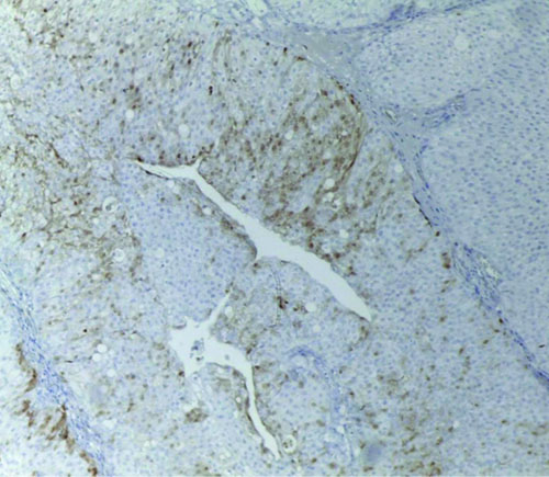
Regarding the association of p16 expression with the grade of urothelial carcinoma, there were 18 cases of high-grade urothelial carcinoma in this study. Among these, 11 cases showed loss of p16 with a negative staining pattern, four cases showed a normal staining pattern, and three cases showed high p16 expression. There were 19 cases of low-grade urothelial carcinoma, 18 showed low/normal p16 expression, while one case exhibited overexpression of p16 [Table/Fig-4,5]. Cases that showed loss of p16 expression were invariably high-grade tumours [Table/Fig-6]. Loss of p16 was not observed in any of the low-grade tumours; most low-grade tumours had low levels of p16 expression corresponding to a normal staining pattern. This difference was found to be statistically significant (p-value <0.001).
High expression of p16 seen in one case of low grade non invasive urothelial carcinoma (10X).
Low positive/normal p16 expression in low-grade non invasive urothelial carcinoma (10X).
Association of p16 expression with grade.
| p16 | Grade | p-value |
|---|
| High | Low |
|---|
| High | 3 (75.0) | 1 (25.0) | <0.001 |
| Low (normal) | 4 (18.2) | 18 (81.8) |
| Negative | 11 (100.0) | 0 |
Fisher’s-exact value- 22.457, p-value <0.05, statistically significant
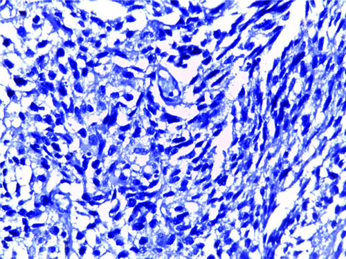
With respect to the association of p16 expression with invasion, there were 14 cases of invasive urothelial carcinoma, among which 11 cases showed loss of p16 with a negative staining pattern [Table/Fig-7]. One case showed normal staining pattern, and two cases showed high p16 expression. Among the 23 cases of non invasive tumours, 21 cases showed normal/low p16 expression, and 2 cases showed high p16 expression. It was found that invasiveness was more common among those with negative p16 expression, while those with low p16 expression were non invasive tumours [Table/Fig-8]. This difference was found to be statistically significant (p-value <0.001). There was no statistically significant relationship between p16 expression and age (p-value=0.906) or gender (p-value=0.621).
Negative p16 expression in high-grade invasive urothelial carcinoma (40X). [Loss of p16-Score-0]
Association of p16 expression with invasion.
| p16 | Invasion | p-value |
|---|
| Invasive | Non invasive |
|---|
| High | 2 (50.0) | 2 (50.0) | <0.001 |
| Low | 1 (4.5) | 21 (95.5) |
| Negative | 11 (100.0) | 0 |
Fisher’s-exact value- 31.254, p-value <0.05, statistically significant
Discussion
Bladder cancer is the most common malignancy involving the urinary system, with urothelial carcinoma being the predominant histologic type. Depending on the grade, it can be classified as low-grade or high-grade carcinoma. Based on invasion, it can be invasive or non invasive, each with its own prognosis. Muscle-invasive urothelial carcinomas typically follow a rapid progression route and metastasise to lymph nodes, bones, lungs, liver, and peritoneum, in that order of frequency. They tend to be aggressive, with usually fatal outcome. On the other hand, non muscle-invasive urothelial carcinomas are less aggressive, but the high frequency of recurrences requires strict follow-up and many revision surgeries [11,12].
Grade and invasion are the two important prognostic factors. Low-grade carcinomas are associated with good prognosis compared to high-grade carcinomas. As further management and prognosis of patients is based on accurate grading of these tumours, this categorisation is of utmost significance. Understanding the expression of p16 can aid in determining the grade of the tumour and predicting the invasive nature of the lesion. This can be more so useful when there are limited biopsy samples, in the absence of deep biopsy, or in cases where muscle tissue is not sampled in the deep biopsy specimen. Moreover, bladder biopsies and transurethral resections are prone to tissue artifacts from cautery and tangential histologic sectioning, further complicating the interpretation of tumour grade. The recognition of lamina propria invasion by urothelial carcinoma is one of the most challenging issues in surgical pathology.
Therefore, present study aimed to determine the usefulness of p16 IHC staining as an adjunctive tool in grading urothelial carcinomas and in assessing their invasive potential. This will help clinicians in prognostication and in determining treatment strategies. In non muscle-invasive disease, treatment is directed at reducing recurrences and preventing progression to a more advanced stage. The goal of therapy in muscle-invasive disease is to determine whether the bladder should be removed or if it can be preserved without compromising survival, to determine if the primary lesion can be managed independently or if patients are at high-risk for distant spread [13-16].
Al Shaikhly AW et al., and Hashmi AA et al., found the mean age of patients to be 60.9±12.55 and 63.43±14.88 years, respectively [4,5]. In present study, there was a male predominance, with 32 (86.5%) of the total 37 participants being male and only 5 (13.5%) being female. This finding was comparable to those in published studies. In present study, the proportion of high-grade and low-grade carcinomas was almost equal. Among the 37 study subjects, 19 (51.4%) had low-grade urothelial carcinoma, while 18 (48.6%) had high-grade tumours. Among the cases included in the study, the number of non invasive tumours was greater than that of invasive tumours. Of the 37 cases of urothelial carcinoma, 14 (37.8%) were invasive tumours, while 23 (62.2%) were non invasive tumours. Among the 14 invasive carcinomas, 12 were high-grade urothelial carcinomas infiltrating into the muscularis, and two were high-grade papillary urothelial carcinomas showing invasion into the lamina propria and among these 14 invasive cases, one case showed low p16,11 cases showed negative p16 and 2 cases showed high p16.
Regarding p16 expression in urothelial carcinoma, present study found that 11 (30%) cases of bladder urothelial carcinoma showed loss of p16 expression; 22 (60%) cases exhibited low p16 expression, corresponding to a normal p16 staining pattern; and four cases showed overexpression of p16.
In the study by Wu WJ et al., homozygous deletion of p16 was detected in 12 cases (20%) of urothelial carcinomas [Table/Fig-9] [17]. Hashmi AA et al., analysed p16 in 121 cases of bladder cancer and its association with tumour grade, lamina propria invasion, muscularis propria invasion, and survival status [5]. Low expression of p16 was noted in 86% of cases [Table/Fig-9].
Data from other studies and present study [4,5,16-18,21].
| Parameters | Al Shaikhly AW et al., [4] (Iraq) | Hashmi AA, et al., [5] (Karachi) | Wu WJ et al., [17] (Taiwan) | Mao X et al., [21] (China) |
|---|
| 2017 | 2019 | 2000 | 2018 |
|---|
| Mean age (years) | 60.9±12.55 | 63.43+14.88 | 68 | 64.2 |
| Sex distribution | Male % | 70.8 | 72.8 | 34.6 | 58.4 |
| Female % | 29.2 | 27.2 | 65.4 | 41.6 |
| High-grade tumours | n (total) | 24 (48) | 71 (151) | 67 (78) | 47 (65) |
| Low-grade tumours | 24 (48) | 80 (151) | 11 (78) | 18 (65) |
| Invasive | 17 (48) | 49 (151) | 54 (78) | 42 (65) |
| Non invasive | 31 (48) | 102 (151) | 24 (78) | 23 (65) |
| p16 immunoreactivity | | 83% of non invasive tumours showed positive p1652% of invasive tumours showed negative p16.Significant association found with invasiveness. | Significant association of high p16 expression was noted with worse long term survival (p-value=0.020) and patients with low p16 expression were at low-risk (HR=0.194) to die of disease as compared to patients with high p16 expression. | High p16 expression was associated low-grade carcinoma p-value=0.046and absence of tumour invasivenessp-value 0.003. | Heterozygous and homozygous loss of p16 was identified in 12 (18.5%) and 23 (35.4%) patients with UC, respectively. |
| Parameters | | da Silva JNL et al., [16] (Brazil) | Krüger S et al., [18] (Leubeck) | Present study (India) |
| 2020 | 2005 | 2024 |
| Mean age (years) | | 63 | 68 | 69.81±10.59 |
| Sex distribution | Male % | 72 | 82.2 | 86.5 |
| Female % | 28 | 17.8 | 13.5 |
| High-grade tumours | n (total) | 39 (93) | - | 18 (37) |
| Low-grade tumours | 54 (93) | - | 19 (37) |
| Invasive | 17 (93) | - | 14 (37) |
| Non invasive | 76 (93) | 63 (63) | 23 (37) |
| p16 immunoreactivity | | p16, altered in 31% of patients associated with presence of recurrence (p-value=0.005) and lower recurrence-free survival (p-value=0.004). | Loss of p16 expression in 54% and concluded that loss of p16 expression of T1 bladder carcinomas correlated with decreased progression-free survivaland worse prognosis of bladder cancer patients. | Cases which showed loss of p16 expression were invariably high-grade and invasive tumours. (100)p-value <0.001. |
p16 expression in invasive tumours: In the present study, there were 14 cases of invasive urothelial carcinoma, among which 11 cases (78%) showed loss of p16 with a negative staining pattern; one case exhibited a normal staining pattern, and two cases showed high p16 expression. Among the 23 cases of non invasive tumours, 21 cases showed normal/low p16 expression, while 2 cases showed high p16 expression. It was found that cases with negative p16 expression were invariably invasive, and among those with low p16 expression, the majority were non invasive tumours. This difference was found to be statistically significant. Thus, retention of low/normal p16 expression can be considered a favourable prognostic factor. A similar observation was noted in the study by Krüger S et al., who noted a significant correlation between loss of p16 expression and tumour progression in patients with minimally invasive bladder cancer [18]. Hashmi AA et al., and Yang CH et al., demonstrated a significant negative association between p16 expression and invasiveness, carcinoma grade, and tumour size [5,19]. Meta-analyses performed by Gan X et al., showed downregulated p16 expression is associated with poor prognosis in patients with bladder cancer [20]. Al Shaikhly AW et al., noted increased p16 expression in non invasive papillary urothelial carcinoma cases and considered p16 a good prognostic factor [Table/Fig-9] [4].
The mean age in the study population was found to be 69.81±10.59 years, which was consistent with published literature [Table/Fig-9].
p16 expression and grade of urothelial carcinoma: Cases which showed loss of p16 expression in present study were invariably high-grade tumours and were muscle invasive carcinomas. Loss of p16 was not seen in any of the low-grade tumours. Most of the low-grade tumours had low levels of p16 expression corresponding to a normal staining pattern. This difference was also found to be statistically significant. Therefore, the loss of p16 is an important mechanism in the pathogenesis of invasive urothelial carcinomas. The authors recommend the use of p16 in assigning grade to urothelial carcinoma and assessing invasion, especially when histological features are borderline and when samples are inadequate [Table/Fig-9] shows comparison of different studies [4,5,16-18,21].
Limitation(s)
The limitations of present study included a limited sample size, and follow-up was not part of the study due to time constraints. Additionally, squamous cell carcinomas and adenocarcinomas were not included in present study.
Conclusion(s)
The p16 is a valuable biomarker that can serve as a useful adjunct to morphology in the diagnosis and categorisation of urothelial carcinoma of the urinary bladder. The loss of p16 is invariably observed in high-grade and invasive urothelial carcinoma. This loss is an important mechanism in the oncopathogenesis of muscle-invasive urothelial carcinomas and plays a role in the progression of urothelial carcinoma of the urinary bladder. Analysing the expression of p16 can assist in determining the grade of the tumour and predicting the invasive nature of the lesion. This can be more so useful when biopsy samples are limited or in the absence of deep biopsy, especially when muscle tissue is not sampled or when tissue artifacts from cautery and tangential histologic sectioning complicate the interpretation of tumour grade and the recognition of lamina propria invasion. Therefore, p16 marker is recommended to determine the grade of the tumour and its invasiveness. This will aid clinicians in prognostication and in determining treatment strategies.
Fisher’s-exact value- 22.457, p-value <0.05, statistically significant
Fisher’s-exact value- 31.254, p-value <0.05, statistically significant