Case Report
A 67-year-old female presented to the emergency department with complaints of left lower limb swelling for the past seven days. The swelling was acute in onset and progressive, associated with severe pain. The pain was sharp and cramping in nature, moderate to severe in intensity, radiating to the left thigh and knee. It was aggravated by movement, with no specific relieving factors. The patient experienced partial loss of sensation in the groin, radiating to the left thigh, accompanied by an inability to flex her left thigh, leading to difficulty in walking. She reported difficulty in sitting, walking, and climbing stairs. These symptoms were suggestive of femoral nerve compression. Additionally, the patient complained of abdominal pain in the left iliac region, which had an acute onset and was dull and aching in nature, mild to moderate in intensity, persisting for the past seven days. This abdominal pain was associated with distention and vomiting. The patient had a history of fever 4-5 days prior, which was acute in onset and accompanied by chills and high-grade fever. She also reported haematuria for the past 2-3 days. There was no history of trauma, chest pain, breathlessness, or joint pain. The patient had a known history of hypertension and diabetes for the past three years and was on the following medications: Tab glimepiride, metformin, and voglibose (1/500/0.2 mg) orally twice a day, and Tab. amlodipine (5 mg) once daily. She had also undergone double valve replacement surgery 20 years ago and was prescribed Tab warfarin (5 mg) orally once daily, as well as Tab digoxin (0.25 mg) orally once daily.
On general examination, her blood pressure was 110/70 mmHg taken in the right arm in a supine position; her pulse was 96 beats per minute, irregularly irregular, and normovolumic. There was no radio-radial or radio-femoral delay, and all peripheral pulses were palpable. The recorded temperature was 98.4° Fahrenheit, with a respiratory rate of 18 breaths per minute and oxygen saturation of 96% on room air. The patient exhibited pallor and pitting oedema, but no icterus, cyanosis, or clubbing was observed. There was no palpable superficial lymphadenopathy. Ecchymosis over the left lower abdomen and left thigh was noted.
On systemic examination, the respiratory system was normal. The cardiovascular examination revealed a metallic click sound in the mitral and aortic areas. During the abdominal examination, multiple ecchymotic patches were observed. Upon palpation, there was generalised tenderness with a local rise in temperature. Percussion showed no evidence of free fluid. The central nervous system evaluation revealed diminished power in the left lower limb, with power rated at 3/5 for left hip flexion and left knee extension. The sensory examination indicated diminished touch sensation over the left anterior thigh. On local examination, there was left lower limb oedema extending from the thigh to the mid-leg, a shiny appearance of the limb, and ecchymosis over the thigh.
After this history and clinical examination, the patient was provisionally diagnosed with left femoral neuropathy. Various differential diagnoses were considered for further evaluation, such as left lower limb cellulitis and left leg Deep Vein Thrombosis (DVT). Initially, the differential diagnosis of cellulitis with sepsis was considered, as the patient had a high-grade fever with elevated leukocyte counts and tenderness over the left thigh, suggesting a probable complicated skin and soft-tissue infection. Therefore, higher antibiotics were considered empirically to cover Gram-positive, Gram-negative, and anaerobic organisms. The patient was started on Inj. Meropenem 1 gm thrice a day and Inj. Linezolid 600 mg twice a day.
Measures to reduce oedema were initiated with magnesium sulfate dressings applied to the left lower limb three times a day and elevation of the left limb. To control blood sugar levels, the patient was administered Human Actrapid Insulin subcutaneously according to a sliding scale prior to meals. Initial laboratory investigations showed deranged prothrombin time and INR values, while other laboratory parameters were within normal limits, as tabulated in [Table/Fig-1] [1].
Day-wise routine investigations with reference range [1].
| Labs | Reference range | Day 1 | Day 3 | Day 5 | (Post-treatment labs) |
|---|
| Haemoglobin (g/dL) | 13-17 | 6.90 | 7.20 | 8.70 | 9.40 |
| TLC (/μL) | 4000-10000 | 15,800 | 11,400 | 7,000 | 6700 |
| Platelets (/μL) | 150000-450000 | 4,24,000 | 3,00,000 | 2,75,000 | 2.83,000 |
| MCV (fL) | 78.2-97.9 | 90.1 | 90.6 | 91.0 | 94.0 |
| Total bilirubin (mg/dL) | 0.2-1.20 | 2.24 | - | | 0.79 |
| Direct bilirubin (mg/dL) | Upto 0.5 | 0.81 | - | | 0.36 |
| Indirect bilirubin (mg/dL) | 0.1-1.0 | 1.43 | - | | 0.43 |
| SGOT (U/L) | 8-48 | 22 | - | | 24 |
| SGPT (U/L) | 7-55 | 33 | - | | 29 |
| Urea (mg/dL) | 17-49 | 21 | - | 18 | 20 |
| Creatinine (mg/dL) | 0.6-1.35 | 0.53 | - | 0.42 | 0.54 |
| aPTT (sec) | 24.7-34.3 | 44.60 | - | 26.40 | 25.30 |
| PT (sec) | 10.24-12.71 | 34.70 | - | 14.0 | 13.20 |
| INR | 0.85-1.15 | 3.88 | - | 2.22 | 1.35 |
| S.Sodium (mmol/L) | 136-145 | 131 | - | 135 | 136 |
| S.Potassium (mmol/L) | 3.50-5.10 | 4.46 | - | 4.1 | 4.6 |
| S.Calcium (mg/dL) | 8.6-10.2 | 7.50 | - | - | - |
| S.Magnesium (mg/dL) | 1.8-2.40 | 1.70 | - | - | - |
| S.Phosphorus (mg/dL) | 2.6-4.7 | 2.50 | - | - | - |
| S.Protein (g/dL) | 6.4-8.3 | 5.20 | - | - | - |
| S.Albumin (g/dL) | 3.5-5.2 | 2.80 | - | - | - |
| HbA1c (%) | 4.0-5.6 | 7.90 | - | - | - |
| Urine Rm (Routine/microscopy) | pH-4.6-8.0Protein-absentGlucose-absentAcetone-absentBile pigment- absentRBCs- 0-2 per hpfPus cells- 0-5 per hpf | Protein: absentglucose: 1+Rest WNL | - | - | - |
TLC: Total leukocyte count; MCV: Mean corpuscular volume; PT: Prothrombin time; aPTT: Activated plasma thromboplastin time; INR: International normalised ratio; PCV: Packed cell volume; SGOT/AST: Aspartate aminotransferase; SGPT/ALP: Alkaline phosphatase; HbA1C: Glycated haemoglobin; WNL: Within normal limit; hpf: High power field
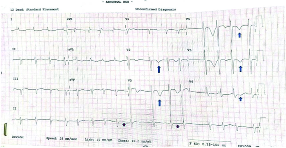
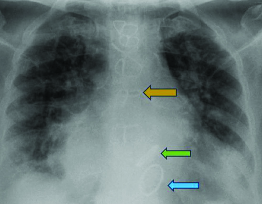
The electrocardiogram showed T wave inversion in leads V2, V3, V4, V5, and V6, as well as absent P waves [Table/Fig-2]. Cardiac biomarkers were sent for analysis and were found to be within normal limits, as summarised in [Table/Fig-3]. The chest radiograph revealed a sternal suture and valve prosthesis in situ [Table/Fig-4].
ECG showing T wave inversions in leads V2-V6 (marked in blue arrow) with irregular rhythm and absent p waves (marked in red arrow).
| Investigations | Value of patient | Reference value |
|---|
| Troponin I (pg/mL) | 10.0 | <15.0 |
| Ckmb (U/lt) | 15.0 | <24.0 |
| NT-proBNP (pg/mL) | 248.50 | </=450 |
X-ray showing intact sternal suture (yellow arrow) with aortic (green arrow) and mitral valve insitu (blue arrow).
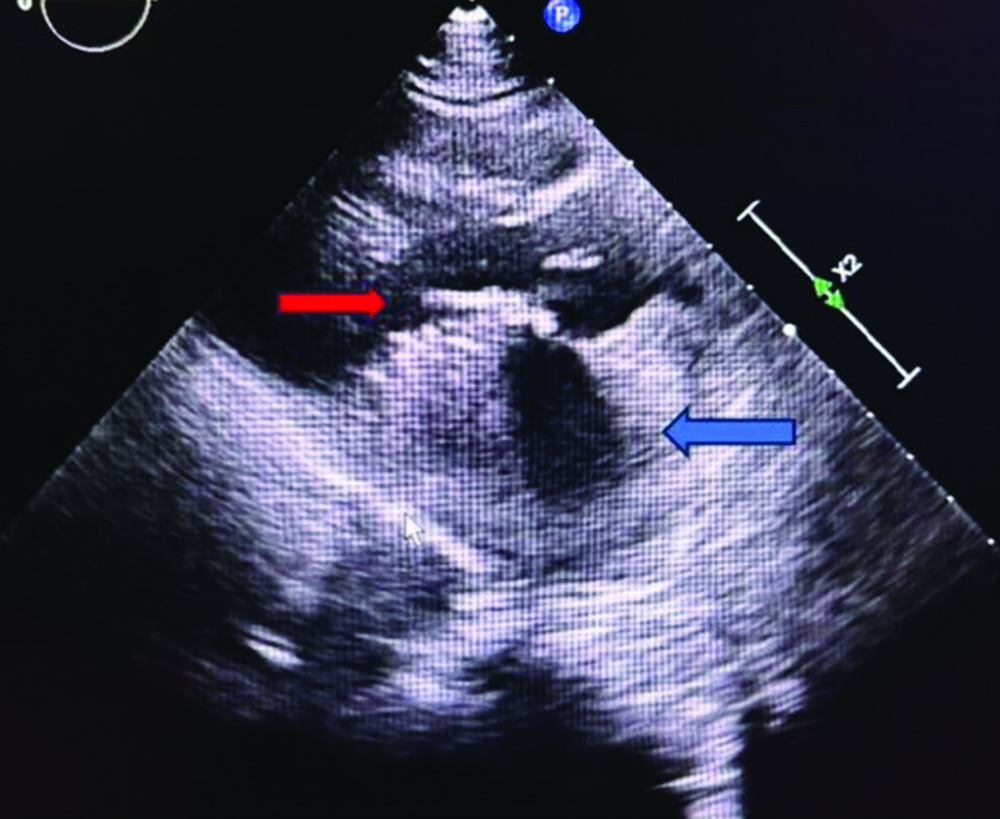
The 2D echocardiogram findings were as follows: dilated left atrium, ejection fraction of 60% [Table/Fig-5], no regional wall motion abnormalities, mitral valve and aortic valve prostheses in situ, severe tricuspid regurgitation, and severe pulmonary artery hypertension. There was no evidence of clot, vegetation, or pericardial effusion. In light of the haematuria, warfarin was withheld, and the patient was started on low molecular weight heparin at a dosage of 0.6 cc, administered subcutaneously twice daily [2], due to the presence of mechanical valves following a cardiology consultation.
A 2D ECHO showing prosthetic valve insitu (red arrow) with dilated left atrium (blue arrow).
Ultrasonography of the abdomen and pelvis revealed a heterogeneous collection with dense internal echoes and debris measuring 111×68×70 mm Craniocaudal, Anteroposterior and Transverse (CC×AP×TR) (approximately, 270 cc) in the region of the left iliacus muscle, with no obvious vascularity on colour Doppler. The differentials considered were a left iliacus muscle abscess or haematoma based on these findings. Subcutaneous oedema was observed in the anterior abdominal wall in the iliac fossa region [Table/Fig-6].
Doppler showing subcutaneous oedema of lower limb (yellow arrow).
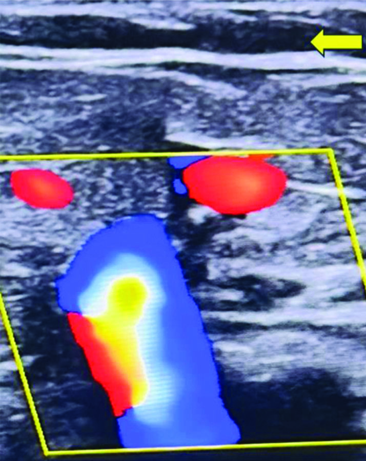
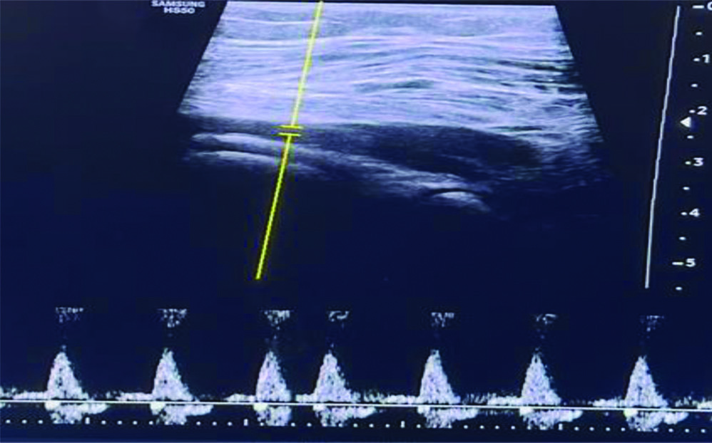
Consequently, a bilateral lower limb arteriovenous doppler study was performed, which showed no evidence of superficial or DVT [Table/Fig-7]. Extensive subcutaneous oedema was noted in the left leg, extending from the proximal thigh to the dorsum of the foot, presenting a cobblestone appearance- changes suggestive of cellulitis.
Lower limb arteriovenous doppler showing no evidence of superficial or deep vein thrombosis.
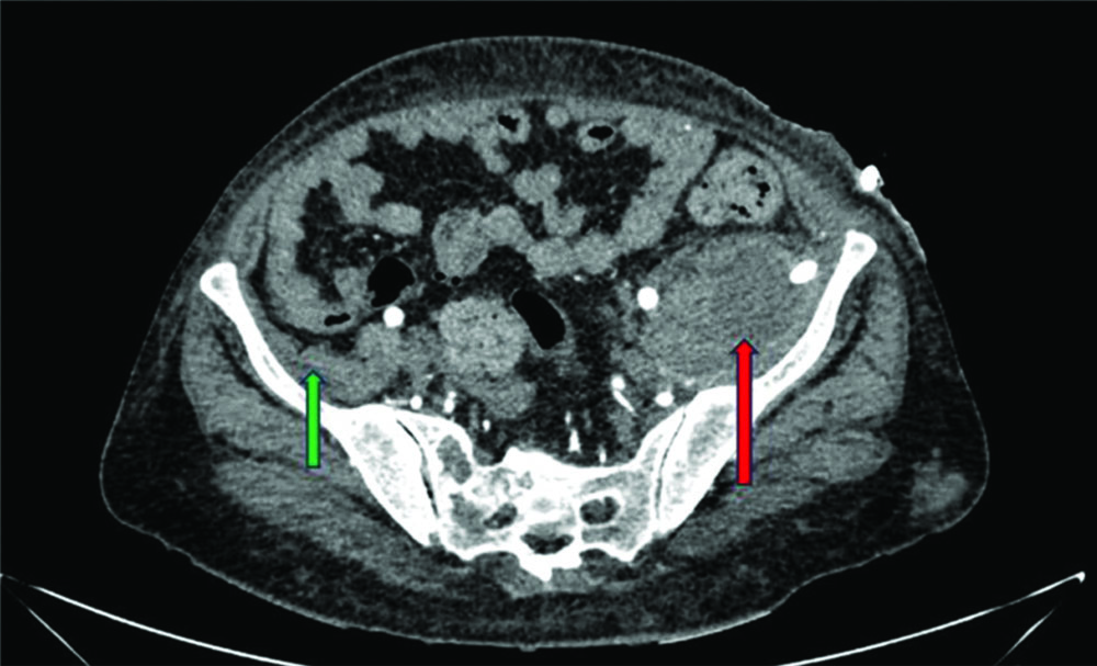
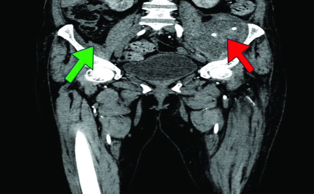
A CT angiography of the lower limb was performed, which revealed a large, well-defined, peripherally enhancing homogeneous collection in the left iliacus muscle. Hyperdense contents within the collection were suggestive of a haematoma, measuring 9.8×5.7×7.2 cm (CC×AP×TR) [Table/Fig-8,9].
(Axial View): CT angiography lower limb showing left iliacus haematoma (red arrow) and normal right sided iliacus muscle haematoma (green arrow).
(Coronal View): Ct angiogram lower limb showing left iliacus muscle haematoma (red arrow) and normal right sided iliacus muscle (green arrow).
Based on the clinical presentation, antecedent history, and radiological imaging, a diagnosis of spontaneous iliacus haematoma compressing the left femoral nerve was made. A conservative approach was taken. Intravenous Vitamin K 30 mg and 4 units of Fresh Frozen Plasma (FFP) were administered immediately to lower the INR levels.
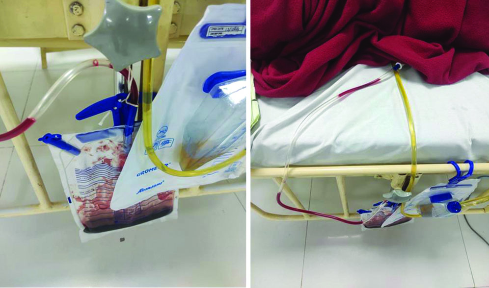
To drain the collection, a pigtail catheter was inserted under ultrasound guidance. The fluid drained was reddish-brown in colour and turbid in appearance [Table/Fig-10], which was subsequently sent for cultures and analysis.
Image of drain showing collections.
Fluid analysis revealed turbid, reddish-coloured fluid with abundant polymorphs, indicating a haematoma. The culture report showed no growth of any organisms, ruling out any infective aetiology, as depicted in [Table/Fig-11].
| Appearance | Reddish, turbid |
| Deposit | Absent |
| Polymorph | 70% |
| Lymphocytes | 20% |
| Red Blood Cells (RBCs) | Many |
The gross and microscopic appearance of the drained fluid was suggestive of a haematoma.
The drain was kept in situ for seven days. Aseptic precautions were taken during daily dressing changes and assessments of the drain. Output was monitored daily, with the highest output noted on day 1 (50 mL) and the lowest on day 7 (10 mL). As the output decreased and the patient gained symptomatic relief, the drain was removed. There was significant relief of pain and an improvement in weakness. The patient was initiated on physiotherapy and encouraged to mobilise. Upon discharge, the patient was mobile, and power in the lower limb had improved. She was discharged on tablet warfarin 5 mg once a day, tablet metoprolol 25 mg once a day, and tablet digoxin 0.25 mg (5/7 days). She was advised to follow-up every month with serial monitoring of PT/INR values.
On follow-up after a month, the patient exhibited significant improvement in weakness, with power in the left hip flexion and knee extension noted to be 5/5. The follow-up PT was 29.2 seconds, with an INR of 2.5. The patient was advised to continue taking tablet warfarin 5 mg once a day, along with a warfarin diet. The patient was counselled to monitor PT/INR periodically and to report immediately in the event of any bleeding manifestations.
Discussion
An iliacus muscle haematoma is usually seen following procedures such as hip arthroplasties, bone grafts, or as a result of trauma [3]. A spontaneous iliacus haematoma is a rare occurrence, typically observed in patients on anticoagulant medication or those with congenital or acquired clotting abnormalities [4]. Patients often experience quadriceps weakness and excruciating discomfort in the affected thigh, hip, and groin [3,4]. Other related signs, such as tachycardia, hypotension, and abdominal pain, may occur depending on the extent of the bleeding [4]. Spontaneous iliacus muscle haematoma with neuropathy is an unusual presentation, and discussions are ongoing regarding the best course of treatment for these patients [5]. The femoral nerve originates from the lumbar plexus, travels laterally to the psoas major, and enters the thigh behind the inguinal ligament. The femoral nerve can be compressed anywhere along its course, but the most common site of compression is within the iliacus muscle compartment [5]. It supplies branches to the psoas and iliacus muscles [6]. It aids in hip flexion and knee extension [6]. The common presenting symptoms are pain in the lower back and groin region [6]. The femoral nerve is more prone to compression due to haematoma or other pathological conditions, as the fascia over the iliacus muscle is not easily stretched. A haematoma of the iliacus muscle does not resolve spontaneously and requires intervention, thereby making the femoral nerve more susceptible to chronic compression and neuropathy [7], as seen in this case.
Patients on anticoagulant therapy, particularly those taking warfarin, are at a higher risk of developing haemorrhagic complications, most commonly in the gastrointestinal, intracranial, and retroperitoneal regions [8,9]. Iliacus muscle haematoma usually occurs secondary to trauma but can also arise from other causes, such as in present case patient- secondary to warfarin therapy [6,10,11]. Femoral neuropathy is characterised by difficulty in hip flexion, knee extension, and reduced patellar reflex [11]. In present case patient presented with similar complaints.
Common imaging modalities used for diagnosis include ultrasonography, MRI, and CT. CT with contrast is the preferred modality to localise the haematoma [11]. Ultrasonography is considered a second-line modality as it cannot effectively visualise deep tissues. In index patient, a USG and CT angiogram of the left lower limb were performed, which revealed an iliacus muscle haematoma measuring 9.8×5.7×7.4 cm. Surgical decompression is preferred in the early stages and in patients with neurological involvement [11]. In haemodynamically stable patients, a non surgical approach can be considered [11]. In present case non surgical approach was opted involving pigtail catheterisation and drainage of the haematoma, which yielded a good outcome. Following drainage, index patient showed improvement in muscle strength and significant resolution of numbness and pain.
It should be noted that a drain is not effective in the later stages after haematoma organisation. Therefore, prompt diagnosis and early intervention are essential.
Conclusion(s)
Iliacus haematoma can mimic conditions such as lower limb cellulitis, DVT, ureteric colic, and lumbar radiculopathy. This diagnosis should always be considered in patients on anticoagulant therapy. A conservative approach should not be dismissed, as it can yield good results, as seen in this case.
TLC: Total leukocyte count; MCV: Mean corpuscular volume; PT: Prothrombin time; aPTT: Activated plasma thromboplastin time; INR: International normalised ratio; PCV: Packed cell volume; SGOT/AST: Aspartate aminotransferase; SGPT/ALP: Alkaline phosphatase; HbA1C: Glycated haemoglobin; WNL: Within normal limit; hpf: High power field
[1]. Bates I, Lewis SR, Reference ranges and normal valuesIn: Practical Hematology 2011 8th EditionUKChurchill Livingstone:08-17. [Google Scholar]
[2]. Tao E, Luo YL, Tao Z, Wan L, A meta-analysis of bridging anticoagulation between low molecular weight heparin and heparinMedicine (Baltimore) 2020 99(3):e18729 [Google Scholar]
[3]. Crawford AM, Guild TT, Striano BM, Von Keudell AG, Spontaneous iliacus hematoma with femoral nerve palsy: An appeal to involve surgical teams earlyBMJ Case Rep 2021 14(1):4 [Google Scholar]
[4]. Al F, Köroglu M, xsözdeş HU, Aslantürk O, Iliacus haematoma causing late femoral nerve palsyClin Case Rep 2024 12(3):e8654 [Google Scholar]
[5]. Kim TH, Lee DJ, Kim W, Do HK, Compressive femoral neuropathy caused by anticoagulant therapy induced retroperitoneal hematoma: A case reportMedicine (Baltimore) [Internet] 2022 101(7):e28876Cited 2024 Jan 23. Available from: https://www.ncbi.nlm.nih.gov/pmc/articles/PMC9282122/#:~:text=Retroperitoneal%20haematomas%20can%20be%20accompanied [Google Scholar]
[6]. Battaglia R, Cerasa A, Pugliese ME, Lucca LF, Tonin P, Iliacus muscle hematoma uncommon complication in a rehabilitation unit: A case report studyHealthcare (Basel) 2022 10(2):297 [Google Scholar]
[7]. Kong WK, Cho KT, Lee HJ, Choi JS, Femoral neuropathy due to iliacus muscle hematoma in a patient on warfarin therapyJ Korean Neurosurg Soc 2012 51(1):51-53. [Google Scholar]
[8]. Asati S, Kundnani VG, Sagane SS, Jain S, Raut S, Iliopsoas hematoma mimicking femoral neuropathyAnn Surg Perioper Care 2021 6(1):1044 [Google Scholar]
[9]. Kim SW, Kim DH, Jung SN, Spontaneous iliopsoas hematoma: A rare complication of anticoagulant useArch Plast Surg 2015 42(04):507-10. [Google Scholar]
[10]. Refai NA, Black AC, Tadi P, Anatomy, bony pelvis and lower limb: Thigh femoral nerve [Internet]. Nih.gov 2023 StatPearls Publishing[cited 2024 Aug 23]. Available from: https://www.ncbi.nlm.nih.gov/books/NBK556065/ [Google Scholar]
[11]. Kim SJ, Park HS, Lee DW, Iliacus hematoma syndrome: A systematic review of recent literature and case reportInjury 2020 51(8):1744-50.10.1016/j.injury.2020.05.028 [Google Scholar] [CrossRef]