Radiological findings of Extramedullary Haematopoiesis of Spleen in Beta Thalassaemia Major: A Case Report
Soumyabrata Debnath1, Pulkit Kumar Santoshi2, Monali Debnath3
1 Senior Resident, Department of Radiodiagnosis, Agartala Government Medical College, Agartala, Tripura, India.
2 Postgraduate Trainee, Department of Radiodiagnosis, Agartala Govt Medical College, Agartala, Tripura, India.
3 Senior Resident, Department of Pathology, ESIC Hospital, New Delhi, India.
NAME, ADDRESS, E-MAIL ID OF THE CORRESPONDING AUTHOR: Dr. Soumyabrata Debnath, Arundhati Nagar, Road No. 14, S.D. Mission, Agartala-799003, Tripura, India.
E-mail: soumyabrata598@gmail.com
Extramedullary Haematopoiesis (EMH) is a compensatory response occurring secondary to inadequate bone marrow function and is observed in many haematological conditions. EMH can occur in many haemoglobinopathies. Extramedullary haemopoiesis favours certain sites such as the liver, the spleen, and the paraspinal regions of the thorax. However, in addition to these common sites of extramedullary haemopoiesis, the process can involve virtually any organ or tissue and can often manifest as a mass mimicking a neoplasm. Although EMH is usually asymptomatic, clinical symptoms can appear due to mass effect. Radiological evaluation can be done using Ultrasonography (USG), Computed Tomography (CT) scan, and T2, T1 weighted Magnetic Resonance Imaging (MRI). Both conservative and surgical treatments are practiced depending on the organ involvement. Here, is a rare case of a young female in her mid-20s, who was a known case of beta thalassaemia major. She presented with a mass insidiously growing in left flank region, which on radiological imaging was found to be focal mass in the spleen. The pathological diagnosis after elective splenectomy was EMH of spleen. Due to chronic transfusions in patients of beta thalassaemia major, EMH is rarely seen. In-fact, the incidence of EMH in beta-thalassaemia major is thought to be less than 1%, which makes the present case a rare entity.
Computed tomography scan, Flank region, Magnetic resonance imaging, Ultrasonography
Case Report
A 24-year-old female presented with pain in the left flank region and a vague flank mass on the left side of the abdomen for the last one year. She also complained of weakness, myalgia, and dyspnea on exertion. The patient was diagnosed case of beta thalassaemia major. There was a history of multiple blood transfusions in the past with 3-5 blood transfusions per year. There was a history of a laparoscopic operation done for ovarian ectopic pregnancy seven-month-back. On general examination, signs of pallor were present. Per abdominal examination showed splenomegaly extending 15 cm below the left rib cage. No significant abnormality was observed in other systemic examinations. Laboratory investigations were done and the patient’s Hb was 3.2 g%. On peripheral smear, RBCs showed microcytic hypochromic blood picture. Other biochemical investigations were within normal limits.
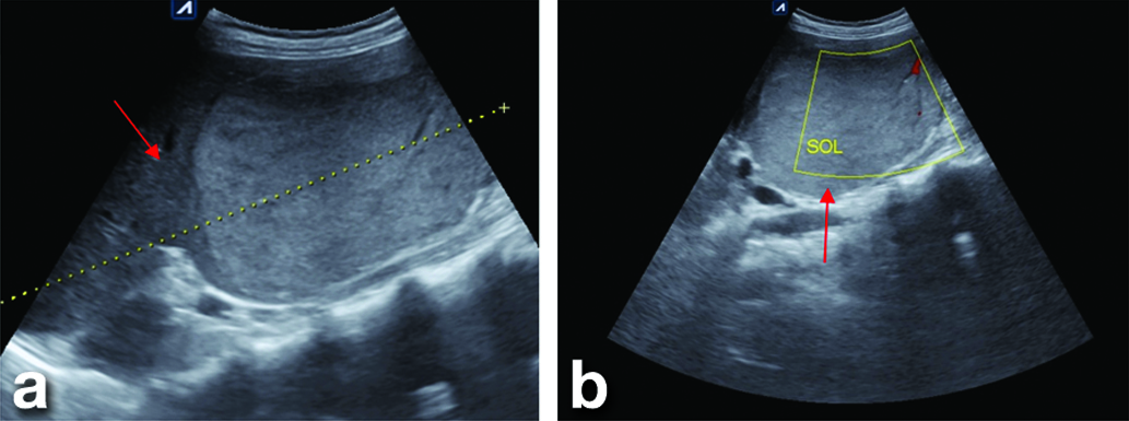
On USG imaging, the spleen was homogeneously enlarged with intermediate to low echo reflectivity. A well-defined hyperechoic round solid lesion was seen in the lower pole of the spleen. On colour doppler examination, no internal vascularity was seen, however, peripheral vascularity was observed. Focal tiny peripheral calcification was seen in the space-occupying lesion. Splenic hilum appearance was normal [Table/Fig-1].
USG abdomen (a) splenomegaly with (b) well-defined hyperechoic mass lesion (arrow) involving the lower pole of spleen with peripheral vascularity. The lesion is effacing the normal isoechoic splenic parenchyma with a surrounding hypoechoic halo.
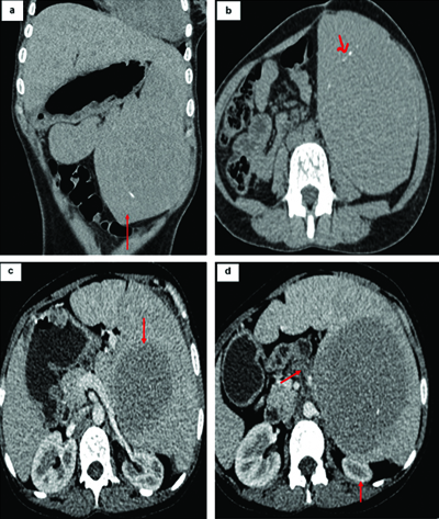
CT scan of abdomen showed massive splenomegaly. Large well-defined iso to hypodense solid mass lesion was seen in the spleen with peripheral thin rim of calcification. In Contrast-Enhanced Computed Tomography (CECT) abdomen, the mass lesion appeared as non-enhancing solid hypodense mass lesion in spleen. Enlarged spleen with this mass lesion was exerting mass effect on adjacent organs (left kidney, bowel) [Table/Fig-2].
CT abdomen: (a) Non-contrast coronal view depicting a massively enlarged spleen (arrow); (b) Non-Contrast axial view showing a well-defined iso-dense mass lesion within the splenic parenchyma with surrounding peripheral rim-like calcifications (arrow); (c) Post-contrast CT showing non-enhancing mass lesion causing (d) mass effect (arrow).
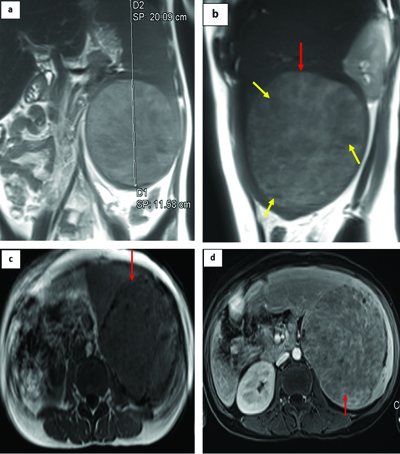
MRI of abdomen showed massive splenomegaly. In T2-weighted image, a large well-defined hyperintense mass was seen in the spleen with internal hypointense areas. In T1 weighted MR images the mass lesion appeared as well-defined mass appearing isointense to splenic parenchyma. In T1-contrast weighted MRI, mass showed heterogenous contrast enhancement with multiple internal hypointense areas [Table/Fig-3].
MR imaging of abdomen showing: (a) splenomegaly (coronal view) with; (b) T2WI (sagittal view) showing well-defined round hyperintense lesion (red arrow) in the lower pole of spleen with multiple internal hypointense areas (yellow arrows); (c) T1WI showing the mass lesion (arrow) is iso-intense to splenic parenchyma; (d) T1 post-contrast image depicting heterogeneous enhancement of mass lesion (arrow).
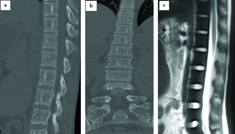
Additional radiological findings were decreased bone density in all the vertebral bodies with diffusely low signal throughout the bony skeleton on T2 sequences. The skeletal aspects of the disease are due to bone marrow proliferation consisting of expansion of the medulla, thinning of cortical bone, and resorption of cancellous bone resulting in a generalised loss of bone density [Table/Fig-4].
NCCT scan of the spine: (a) Sagittal and (b) Coronal view showing decreased done density in all vertebral bodies; (c) T2WI showing diffuse low signal in all vertebral bodies.
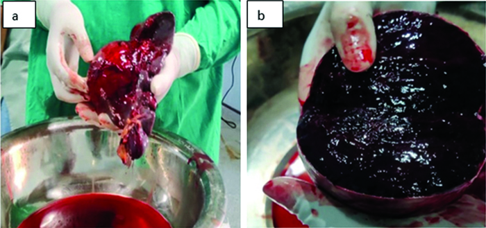
After considering the history of beta-thalassaemia major and detailed radiological findings, a probable diagnosis of EMH presenting as a space occupying lesion in the spleen was made. Patient underwent elective open splenectomy [Table/Fig-5]. Prior to OT, multiple blood transfusions and prophylactic antibiotics were administered to the patient. The specimen was sent to pathology department for histopathological examination.
a) Post splenectomy specimen showing splenic mass surrounded by normal splenic tissue; b) On serial sectioning of spleen, a well-defined, encapsulated reddish-brown mass identified measuring (10.2×6.8×3.5) cm.
On histopathology, multiple sections studied from spleen showed large areas of haemorrhage, sinusoidal dilation and congestion, expansion of red pulp, fibrous bands with haemosiderin pigments and calcium deposits and evidence of EMH.
Following surgical management and histopathological confirmation of diagnosis, the patient was discharged after five days of uneventful postoperative care and advised to continue regular blood transfusions, owing to beta thalassaemia major. The patient visited surgery OPDs on regular basis. Radiological evaluation was advised after six months to assess recurrence of mass lesion. However, the patient failed to follow-up for the same.
Discussion
Haematopoiesis is the process by which blood cells are formed, developing from Haematopoietic Stem Cells (HSCs) through Haematopoietic Progenitor Cells (HPCs) to specialised blood cells [1]. EMH occurs when mature erythroid and myeloid progenitor cells are produced outside the bone marrow [2].
The primary mechanism driving EMH involves regulatory feedback within erythropoietic cells. Chronic anaemia leads to hypoxic conditions, resulting in increased Erythropoietin (EPO) production. This elevation activates the EPO Receptor Janus Kinase-2 Pathway (EPOR-JAK2), aimed at enhancing erythrocyte production. However, in cases of ineffective erythropoiesis, EMH acts as a compensatory mechanism in response to heightened signalling [3]. EMH predominantly affects the mononuclear phagocytic system, including the liver, spleen, and lymph nodes, with the liver and spleen being the most common abdominal sites. Typically, this condition presents as diffuse microscopic involvement, although focal masses can occasionally be observed [4].
In the context of beta thalassaemia, EMH is primarily seen in the minor and intermedia subtypes. It is generally not expected in patients with beta thalassaemia major due to chronic transfusion therapy, making such cases rare. The incidence of EMH in beta thalassaemia major is estimated to be less than 1% [3]. Ultrasonographic features of focal splenic EMH can vary widely, ranging from simple splenomegaly to solitary or multiple hypo- or hyperechoic solid lesions [5]. On Contrast-Enhanced CT (CECT) scans, these lesions typically appear hypodense with heterogeneous enhancement. MRI findings can also differ; active lesions may appear isointense on T1-weighted images, hyperintense on T2-weighted images, and show enhancement following intravenous contrast administration [6].
Similar cases of focal EMH in the spleen have been reported in the literature, including one by Hosoda K et al., involving a 68-year-old male with essential thrombocytopenia, and another by Bouktib Y et al., where a 51-year-old patient’s chronic anaemia had an undetermined cause [7,8]. A systematic review by Subahi AE et al., analysed data from 253 beta thalassaemia major cases, revealing that only one case identified the spleen as the site of EMH [3].
Due to the non-specific nature of these radiological findings, distinguishing EMH from focal splenic tumours- such as haemangiomas, hamartomas, lymphangiomas, inflammatory pseudotumours, malignant lymphomas, metastases, or haemangiosarcoma’s- can be challenging based solely on imaging studies [7]. The characteristics observed on CT and MRI can vary depending on the state of EMH, making histopathological examination essential for accurate diagnosis.
Conclusion(s)
EMH in a patient of beta thalassaemia major is a rare entity. It can involve different organ systems and have varied clinical presentation. Along with clinical evaluation, different radiological modalities are used to guide the diagnosis; however, histopathology can give the definitive diagnosis.
Author Declaration:
Financial or Other Competing Interests: None
Was informed consent obtained from the subjects involved in the study? Yes
For any images presented appropriate consent has been obtained from the subjects. Yes
Plagiarism Checking Methods: [Jain H et al.]
Plagiarism X-checker: May 25, 2024
Manual Googling: Jul 09, 2024
iThenticate Software: Jul 22, 2024 (7%)
[1]. Cenariu D, Iluta S, Zimta AA, Petrushev B, Qian L, Dirzu N, Extramedullary hematopoiesis of the liver and spleenJ Clin Med 2021 10(24):5831 [Google Scholar]
[2]. Cailleteau A, Agbetsivi K, Guimas V, Supiot S, Ollivier L, Extramedullary hematopoiesis causing spinal cord compression in polycythemia vera: A case report and literature reviewClin Transl Radiat Oncol 2022 38:43-46.PMCID: PMC963871910.1016/j.ctro.2022.09.00936353654 [Google Scholar] [CrossRef] [PubMed]
[3]. Subahi AE, Ata F, Choudry H, Iqbal P, AlHiyari MA, Soliman AT, Extramedullary haematopoiesis in patients with transfusion dependent β-thalassaemia (TDT): A systematic reviewAnn Med 2022 54(1):764-74.PMCID: PMC894194810.1080/07853890.2022.204806535261317 [Google Scholar] [CrossRef] [PubMed]
[4]. Sahli H, Boularab J, Mandour JE, Allali N, Chat L, Haddad SE, Multifocal extramedullary hematopoiesis in a 15-year-old girl with beta thalassaemia: A case reportRadiol Case Rep 2022 17(7):2315-19.PMCID: PMC909566410.1016/j.radcr.2022.03.08535570880 [Google Scholar] [CrossRef] [PubMed]
[5]. Bradley MJ, Metreweli C, Ultrasound appearances of extramedullary hematoiesis in the liver and spleenBr J Radiol 1990 63:816-18. [Google Scholar]
[6]. Rajesh S, Agrawal N, Bihari C, Pargewar SS, Deogaonkar G, Desai SN, Focal intrasplenic extramedullary hematopoiesis masquerading as a tubercular abscessTropical Gastroenterology 2016 :37 [Google Scholar]
[7]. Hosoda K, Shimizu A, Kubota K, Notake T, Sugenoya S, Yasukawa K, A focal extramedullary hematopoiesis of the spleen in a patient with essential thrombocythemia presenting with a complicated postoperative course: A case reportSurg Case Rep 2021 7(1):33PMCID: PMC783822910.1186/s40792-021-01119-533496866 [Google Scholar] [CrossRef] [PubMed]
[8]. Bouktib Y, Hajjami AE, Odongo S, Boutakioute B, Ouali M, Idrissi NEG, Focal extramedullary hematopoiesis in the spleen: Case reportPAMJ Clinical Medicine 2024 15:2210.11604/pamj-cm.2024.15.22.43795 [Google Scholar] [CrossRef]