Sebocystomatosis of the Axillae: Unveiling a Rare Entity
Garima Chandrakar1, Simran Khan2
1 Junior Resident, Department of Pathology, Jawaharlal Nehru Medical College, Sawangi Meghe, Wardha, Maharashtra, India.
2 Junior Resident, Department of Pathology, Jawaharlal Nehru Medical College, Sawangi Meghe, Wardha, Maharashtra, India.
NAME, ADDRESS, E-MAIL ID OF THE CORRESPONDING AUTHOR: Dr. Garima Chandrakar, Shalinata P.G Girl’s Hostel, Jawaharlal Nehru Medical College, Sawangi Meghe, Wardha-442001, Maharashtra, India.
E-mail: garimachandrakar27@gmail.com
Autosomal dominant, Histopathology, Keratin 17, Steatocystoma multiplex
A 32-year-old female presented to the Outpatient Department of Surgery with complaints of swelling in the right breast and similar complaints in the right axillary region for the past three months. The patient was fine three months ago but developed swelling in the upper outer quadrant of the right breast that gradually increased, along with swelling in the right axilla and multiple small nodules in it. On local examination, the swelling in the right breast was initially small, painless, and gradually increasing in size, with a soft to firm consistency. The right axillary swelling was soft in consistency, non tender on palpation, and had multiple small skin-coloured to yellow nodules.
Routine and specific investigations were performed based on the signs and symptoms, including a complete blood count, sonomammography, and mammography. Systemic investigations included blood culture tests to rule out septicaemia. Laboratory parameters such as liver function tests for any hepatic dysfunction and lipid panel evaluation to rule out hypertriglyceridaemia were also conducted, and both these parameters were normal. Sonomammography revealed a well-circumscribed hypoechoic mass lesion at the 1 o’clock position peripherally with foci of calcification in the right breast, while the right axilla showed evidence of two well-circumscribed, hypoechoic lesions in detected nodules with posterior enhancement and were confined to the dermis.
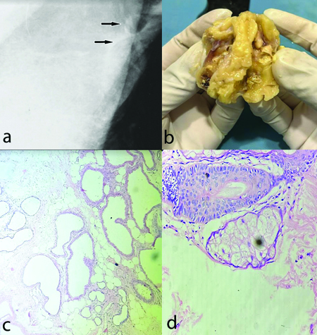
The mediolateral oblique mammogram showed evidence of well-circumscribed radiolucent oily cysts with a peripheral continuous rim. The nodules were usually round with a diameter of <1 cm [Table/Fig-1a]. After investigating the results, excision of lumps in the right breast and right axilla was performed, and specimens were sent for histopathological diagnosis (H&E). Grossly, the lump in the right breast appeared irregular, globular in structure measuring 6×4.5×2.8 cm. On cut section, white homogeneous areas with a single calcified area were noted. The right axillary lump appeared grayish-yellow, irregular with skin and hair attached, measuring 5.2×3.5 cm. On cut section, whitish cheesy material with areas of haemorrhage was identified below the skin margin [Table/Fig-1b]. Microscopic examination confirmed the diagnosis of sebocystomatosis of the right axillary lump, showing an epithelial cyst lining in a wavy, homogeneous, eosinophilic horny layer with stratification of collapsed epithelial cells around the cystic space. The flattened sebaceous gland lobules appear to be embedded or lying close within the cyst wall [Table/Fig-1c,d]. Histologically, the right breast lump showed only fibrocystic changes.
a) Mammogram- revealed radiolucent oily cysts with peripheral continuous rim; b) Grossly (cut section)- yellowish irregular lump with small nodules, whitish cheesy material; c) Microscopic examination (H&E, 40x): multiple folded cysts lined by stratified squamous epithelial lining with eosinophilic cuticles; d) Presence of sebaceous glands in close proximity to the cyst wall (H&E, 100x).
Sebocystomatosis (steatocystoma multiplex) is a hamartomatous malformation of the pilosebaceous duct junction [1]. It is an uncommon cutaneous disorder characterised by multiple intradermal cysts, primarily distributed over the trunk, axilla, neck, and groin region [2,3]. It affects both genders equally and is commonly diagnosed in adolescence or early adulthood [2]. It is often inherited as an autosomal dominant trait associated with a mutation in the gene coding for Keratin 17 (KRT17), but sporadic cases have also been reported [2]. Pachyonychia congenita type 2 also results from a mutation in KRT17, but since it is associated with more severe nail dystrophy, palmoplantar keratoderma, follicular keratosis, and leukoplakia, it is ruled out from the differentials of steatocystoma multiplex [1].
The absence of sebaceous glands lying in close proximity to the cyst wall, as well as abundant lamellated keratin and vellus hair shafts within the cyst, excludes its closest differential of eruptive vellus hair cyst. Other differential diagnosis include acne conglobata, epidermoid cysts, milia, xanthomatosis, pseudofolliculitis, and nodular acne [4]. Steatocystoma multiplex clinically presents as asymptomatic, multiple, smooth, cystic papules, and nodules. The lesions were uniform and skin to yellow-coloured. On puncture of the nodule, odourless, oily material oozed out [4-6].
In this case, the patient refused a surgical approach to intervention. She was put on oral isotretinoin (1 mg/kg/day) for one month. This drug was started initially as it causes shrinkage of inflamed cysts and decreases sebaceous gland activity of sebum production. However, after one month of complete dosage and duration of isotretinoin, the patient experienced episodes of inflammation with suppuration. Subsequently, oral rifampicin (600 mg/day) and clindamycin (500 mg/day) were added with isotretinoin. After four months of subsequent follow-up, these drugs were stopped as she was clinically stable with remission of signs and symptoms. Later, after two months of discontinuation, flare and recurrence of lesions occurred. Finally, a surgical approach was performed. This included incision followed by expression of cyst contents and forceps-assisted removal of the cyst wall. There has been no evidence of recurrence up to the present six months of follow-up, indicating a good prognosis/outcome after surgical treatment of the lesions.
In this case, histopathological diagnosis with radiographic findings played a pivotal role in the confirmation of sebocystomatosis. Since it is asymptomatic and presents with unusual symptoms mimicking lesions of acne conglobata or hidradenitis suppurativa, timely recognition is important to prevent complications and misdiagnosis.
Author Declaration:
Financial or Other Competing Interests: None
Was informed consent obtained from the subjects involved in the study? Yes
For any images presented appropriate consent has been obtained from the subjects. Yes
Plagiarism Checking Methods: [Jain H et al.]
Plagiarism X-checker: May 09, 2024
Manual Googling: May 17, 2024
iThenticate Software: May 24, 2024 (8%)
[1]. Santana CN, Pereira D do, Lisboa AP, Leal JM, Obadia DL, Silva RS, Steatocystoma multiplex suppurativa: Case report of a rare conditionAn Bras Dermatol 2016 91(5 suppl 1):51-53.10.1590/abd1806-4841.20164539 [Google Scholar] [CrossRef]
[2]. Shin NY, Kang JH, Kim JE, Symkhampa K, Huh KH, Yi WJ, Steatocystoma multiplex: A case report of a rare entityImaging Sci Dent 2019 49(4):31710.5624/isd.2019.49.4.31 [Google Scholar] [CrossRef]
[3]. Kamra HT, Gadgil PA, Ovhal AG, Narkhede RR, Steatocystoma multiplex- A rare genetic disorder: A case report and review of literatureJ Clin Diagn Res 2013 7(1):166-68.10.7860/jcdr/2012/4691.2698 [Google Scholar] [CrossRef]
[4]. Sharma A, Agrawal S, Dhurat R, Shukla D, Vishwanath T, An unusual case of facial steatocystoma multiplex: A clinicopathologic and dermoscopic reportDermatopathology 2018 5(2):58-63.10.1159/000488584 [Google Scholar] [CrossRef]
[5]. Hyun DN, Won JH, Park JS, Chung H, A case of steatocystoma simplex involving the scalpAnn Dermato 2008 20(4):23010.5021/ad.2008.20.4.230 [Google Scholar] [CrossRef]
[6]. Rainamira A, Krisnanti IA, Rihatmadja R, Suprapto N, Surya D, Steatocystoma multiplex suppurativa: A case reportBali Dermatology Venereology and Aesthetic Journal 2023 6(2):36-38.10.51559/rzd0ak48 [Google Scholar] [CrossRef]