The world of pathology encompasses a broad spectrum of diseases beyond the boundaries of specialties, with each disease having specific and interesting gross and microscopic features. Some of these features are pattern-based, while others are eponyms that compare them to objects, food, animals, etc., due to their striking resemblance and quick recall. Medical nomenclature is of vital importance, and it has significantly evolved historically, with etymological roots from Latin, Greek, and Roman languages of ancient times to the current internationally uniform codes of English and other modern languages. Eponyms are valuable medical literary epithets that have been used across specialties and include animal names, food names, discoverer names, geographic references, and more. The subject of pathology, in particular, has immense use of eponyms as they are valuable tools for assisting in adult learning, or andragogy. The specialty of pathology is unique in its complex patterns and diagnostic algorithm, always in need of alternate systems to arrive at quick and accurate diagnosis. Also known as intuitive thinking or reflex thinking, pattern viewing and eponyms trigger a reflex recognition system, reducing recall time and aiding in precise diagnosis. The present article aimed to review the terminological phenomenon of animal eponym usage in the context of pathological diagnosis.
Background of Study
An eponym, by definition, is a place, person, or thing after which something is named or thought to be named. It holds a privilege of its own in various specialties, including pathology. Eponyms are valuable tools for initiating pattern recognition and reflex/intuitive thinking to aid in adult learning, as well as facilitating easy recall and precise diagnosis [1]. An attempt has been made to incorporate known eponyms into the world of pathology after an extensive literature search.
Antler horn pattern in skin-Delicate digitate downgrowth of the epidermal layer with melanocytic hyperplasia at the tip, giving the resemblance of an antler-like pattern seen in Dowling-Degos disease [2].
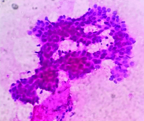
Antler horn pattern in breast cytology-Sheets and branching clusters of cohesive ductal epithelial cells with an antler horn pattern seen in fine-needle aspiration cytology of fibroadenoma [Table/Fig-1] [3].
Fine needle aspirate cytology showing benign ductal epithelial cell clusters branching in antler horn pattern in a case of fibroadenoma (H&E stain 40x).
(Swarm of) Bees-Perifollicular inflammation predominantly, peribulbar infiltrates of lymphocytes seen around the anagen hair follicles in alopecia areata [4].
Bull’s eye inclusions-Central Periodic Schiiff’s Reagent (PAS)-positive round inclusions in the cells seen in malignant effusions, giving a target or bull’s eye appearance [5].
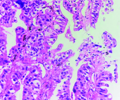
Butterfly sitting on a fence appearance-Arrangement of neoplastic cells on either side of the alveolar septa resembling a butterfly sitting on a fence, seen in adenocarcinoma lung; lepidic variant (earlier called bronchoalveolar carcinoma) [Table/Fig-2] [6].
Neoplastic cells in lung adenocarcinoma arranged along the alveolar septa giving a "Butterfly sitting on a fence" appearance (H&E stain 40x).
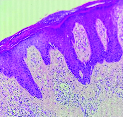
Camel foot-In chronic plaque psoriasis, rete pegs elongate into the dermis and take a plunge to accommodate the increasing basal cell population, giving rise to a camel foot appearance to dermal rete ridges [Table/Fig-3] [7].
Skin showing rete pegs elongate into the dermis and take a plunge to accommodate the increasing basal cell population giving rise to camel foot appearance in chronic plaque psoriasis (H&E stain 40x).
Caterpillar chromatin-Macrophage with an ovoid nucleus and chromatin condensed toward the center of the nucleus in a wavy rod-like pattern that resembles a caterpillar, seen in rheumatic heart disease [8].
Chicken fat clot-The yellowish part of the postmortem clot which is cell-free [9].
Chicken wire calcification-Thin lacy chicken wire-like calcification seen in chondroblastoma [10].
Chicken wire vasculature-Thin chicken wire-like arrangement of small-sized thin-walled vessels in oligodendroglioma [11].
CLAM-Clustering- constrained- attention multiple- instance learning (CLAM) uses attention-based learning to identify subregions of high diagnostic value to accurately classify whole slides in Whole Slide Imaging (WSI) [12].
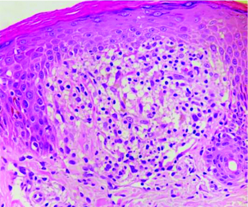
Claw clutching ball-Well-circumscribed granulomatous infiltrate composed of lymphocytes, epithelioid cells, and occasional Langhans’ giant cells that are clutched by surrounding claw-shaped hyperplastic rete ridges, seen in Lichen Nitidus [Table/Fig-4] [13].
Hyperplastic rete ridges clutching the superficial dermal infiltrate of lymphocytes and epithelioid cells (H&E stain,40x) resembling a Claw clutching a ball.
CRAB criteria-Multiple myeloma defining events comprising Hypercalcemia, Renal insufficiency, Anaemia, and Bone lytic lesions (CRAB).
Crab-Crab-like streaks of chalky white elastic stroma penetrating surrounding stroma seen in the breast in cases of Infiltrating ductal carcinoma of no special type [Table/Fig-5] [14].
Crab-like white streaks of infiltrating duct carcinoma, invading into the surrounding elastotic stroma of the breast.
Cor bovinum-Heart with massive left ventricular hypertrophy seen in tertiary syphilis, chronic aortic regurgitation, and ischemic heart disease [15].
DOG1-Discovered on GIST 1-It is a sensitive and specific marker for gastrointestinal stromal tumour [16].
Feathery growth-Spindle stellate cells arranged in a loose fascicular to storiform pattern in nodular fasciitis [17].
Feline oesophagus-Trachealisation of the oesophagus on gross examination, characterised by concentric rings on the mucosal side of the oesophagus. It is seen in association with gastroesophageal reflux disease and hiatus hernia [18].
Fishnet appearance-Intercellular Immunoglobulin (Ig)G and C3 deposits in the epidermis seen in direct immunofluorescence in cases of pemphigus vulgaris [19].
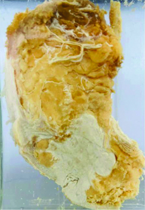
Fish flesh appearance-Smooth, slightly bulging, pale tan to grey-white colour on the cut surface in lymphomas and soft-tissue sarcomas [Table/Fig-6] [20].
Cut surface of the lymphnode showing homogenous, bulging fish flesh appearance, in a case of lymphoma.
(School of) Fish-Spindle cell proliferation of endothelial cells in a fascicular pattern in Kaposi sarcoma has been likened to a school of fish. The spindle cells infiltrate through the collagen, forming slit-like spaces, especially towards the periphery of the lesions [21].
(School of) Fish-Enlarged cohesive sheets of cells with a “school of fish” architecture in reactive/regenerative changes in cervical cytology [22].
Fish mouth appearance-Narrowing of the mitral valve in rheumatic heart disease seen as a fish mouth deformity when viewed from the ventricular aspect into the atrium [23].
Flea-bitten kidney-Pinpoint haemorrhages on the external surface of the kidney in malignant nephrosclerosis and hyperacute rejection [24].
Hedgehog signaling pathway-First identified in Drosophila, the common fruit fly, represents a highly conserved evolutionary pathway of signal transmission from the cell membrane to the nucleus, playing a major role in carcinogenesis [25].
Herringbone pattern-Arrangement of neoplastic spindle cells in long sweeping fascicles forming a fish skeleton pattern in fibrosarcoma [26].
Hippo signaling-Is an evolutionarily conserved network that plays a central role in regulating cell proliferation and cell fate to control organ growth and regeneration [27].
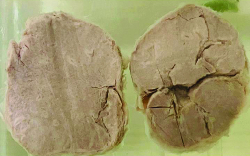
Horseshoe kidney-A common renal fusion anomaly characterised by the fusion of two kidneys at one of the poles due to abnormal migration of nephrogenic rests [Table/Fig-7] [28].
Specimen of bilateral kidneys showing abnormal fusion in the lower pole with horseshoe appearance.

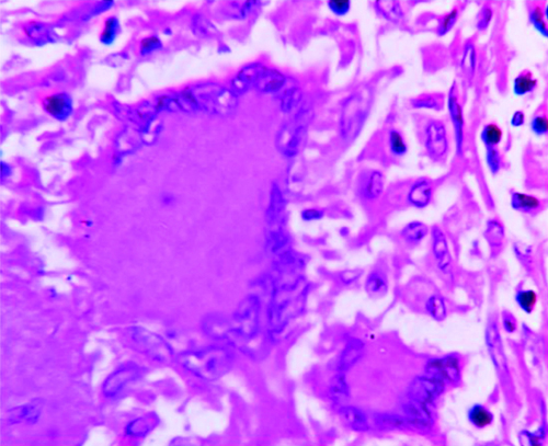
Horseshoe nucleus-Langhans type multinucleate giant cell showing nuclei arranged peripherally in a horseshoe shape, seen in tuberculosis [Table/Fig-8] [29].
Langhan type of multinucleate giant cell with nuclei arranged along the periphery in a horseshoe shape (H&E stain 40x).
LAMB syndrome-Includes Lentigines, Atrial myxoma, Mucocutaneous myxoma, and Blue nevi (LAMB) [30].
Leopard skin-Like colonic mucosa- Gross appearance of colonic mucosa in chronic granulomatous disease due to pigment-laden macrophages giving rise to brown dots across oedematous yellow colonic mucosa [31].
Moth-eaten appearance-Destruction of soft tissue or bone giving rise to a moth-eaten appearance in alopecia (secondary syphilis) and lytic lesions of bone [32].
Octopus sign-Fibrosis in pulmonary Langerhans cell histiocytosis histologically comprises a central scar with septal strands and associated airspace enlargement that give rise to an octopus-like appearance [33].
Owl eye inclusions-Large ovoid or pleomorphic nucleus with basophilic intranuclear inclusions (Cowdry bodies) surrounded by a clear halo [34].
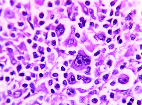
Owl eye nuclei-Reed-Sternberg cell showing a bilobed nucleus with a prominent eosinophilic nucleolus and perinucleolar halo giving it an owl-eye appearance [Table/Fig-9] [35].
Reed-Sternberg cells of Hodgkin’s Lymphoma showing mirror image binucleation with prominent eosinphilic nucleoli and perinucleolar halo, giving a striking resemblance to owl eye (H&E stain 40x).
Pachydermia (elephant skin)-Abnormal thickening of the skin like that of a pachyderm (a tough-skinned animal like an elephant), seen in pachydermatoperiostosis and elephantiasis [36].
Phrynoderma (Toad skin)-Follicular hyperkeratosis with keratin plugs accompanied by sebaceous atrophy due to nutritional deficiency giving rise to toad-like skin [36].
Pulmonary Interstitial Glycogenosis (PIG) syndrome-A form of pulmonary interstitial disease characterised by diffuse expansion of the alveolar interstitium by glycogenated mesenchymal cells, seen in babies [37].
Salmon pink-Salmon-pink coloured amorphous acellular deposits of amyloid in Congo red stain in amyloidosis [38].
Snail expression-Zinc finger protein SNAI1, a family of transcription factors that promote the repression of the adhesion molecule E-cadherin to regulate Epithelial to Mesenchymal Transition (EMT) during embryonic development [39].
Snail track ulcer-Painless or painful erythematous lesions, greyish-white mucous patches, or irregular linear erosions coalescing to form snail track ulcers in syphilis [15].
Snake-like fashion-Renal cell carcinoma invading the renal vein and ascending up the inferior vena cava in a snake-like pattern [40].
Spider angioma-A vascular lesion characterised by anomalous dilatation of end vasculature found just beneath the skin surface. The lesion contains a central red spot and reddish extensions that radiate outward like a spider’s web, seen due to the vasodilatory effects of alcohol and hyperestrogenism in cirrhosis [41].
Staghorn nucleus- Megakaryocytes in essential thrombocythemia with dysplastic staghorn-like branching nuclei [42].
Staghorn vasculature-Capillaries or small-sized thin-walled vessels branching in a staghorn pattern in solitary fibrous tumours and hemangiopericytoma [42].
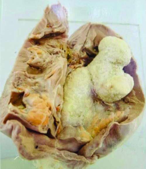
Staghorn calculus-Staghorn renal stones are large kidney stones that fill the renal pelvis and atleast one renal calyx. They are also called triple phosphate stones and are composed of struvite (magnesium ammonium phosphate) [Table/Fig-10].
A large stone in the renal pelvis which is hard and creamy white in color branching along the renal pelvicalyceal architecture giving a stag horn appearance.
Tadpole cells-Neoplastic squamous cells with a tadpole shape seen in cervical cytology in squamous cell carcinoma of the cervix [43].
Tadpole pattern-In syringoma, neoplastic strands of eccrine glands arranged in tadpole-shaped strands are best appreciated at low power [44].
Tigered effect-Describes the fatty change seen in the liver and myocardium. Microscopically and grossly, the lipid accumulation leads to normal-appearing tissue parenchyma interspersed with yellowish areas showing intracellular lipid. It is known to be seen in the liver in steatosis and in the myocardium due to hypoxia [45].
Tigroid background-Seen in cytology smears of seminoma characterised by the presence of a relatively granular, reticulated material in the background, giving it a foamy, lazy tiger-striped appearance [46].
Ugly duckling sign-Nevi in an individual generally tend to share a similar appearance, so one that does not share the same characteristics should be considered for biopsy [30].
Wolf-like/butterfly rash-Facial symmetric erythematous rash seen in systemic lupus erythematosus resembling a wolf bite/butterfly wings [47].
Zebra bodies-Deposits of lamellate, lipid-like, electron-dense material forming concentric bodies in the cytoplasm of podocytes and endothelial cells in glomerular and peritubular capillaries-in Fabry disease [48].
Conclusion(s)
Constant training of one’s neural networks in recognising pathways and patterns in pathology can be an uphill task. Employing the use of literary tools like eponym usage can prove to be an interesting methodology in acquiring reflexive thinking in adult learning. The present article integrates and highlights the role of pattern recognition using animal eponyms. Students and young consultants alike will benefit from the usage of these eponyms during routine reporting and medical education.
[1]. Tay SW, Ryan P, Ryan CA, Systems 1 and 2 thinking processes and cognitive reflection testing in medical studentsCan Med Educ J 2016 7(2):e97-103. [Google Scholar]
[2]. Arundhathi S, Rajagopal P, Gopinath H, Rupa Ramani J, Follicular Dowling-Degos disease camouflaged as comedones: A case report and literature reviewCureus 2022 14(6):e26078Available from: https://dx.doi.org/10.7759/cureus.26078 [Google Scholar]
[3]. Bhatia A, Phulware RH, Ahuja A, Kaushal M, Hamartomas of the breast: A mimic of fibroadenoma and cytological pitfallJ Cytol 2020 37(4):210-11.Available from: https://dx.doi.org/10.4103/JOC.JOC_138_20 [Google Scholar]
[4]. Amin SS, Sachdeva S, Alopecia areata: A reviewJournal of the Saudi Society of Dermatology & Dermatologic Surgery 2013 17(2):37-45. [Google Scholar]
[5]. Kumar PV, Eqbali S, Monabati A, Talei AR, Bull’s eye (target) inclusions in neoplastic cells in malignant serous effusions. A study of 289 casesActa Cytol 2000 44(4):543-46.Available from: https://dx.doi.org/10.1159/000328527 [Google Scholar]
[6]. Dalpiaz G, Cancellieri A, Case-based glossary with tips and tricksIn: Atlas of Diffuse Lung Diseases: A Multidisciplinary Approach 2016 ChamSpringer International Publishing:251-89. [Google Scholar]
[7]. Madke B, Doshi B, Khopkar U, Dongre A, Appearances in dermatopathology: The diagnostic and the deceptiveIndian J Dermatol Venereol Leprol 2013 79(3):338-48. [Google Scholar]
[8]. Satoh F, Tsutsumi Y, Anitschkow cells in the human heartPathol Int 1999 49(1):85-87.Available from: https://dx.doi.org/10.1046/j.1440-1827.1999.00827.x [Google Scholar]
[9]. Uekita I, Ijiri I, Nagasaki Y, Haba R, Funamoto Y, Matsunaga T, Medico-legal investigation of chicken fat clot in forensic cases: Immunohistochemical and retrospective studiesLeg Med (Tokyo) 2008 10(3):138-42.Available from: https://dx.doi.org/10.1016/j.legalmed.2007.11.004 [Google Scholar]
[10]. Serafino DM, Gioioso M, Severino R, Lisanti F, Rocca R, Sorbo P, The idiopathic localized tumoural calcinosis: The “chicken wire” radiographic patternRadiol Case Rep 2017 12(3):560-63. [Google Scholar]
[11]. Rani SB, Mahadevan A, Anilkumar SR, Raju TR, Shankar SK, Expression of nestin - A stem cell associated intermediate filament in human CNS tumoursIndian J Med Res 2006 124(3):269-80. [Google Scholar]
[12]. Lu MY, Williamson DFK, Chen TY, Chen RJ, Barbieri M, Mahmood F, Data-efficient and weakly supervised computational pathology on whole-slide imagesNat Biomed Eng 2021 5(6):555-70.Available from: https://dx.doi.org/10.1038/s41551-020-00682-w [Google Scholar]
[13]. Podder I, Mohanty S, Chandra S, Gharami RC, Isolated Palmar lichen nitidus-A diagnostic challenge: First case from eastern IndiaIndian J Dermatol 2015 60(3):308-09.Available from: https://dx.doi.org/10.4103/0019-5154.156398 [Google Scholar]
[14]. Invasive breast cancer of no special type (NST) [Internet]Pathologyoutlines.com. [cited 2024 Apr 2]. Available from: https://www.pathologyoutlines.com/topic/breastmalignantductalnos.html [Google Scholar]
[15]. Fluri S, Gebbers JO, Cor bovinum: Abnehmende Häufigkeit in den letzten 20 Jahren--ein Therapieerfolg? In: Cor bovinum: Decreased incidence over the lastPraxis (Bern 1994) 2001 90(45):1964-72. [Google Scholar]
[16]. Lopes LF, West RB, Bacchi LM, van de Rijn M, Bacchi CE, DOG1 for the diagnosis of gastrointestinal stromal tumour (GIST): Comparison between 2 different antibodiesAppl Immunohistochem Mol Morphol 2010 18(4):333-37.Available from: https://dx.doi.org/10.1097/PAI.0b013e3181d27ec8 [Google Scholar]
[17]. Murugesan D, Mathew M, Kudva A, Solomon M, Oral nodular fasciitis- A case report with a diagnostic schemaJ Oral Med Oral Surg 2019 25(2):21 [Google Scholar]
[18]. Artul S, Shkara HA, Khoury R, Habib G, Human feline oesophagusBMJ Case Rep 2014 2014:bcr2013203061-bcr2013203061.Available from: https://dx.doi.org/10.1136/bcr-2013-203061 [Google Scholar]
[19]. Shetty VM, Subramaniam K, Rao R, Utility of immunofluorescence in dermatologyIndian Dermatol Online J 2017 8(1):01-08.Available from: https://dx.doi.org/10.4103/2229-5178.198774 [Google Scholar]
[20]. Vimal M, Nishanthi A, Food eponyms in PathologyJ Clin Diagn Res 2017 11(8):EE01-06.Available from: https://dx.doi.org/10.7860/jcdr/2017/28375.10360 [Google Scholar]
[21]. Schmidt BM, Holmes CM, Classic solitary Kaposi sarcoma of the foot in an immunocompetent patient: A case reportWounds 2016 28:E35-40. [Google Scholar]
[22]. Colgan TJ, Woodhouse SL, Styer PE, Kennedy M, Davey DD, Reparative changes and the false-positive/false-negative Papanicolaou test: A study from the College of American Pathologists Interlaboratory Comparison Program in Cervicovaginal CytologyArch Pathol Lab Med 2001 125(1):134-40.Available from: https://dx.doi.org/10.5858/2001-125-0134-RCATFP [Google Scholar]
[23]. Han J, Xiang H, Ridley WE, Ridley LJ, Fish mouth appearance: Mitral valveJ Med Imaging Radiat Oncol 2018 62(Suppl 1):28Available from: https://dx.doi.org/10.1111/1754-9485.15_12785 [Google Scholar]
[24]. Omenai SA, Ajani MA, Nwadiokwu JI, Okolo CA, Histomorphological assessment of non-neoplastic renal diseases at autopsy: An institutional experience in Southwestern NigeriaMalawi Med J 2021 33(4):281-86.Available from: https://dx.doi.org/10.4314/mmj.v33i4.9 [Google Scholar]
[25]. Skoda AM, Simovic D, Karin V, Kardum V, Vranic S, Serman L, The role of the Hedgehog signaling pathway in cancer: A comprehensive reviewBosn J Basic Med Sci 2018 18(1):08-20.Available from: https://dx.doi.org/10.17305/bjbms.2018.2756 [Google Scholar]
[26]. Kaur H, Gupta V, Mishra D, Yadav VS, Fibrosarcoma: Origin, differential diagnosis, and report of a case in the mandibleJ Indian Soc Periodontol 2022 26(2):169-77.Available from: https://dx.doi.org/10.4103/jisp.jisp_188_21 [Google Scholar]
[27]. Misra JR, Irvine KD, The Hippo signaling network and its biological functionsAnnu Rev Genet 2018 52(1):65-87.Available from: https://dx.doi.org/10.1146/annurev-genet-120417-031621 [Google Scholar]
[28]. Bhandarkar KP, Kittur DH, Patil SV, Jadhav SS, Horseshoe kidney and associated anomalies: Single institutional review of 20 casesAfr J Paediatr Surg 2018 15(2):104-07.Available from: https://dx.doi.org/10.4103/ajps.AJPS_55_17 [Google Scholar]
[29]. Kumar SN, Prasad TS, Narayan PA, Muruganandhan J, Granuloma with langhans giant cells: An overviewJ Oral Maxillofac Pathol 2013 17(3):420-23.Available from: https://dx.doi.org/10.4103/0973-029X.125211 [Google Scholar]
[30]. Jindal N, Jindal P, Kumar J, Gupta S, Jain VK, Animals eponyms in dermatologyIndian J Dermatol 2014 59(6):631Available from: https://dx.doi.org/10.4103/0019-5154.143573 [Google Scholar]
[31]. Obayashi N, Arai K, Nakano N, Mizukami T, Kawai T, Yamamoto S, Leopard skin-like colonic mucosa: A novel endoscopic finding of chronic granulomatous disease-associated colitisJ Pediatr Gastroenterol Nutr 2016 62(1):56-59.Available from: https://dx.doi.org/10.1097/MPG.0000000000000905 [Google Scholar]
[32]. Qiao J, Fang H, Moth-eaten alopecia: A sign of secondary syphilisCMAJ 2013 185(1):61Available from: https://dx.doi.org/10.1503/cmaj.120229 [Google Scholar]
[33]. Poellinger A, Berezowska S, Myers JL, Huber A, Funke-Chambour M, Guler S, The octopus sign-A new HRCT sign in pulmonary langerhans cell histiocytosisDiagnostics (Basel) 2022 12(4):937 [Google Scholar]
[34]. Mattes FM, McLaughlin JE, Emery VC, Clark DA, Griffiths PD, Histopathological detection of owl’s eye inclusions is still specific for cytomegalovirus in the era of human herpesviruses 6 and 7J Clin Pathol 2000 53(8):612-14.Available from: https://dx.doi.org/10.1136/jcp.53.8.612 [Google Scholar]
[35]. Li SJ, Vercauteren SM, Nodular sclerosis Hodgkin lymphoma with classic reed-Sternberg cellsBlood 2014 124(7):997Available from: https://dx.doi.org/10.1182/blood-2014-04-571745 [Google Scholar]
[36]. Maronn M, Allen DM, Esterly NB, Phrynoderma: A manifestation of vitamin A deficiency? The rest of the storyPediatr Dermatol 2005 22(1):60-63.Available from: https://dx.doi.org/10.1111/j.1525-1470.2005.22113.x [Google Scholar]
[37]. Deutsch GH, Young LR, Histologic resolution of pulmonary interstitial glycogenosisPediatr Dev Pathol 2009 12(6):475-80.Available from: https://dx.doi.org/10.2350/08-12-0575.1 [Google Scholar]
[38]. Suresh MS, Guruprasad P, Sambangi J, Karri SBS, Primary systemic amyloidosis with multisystem involvement: A case reportIndian Dermatol Online J 2023 15(2):316-18. [Google Scholar]
[39]. Wu Y, Zhou BP, Snail: More than EMTCell Adh Migr 2010 4(2):199-203.Available from: https://dx.doi.org/10.4161/cam.4.2.10943 [Google Scholar]
[40]. Vinay AK, Abbas JC, Aster JA, Perkins SL, Robbins basic pathology 2018 10th edPhiladelphia, PAElsevier [Google Scholar]
[41]. Samant H, Kothadia JP, Spider Angioma 2023 StatPearls Publishing [Google Scholar]
[42]. Sun Z, Li F, Cai X, Jiang Z, Intracranial primary malignant solitary fibrous tumour/hemangiopericytoma masquerading as meningioma: Report of a rare caseInt J Gen Med 2020 13:963-67.Available from: https://dx.doi.org/10.2147/IJGM.S279483 [Google Scholar]
[43]. Philipose TR, Somayaji BV, Pai MR, Monteiro FN, Bhagavath P, Rai BR, The spectrum of cervical cytological abnormalities in Kuwait using the revised 2001 Bethesda systemJ Evol Med Dent Sci 2014 3(39):9907-13.Available from: https://dx.doi.org/10.14260/jemds/2014/3283 [Google Scholar]
[44]. Tiwary D, Mukherjee M, Roy AD, Mondal H, Clinicopathological spectrum of syringoma: A report of 50 cases from a tertiary care hospital in eastern IndiaCureus 2022 14(12):e32694Available from: https://dx.doi.org/10.7759/cureus.32694 [Google Scholar]
[45]. Parameswaran AC, Cheong BY, Tiger heart: A variant of isolated left ventricular noncompaction?Tex Heart Inst J 2012 39(3):444-45. [Google Scholar]
[46]. Jiménez-Heffernan JA, Rodríguez-García AM, Cima L, Gordillo CH, López-Ferrer P, Vicandi B, A comprehensive review of the “tigroid” background cytological concept: What, when, where and why?Pathologica 2022 114(2):121-27. [Google Scholar]
[47]. Gupta S, Kaplan MJ, Bite of the wolf: Innate immune responses propagate autoimmunity in lupusJ Clin Invest 2021 131(3):e144918Available from: https://dx.doi.org/10.1172/JCI144918 [Google Scholar]
[48]. de Menezes Neves PD, Machado JR, Custódio FB, dos Reis Monteiro ML, Iwamoto S, Freire M, Ultrastructural deposits appearing as “zebra bodies” in renal biopsy: Fabry disease? - Comparative case reportsBMC Nephrol 2017 18(1):01-07.Available from: https://dx.doi.org/10.1186/s12882-017-0571-0 [Google Scholar]