Identification of an individual is a universal process implying scientific principles and has become an important aspect in forensic investigations [1]. The identity of an individual can be estimated by various methods like anthropometry, fingerprints, gender determination, age estimation, and measurement of height, differentiation by blood groups, Deoxyribose Nucleic Acid (DNA) analysis and odontology [2]. Now-a-days, forensic odontology has been emerging as an important tool for identification as the mouth allows for a myriad of possibilities [3]. Teeth being the central component of the masticatory apparatus of the skull are good sources of material for civil and medicolegal identification [4].
Sexual dimorphism denotes the differences in the shape or size between individuals of different sexes in the same species [7]. It is a well-established fact that teeth exhibit sexual dimorphism but in order to help in identification, it is necessary to determine specific population values as baseline data for comparison as various odontometric parameters show differences in specific populations as well as within same population [7]. On extensive literature search, very few studies [12,13] have been found on gender determination among population of Haryana. Furthermore, no study has been found considering the mesiodistal dimensions of teeth among them. Therefore, the aim of the present study was to determine the gender through mesiodistal width as odontometric measure in population of Haryana.
Materials and Methods
The present cross-sectional study was conducted in the Department of Oral and Maxillofacial Pathology, Post Graduate Institute of Dental Sciences, Rohtak, Haryana on 300 subjects (150 males and 150 females) of population from Haryana origin during January 2017 to May 2017. The study was conducted in accordance with the guidelines of Declaration of Helsinki.
The selected subjects were in age group ranging from18-24 years because growth in width of the jaws including the width of the dental arches and eruption of canines are completed before the adolescent growth changes [14].
Sample size calculation: To detect a significant 10% difference between the two groups by using the chi-square test, based on α=0.05 and 80% power, a total of 288 subjects were required in the two groups. With the estimation of the observed correlation among the anterior teeth to be 0.1, the calculated number was increased to 300 [15].
Inclusion criteria: Subjects of origin from Haryana, with permanent teeth without any physiological wear and pathological abnormality, along with healthy gingiva and periodontium were included. Ethnicity of subjects was determined on the basis of their family history, if confirms their birth on land of Haryana.
Exclusion criteria: Subjects with supernumerary, deciduous, abraded, fractured and restored teeth with any developmental abnormality were excluded from the study.
The study was carried out after obtaining written consent from the subjects. The whole maneuver had been explained to the subjects and any unexpected risks that may appear during the course of the research had been declared to participants.
Study Procedure
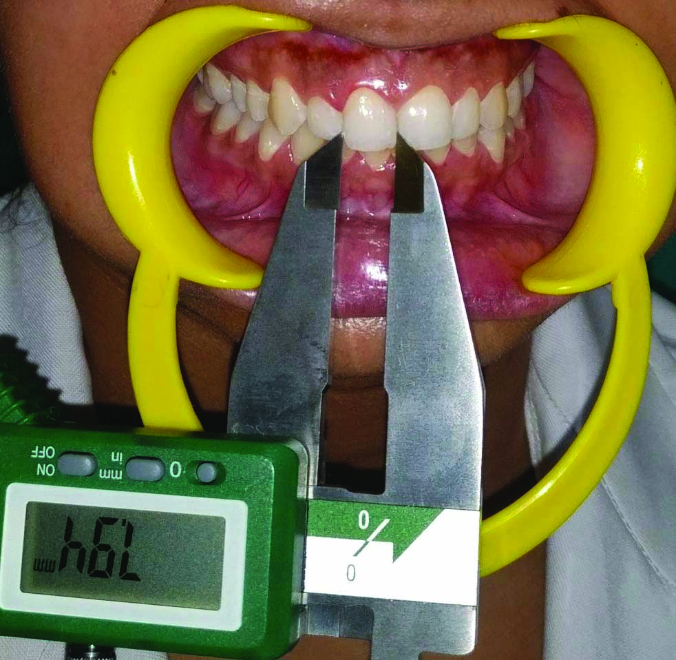
After obtaining informed consent, measurements of mesiodistal crown width of the six permanent maxillary anterior teeth (central incisor, lateral incisor and canine of both upper quadrants) were recorded with the help of calibrated (0-200 mm/0-8″) digital vernier calliper (Insize precision measurement, India). The greatest mesiodistal crown width was measured between the anatomic contact points of each tooth on either side of the jaw, with the vernier calliper held parallel to the occlusal plane to an accuracy level of 0.1 mm [Table/Fig-1]. To reduce the error in the study, each measurement was recorded in millimetres on a proforma three times by a single observer and the average of all the three values was taken as the final value.
Digital vernier calliper being held parallel to the occlusal plane and measurement being taken.
To determine sex, formulas were developed after application of several stepwise discriminant function statistics. The group centroids signify the average discriminant scores for each gender. Sex was determined by multiplying tooth dimensions with the respective unstandardised coefficients. The obtained value was then added to the constant by using following formula [15]:
y=a+b (p1)+b (p2)+b (p3)+b (m1)+b (m2)+b (m3)
a=constant of function between the right and left maxillary central incisors, lateral incisors and canines:
b=unstandardised coefficient of that specific tooth.
(p1, p2, p3 were measurement of teeth 11, 12, 13 and m1, m2, m3 were measurements of teeth 21, 22 and 23, respectively)
The percentage dimorphism can be defined as the percentage by which the tooth size of males exceeds that of females. The percentage of dimorphism for each tooth was calculated with the help of the following formula [16]:
Percentage of dimorphism={(Xm/Xf)-1}×100
Xm=Mean dimension of the male tooth
Xf=Mean dimension of the female tooth
The data collected was subjected to the statistical analysis.
Statistical Analysis
The mean, range, and standard deviation were calculated for the size of the teeth. A two-sample t-test was used to test for statistical difference between means [16]. For data evaluation, discriminant statistical analysis was used with Statistical Package for Social Sciences (SPSS) software version 17.0. To calculate sexual dimorphism, discriminant statistical analysis along with student’s t-test was used. Results were considered significant if p-value comes out to be <0.05%.
Results
The present study comprised of 300 individuals, of which 150 were males and 150 females between the age group of 18-24 years with mean age of 20.81 years and 20.54 years for male and females respectively.
[Table/Fig-2] shows descriptive statistics, t-values and p-values of each tooth selected for study. Mean mesiodistal dimension for each tooth was greater in males when compared with females and this difference was statistically significant with regard to both the canines (p=0.003 and 0.001 for right and left canine respectively) and right lateral incisor (p=0.019).
Mean and standard deviation odontometric parameters according to gender.
| Variable | Female (n=150) | Male (n=150) | ‘t’ value | p-value |
|---|
| Mean (mm) | SD (mm) | Mean (mm) | SD (mm) |
|---|
| Right central (11) | 8.404 | 0.560 | 8.504 | 0.907 | 1.146 | 0.253 |
| Right lateral (12) | 6.700 | 0.715 | 6.891 | 0.693 | 2.355 | 0.019* |
| Right canine (13) | 7.703 | 0.594 | 7.914 | 0.625 | 3.008 | 0.003* |
| Left central (21) | 8.408 | 0.562 | 8.543 | 0.918 | 1.541 | 0.124 |
| Left lateral (22) | 6.749 | 0.700 | 6.887 | 0.679 | 1.741 | 0.083 |
| Left canine (23) | 7.678 | 0.580 | 7.921 | 0.626 | 3.494 | 0.001* |
Student ‘t’ test: *p<0.05; Significant
According to findings of present study, if value obtained is greater than 0.211 then individual was considered as male and less than 0.211 indicated females [Table/Fig-3].
Depicts canonical discriminant function in the present study.
| Tooth | Standardised coefficient | Structure matrix | Unstandardised coefficient | Raw coefficient (Constant) | Group coefficient |
|---|
| Female | Male |
|---|
| 11 | -0.123 | 0.313 | -0.163 | -13.274 | -0.211 | 0.211 |
| 12 | 0.396 | 0.643 | 0.563 |
| 13 | 0.167 | 0.821 | 0.275 |
| 21 | 0.083 | 0.421 | 0.108 |
| 22 | -0.152 | 0.475 | -0.220 |
| 23 | 0.717 | 0.954 | 1.187 |
In the present study, when combination of all the teeth under study were taken into consideration, 62.7% females and 58% males were accurately identified. On cross-validation, 58.7% females and 52.7% males were accurately identified. On further analysis, it was seen that maxillary left canine is statistically significantly (p=0.001) useful as a tool for gender determination. Using dimensions of maxillary left canine alone, 61.3% females and 54% males were correctly identified. First gender determination was done using all the teeth in combination and result was discussed. Then further analysis was done and it was found that only maxillary left canine was statistically significant in determination of gender. Therefore, only maxillary left canine was used as a variable in determination of accuracy of gender. As other teeth were not statistically significant in gender determination hence further analysis had not been carried out using them as separate variable for determination of accuracy of gender.
Left maxillary canine showed highest percentage (3.164%) of sexual dimorphism whereas right maxillary central incisor showed lowest (1.189%) [Table/Fig-4].
Depicts percentage dimorphism for each tooth under study.
| Tooth | Percentage dimorphism |
|---|
| 11 | 1.189 |
| 12 | 2.859 |
| 13 | 2.739 |
| 21 | 1.605 |
| 22 | 2.044 |
| 23 | 3.164 |
Discussion
The present study demonstrates the perseverance of a definite statistically significant sexual dimorphism in left maxillary canine. However, dentition is considered to be a useful adjunct in determining sex since long time. Ditch L and Rose J proved for the first time that teeth dimensions can be used in determining sex in cases where sex could not be identified either because of fragmented or poorly preserved skeletal remains in archaeology [16-19].
It has been found that odontometric parameters show variability not only in specific populations but also within the same population [Table/Fig-5] [7,12-16,19,20-33] so there is a need to find out some specific values in particular areas in order to determine the sex of that very particular region or population [4]. So considering the above mentioned facts and also to develop population specific standards regarding sexing of an individual in forensic identification, in the present study, the authors included the subjects from Haryanvi population and ethnicity was determined on the basis of family history of the participants.
Comparison of odontometric studies done across the world on sexual dimorphism [7,12-16,19,20-33].
| Authors and Year | Region | Sample size | Teeth | Parameters |
|---|
| Abdullah MA (1998) [20] | Saudi Arabia | 100 | All teeth except third Molars | BL dimensions |
| Vodanovic M et al., (2006) [16] | Croatia | 86 | All teeth from skulls | MD, BL, robustness |
| Acharya BA (2008) [21] | Nepal | 123 | All teeth except third Molars | MD, BL dimensions |
| Agnihotri G and Gulati M, (2008) [12] | Faridabad, Haryana (India) | 100 | Permanent maxillary molar and premolar | Premolar and molar indices |
| Pratibha RM et al., (2009) [19] | Mysore (India) | 99 | Permanent maxillary and mandibular teeth except third molars | BL dimensions |
| Boaz K and Gupta C, (2009) [22] | Karnataka (India) | 100 | Canines | MD, BL dimensions |
| Sonika V et al., (2011) [13] | Ulana, Haryana (North India) | 200 | Permanent maxillary first molars | MD, BL dimensions |
| Khangura RK et al., (2015) [7] | Modinagar, North India | 100 | Maxillary incisors and canines | MD dimensions |
| Narang RS et al., (2014) [23] | Punjab, North India | 150 | First molar | BL |
| Sharma P et al., (2013) [24] | Muradnagar, Uttar Pradesh (India) | 200 | Left permanent maxillary first and second molars | MD, BL, MD-DL, DB-ML dimensions |
| Filipovic G et al., (2013) [25] | Serbia | 200 | Maxillary and mandibular permanent canines | MD and BL dimensions |
| Srivastava R et al., (2014) [15] | Uttar Pradesh, India | 300 | Maxillary anterior teeth | MD width |
| Gupta S et al., (2014) [14] | Lucknow, Uttar Pradesh | 180 | Right and left maxillary canines | MD diameter and ICW |
| Gupta A et al., (2014) [26] | Uttar Pradesh, India | 60 | Maxillary anterior teeth | MD width |
| Grewal DS et al., 2017 [27] | Punjab, India | 200 | Maxillary anterior teeth | ICW, IPW, AL and CW |
| Harshala S and Uddhav Y, (2017) [28] | Maharashtra (India) | 70 | Permanent Maxillary anterior teeth | CW, ICW and IPW |
| Mukesh F and Kuldeep B, (2017) [29] | Rajasthan Bikaner | 200 | Permanent Maxillary and mandibular canines | Intercanine distance |
| Banerjee A et al., (2016) [30] | Banglore | 100 | Permanent maxillary central incisor, canine, first premolar and the first molar | BLW, MDW, CL and CA |
| Kazzazi SM and Kranioti EF, (2017) [31] | North-western Iran | 282 | Permanent maxillary and mandibular molars | MD and BL dimensions |
| Davoudmanesh Ze et al., (2017) [32] | Iran | 220 | Permanent canine | MD and BL dimensions |
| Dash KC et al., (2018) [33] | Odisha (India) | 200 | Maxillary and mandibular permanent teeth except third molars | MD and BL dimensions |
| Present study | Haryana (India) | 300 | Permanent maxillary incisors and canines | MD dimensions |
Mesiodistal (MD), Buccolingual (BL), Combined Mesiodistal Width (CW), Intercanine Width (ICW), maxillary Interpremolar Width (IPW), Arch Length (AL), clinical Crown Length (CL), Cervical Angulation (CA), Diagonal Mesiobuccal-Distolingual (MD-DL), Distobuccal-mesiolingual (DB-ML) Mesio Buccal (MB), Distolingual (DL)
Males and females can be characterised on the basis of different features of teeth like morphology, crown size, root length etc., [33]. The crown of permanent teeth is formed at an early stage which can be related to very little change in their dimension during further growth and development. However, their dimensions could be altered because of pathology or nutrition. In this study, subjects were within the age range of 18-24 years. It is in accordance with the studies conducted by [7,27] where subjects range from 20-30 years. As the permanent dentitions of early adulthood show less mutilation and attrition in most individuals, they can be the best sample for tooth size measurements [34].
By measuring the mesiodistal and buccolingual dimensions, teeth can be used as tool for distinguishing gender. Vodanovic M et al., and Lakhanpal M et al., revealed that mesiodistal dimensions have better sex discriminatory ability as compared to buccolingual dimensions of maxillary dentition [16,35]. Previous studies have indicated that there is significantly greater sexual dimorphism in the upper and lower front teeth than in the other permanent teeth [36,37]. Further, it has also been established that sexual dimorphism is more pronounced in the maxillary than in the mandibular canine [25]. Therefore, in this study, the authors took into consideration mesiodistal dimension of crown of permanent maxillary anterior teeth.
In earlier studies, it has been reported that men have larger tooth crowns, although the degree of sexual dimorphism varies among different populations [25,38] which is in collaboration with the findings of present study. The difference in the magnitude of dimorphism in various populations could be explained on the basis of environmental influences (variation in food resources), cultural factors with biological forces and genetic factors on tooth size [7]. This larger crown size in males can be explained on the fact that amelogenesis for both deciduous and permanent dentitions in males occur for a longer period of time [39]. According to Acharya BA, sexual dimorphism in dental measurements could be contributed to Y chromosome producing slower male maturation [21].
In the present study, authors found mesiodistal dimensions of all teeth under study were larger in males than females but a statistically significant difference was observed only in canines and right lateral incisor. Srivastava R et al., found a statistically significant difference in maxillary central incisor and canine in Kanpur, Uttar Pradesh Population [15]. Similarly Khangura RK et al., reported that maxillary canines showed statistically significant difference in Modinagar (Uttar Pradesh) population [7]. Whereas in Odisha population, MD dimension of the maxillary right and left canines and premolars showed statistical significant differences [33]. Rai B and Annand SC, in India, Haryana suggested greatest sexual dimorphism in mandibular canine however they used diagonal parameters like distobuccal and mesiolingual [40]. Contrastingly, Parekh DH et al., in the Gujarat population of India indicated that the upper canine shows the greatest sexual dimorphism [41]. In the present study, it was observed that mesiodistal diameter of maxillary left permanent canine was helpful in determining the gender in population of Haryana. Vodanovic M et al., suggested that mesio-distal diameter of the crown of the upper canine was the variable providing the best sex discrimination among Croatian population [16].
The level of accuracy of sex determination when all the teeth under study were taken into consideration was 62.7% of females and 58% of males. Srivastava R et al., in their study found that 62% of males and 58.7% of females were correctly classified using right and left maxillary central incisor and canine [15]. Narang RS et al., reported that the overall accuracy of sex determination by mandibular canine index and mandibular molar odontometrics ranged from 56% to 84% [23].
Limitation(s)
There are certain limitations of the study such as all the measurements were done directly on the dentition of the participants. However, measurements could have been done on study cast models and variation between two methods could have been evaluated. Another shortcoming of the present study was smaller sample size.
Conclusion(s)
Sexual dimorphism in tooth size may differ among different populations and the population specific data is required. This study concludes that maxillary left permanent canine can aid in sex determination in Haryanvi population in forensic practice when fragmentary remains are encountered in mass disasters. More researches with larger sample size documenting the similar observations will enable the use of linear measurement independently in odontometric sex differentiation. More future studies with larger sample size should be conducted to collect the data in routine practice so as to provide basic registry for forensic odontology.
Student ‘t’ test: *p<0.05; Significant
Mesiodistal (MD), Buccolingual (BL), Combined Mesiodistal Width (CW), Intercanine Width (ICW), maxillary Interpremolar Width (IPW), Arch Length (AL), clinical Crown Length (CL), Cervical Angulation (CA), Diagonal Mesiobuccal-Distolingual (MD-DL), Distobuccal-mesiolingual (DB-ML) Mesio Buccal (MB), Distolingual (DL)