Elemental Analysis of Natal Teeth in a Monoidentical Twin using Scanning Electron Microscopy and Energy Dispersive X-ray Spectroscopy- A Case Report
Jayachandran Sadaksharam1, Vetriselvi Vimalesan2, Sophia Jeba Priya3
1 Professor and Head, Department of Oral Medicine and Radiology, Tamil Nadu Government Dental College and Hospital, Chennai, Tamil Nadu, India.
2 Postgraduate, Department of Oral Medicine and Radiology, Tamil Nadu Government Dental College and Hospital, Chennai, Tamil Nadu, India.
3 Assistant Professor, Department of Oral Medicine and Radiology, Tamil Nadu Government Dental College and Hospital, Chennai, Tamil Nadu, India.
NAME, ADDRESS, E-MAIL ID OF THE CORRESPONDING AUTHOR: Dr. Vetriselvi Vimalesan, Tamil Nadu Government Dental College and Hospital (Affiliated to the Tamil Nadu Dr. M G R Medical University), Chennai-600003, Tamil Nadu, India.
E-mail: vetrivimal360@gmail.com
Dental eruption is a normal physiological process that begins with eruption of primary lower central incisors when a child is six-month-old. Natal teeth are teeth present at birth while neonatal teeth are teeth that erupt within the first month of life. Primary teeth erupting prematurely are referred to as congenital teeth, predeciduous teeth, foetal teeth, and dentitia praecox. The exact aetiology is not known. Natal teeth are three times more common than neonatal teeth. However, natal or neonatal teeth is quite rare with incidence of 1:1000 to 1:30,000 which usually erupts during birth or within one month. This case reports a 24-day-old male neonate with two teeth in the mandibular alveolar region. Since one of the teeth were immature and hypermobile, it was planned for removal fearing aspiration. The extracted natal tooth was quantitatively and qualitatively assessed for the elements present in it by Energy Dispersive X-Ray Spectroscopy (EDX) and Scanning Electron Microscopy (SEM) at the level of middle third, both on the internal and external surfaces. Carbon and oxygen were predominantly present in natal teeth when compared to permanent teeth, which mainly composed of calcium and phosphorus. The natal tooth caused mild problem in feeding, as it was mobile. So, it was extracted to avoid aspiration. The aim of this case report is to discuss a rare case of occurrence of two natal teeth in one of the premature monoidentical twins (male babies) and also to analyse the elemental components present in the natal tooth using SEM and EDX.
Carbon and oxygen, Hypomineralisation teeth, Preterm birth
Case Report
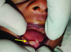
A 24-day-old male (twin-1) preterm baby born to a mother who gave birth to monoidentical twins was referred from Department of Neonatology, to the Department of Pedodontics with a complaint of presence of two teeth in the lower jaw of one of the twins since birth. Other twin is apparently normal with absence of natal teeth. Patient’s mother complained of difficulty in feeding the baby as one of the tooth was mobile. No other relevant medical history was recorded. History of second degree consanguinity was recorded. On examining there were two teeth in mandibular anterior alveolus region [Table/Fig-1] which were white opaque in colour and one of the teeth was grade III mobile and another one was firm. The crown size of one tooth was very small (around 3-4 mm) and two-thirds of crown was covered by gingiva. Adequate coverage of gingiva was seen over the other tooth. Previously, a similar case was reported with occurrence of two natal teeth in premature dizygotic twin females, which has very rarely been reported [1].
Diagnosis was given as natal teeth. Since extraction was the treatment of choice, vitamin k (0.5-1.0 mg) was administered intramuscularly as part of immediate medical care to prevent haemorrhage [2,3]. Informed consent was obtained from the parents before extracting the tooth. Since one of the natal tooth had grade III mobility (According to Spouge and Feasby (1966) and it was covered by gingiva, preference for extraction was given only to that natal tooth [4]. The other natal tooth was firm and immobile, and parents were not willing for removal of the firm tooth. Extraction was done under topical anaesthesia in the Department of Pedodontics and the baby tolerated well. Bleeding was arrested after extraction by asking the mother to feed immediately and paracetamol suspension (120 mg)-1.5 mL was prescribed. The patient was reviewed after three days. Healing was satisfactory.
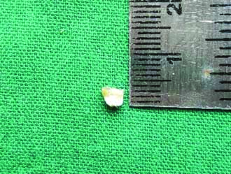
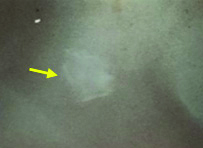
The extracted tooth had a crown without root formation covered by soft tissue roughly measured about 4 mm [Table/Fig-2]. Radiograph of natal teeth was taken which revealed a radiopaque tooth like structure without root formation [Table/Fig-3]. The tooth was collected in a sterile bottle with 10% neutral buffered formalin stored for about 24 hours. After 24 hours, the teeth was rinsed with distilled water and air dried. Dried tooth sample was collected in sterile container.
Arrow represents the mobile natal/neonatal tooth.
Extracted natal tooth measured 4 mm.
Arrow shows Radiograph of extracted natal tooth shows ill-defined radiopaque tooth like structure;
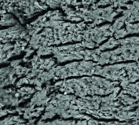
The elemental analysis was done using EDX spectroscopy and surface morphology was analysed using SEM. The ultrastructural analysis was done using FEI-QuantaTM FEG 200F SEM (Oregon, USA) which is suitable for surface morphology research and the elemental distribution and the mean values of the atomic weights were measured by EDX. Natal tooth kept for SEM and EDX analyses were coated with a thin film of evaporated gold to get better electrical conduction and examination was at an accelerating voltage of 20 kV with extended vacuum (10-4000 Pascal (Pa)) mode of 1.5 nm and a working distance of 20 mm [5]. After calibration setting, mid portion were analysed on the enamel and dentinal surface of the natal teeth [5]. Then series of photo micrographs [Table/Fig-4,5] were obtained for viewing surface morphology of the tooth using SEM. The elemental distribution and the mean values of atomic weights were measured by EDX.
Scanning Electron Microscopy (SEM) at magnification of 1000x shows- enamel rods with cracks present on the surface. The enamel surface is rough, with surface defects wide fissures and niches, covered with different size and number particles;
Scanning Electron Microscopy (SEM) at magnification of 1000x taken at adjacent side shows-Dentinal tubules with inter-tubular microporosities.
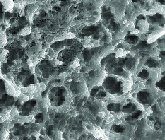
The SEM analysis in the external surfaces of natal tooth at 1000X magnificaiton [Table/Fig-4] revealed enamel rods with cracks present on the surface with wide fissures and rough surface several micropores indicating open enamel prisms. The morphological details of dentin showed irregular and rough surface covered by an amorphic layer indicating the presence of smear layer. The dentinal tubules were patent and inorganic dentin debris were detected. The dentin surface possessed intertubular microporosity [Table/Fig-5].
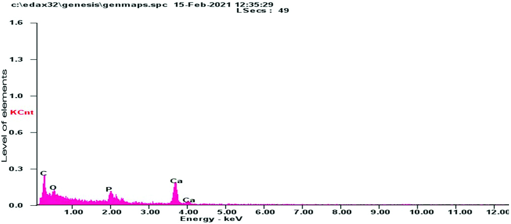
A total of four trace elements ((Calcium (Ca), Phosphorus (P), Carbon (C), Oxygen (O)) were detected in the natal tooth sample by EDX. Analysis taken at two different midportion of natal teeth are shown in [Table/Fig-6,7].
Shows elements present in natal tooth with molecular and atomic weight in percentage.
| Element | Wt% | At% |
|---|
| Carbon | 61.95 | 70.89 |
| Oxygen | 30.65 | 26.33 |
| Phosphorus | 02.35 | 01.04 |
| Calcium | 05.05 | 01.73 |
| Matrix | Correction | ZAF |
Z- atomic number, A- absorption, F- fluorescence; ZAF matrix correction- is used for quantitative analysis of target elements. As a specimen is thicker (several 10nm or thicker through depending on measured elements), the intensity of the emitted characteristic X-rays is influenced by the atomic number effect, the absorption effect, and the fluorescence excitation effect
Shows elements present in natal tooth with molecular and atomic (At%) weight (Wt%) in percentage.
| Element | Wt% | At% |
|---|
| Carbon | 59.62 | 69.03 |
| Oxygen | 31.99 | 27.81 |
| Phosphorus | 02.46 | 01.10 |
| Calcium | 05.94 | 02.06 |
| Matrix | Correction | ZAF |
Z- atomic number, A- absorption, F- fluorescence; ZAF matrix correction- is used for quantitative analysis of target elements. As a specimen is thicker (several 10nm or thicker through depending on measured elements), the intensity of the emitted characteristic X-rays is influenced by the atomic number effect, the absorption effect, and the fluorescence excitation effect
It was found that the concentration of organic and inorganic content varied according to tissue (enamel, dentin) and also depends on the tooth region. Spectral Data 1 and 2 shows increase in organic content of carbon and oxygen [Table/Fig-8,9].
Average spectral data-1: showing higher level of carbon in natal tooth.
C: Carbon; O: Oxygen; P: Phosphorus; Ca: Calcium
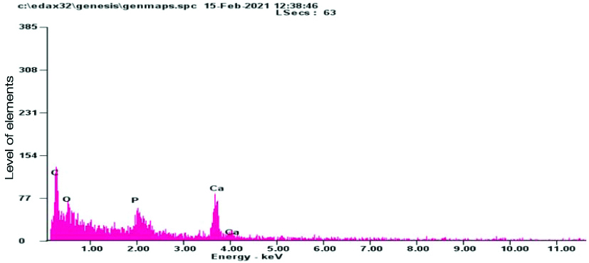
Average spectral data-2: showing higher level of carbon in natal tooth.
Discussion
Natal teeth are also referred as dentition praecox, foetal teeth, or early infancy teeth. Syndromes associated with natal teeth are cleft palate, chondroectodermal dysplasia, Hallermann-Streiff syndrome, Ellis-Van Creveld syndrome. The incidence of natal and neonatal teeth ranges from 1: 2,000 to 1: 3,500. Natal and neonatal teeth are commonly found in the mandibular incisor region with a 66% predilection in females [4]. Most of the natal/neonatal tooth are considered to be early eruption of normal deciduous teeth around 70-90% and incidence of supernumerary teeth is around 10% [2-4]. In addition, it is difficult for the mother to breastfeed because she may be injured or unable to encourage lactation and sometimes the baby may develop a pathology called Riga-Fede ulceration on the ventral surface of the tongue or on the inner surface of the lower lip [6,7]. But this case was no longer related to any syndrome. The natal tooth present in this case was not withstanding normal basic structure [8]. In this case, despite the fact that the natal teeth precipitated moderate trouble in feeding, It showed mobility. So, It was extracted to avoid aspiration of the tooth [9].
Histological variations have been reported in natal teeth when compared with primary teeth in lack of root formation, Hertwig’s sheath and cementum formation, enlarged pulp chambers with an increase in quantity of dilated blood vessels within the pulpal tissue and between the odontoblasts, irregular dentin and cell inclusions in the cervical region and interglobular dentin in the coronal region [10]. Biomineralisation of teeth is a continuous and lifelong process through which inorganic nanocrystal precipitations occur within the organic matrices to form hybrid biological tissues [11]. And additionally in SEM the presence of few fibrous structures within the dentinal tubules and the circular orientation of the peritubular collagen network is suggestive of residual mineral deposit [12]. Natal & neonatal teeth are often associated with hypomineralization of enamel, decrease in thickness, dysplastic and covering only the two-thirds of the crown. These teeth have the capacity to incorporate chemical elements into their structure, the maximum being calcium, phosphorus, magnesium and sodium [11]. Most of the studies, indicates that scanning electron microscope was found to be very useful in the evaluation of the developmental defects of enamel surface [12-14]. It is a type of electron microscope that images the sample surface by scanning it with a high-energy beam of electrons in a raster scan pattern [14]. A broad range of elements are present in dental structures. EDX analysis of permanent teeth samples showed the increase in Ca, O and also presence of C, P [15]. A study using EDX analysis of human permanent teeth shows increased concentration of calcium [15]. Another study done in natal tooth using Laser Ablation Inductively Coupled Plasma Mass Spectrometry (LA-ICP-MS) method shows nine trace elements (Calcium, Cadmium, Copper, Magnesium, Manganese, Nickel, lead, Strontium and Zinc) with increased amount of calcium [10]. In this case, an increase in organic content carbon and oxygen was seen.
Early diagnosis and treatment of natal teeth are often important in lowering the aesthetic and functional problems of the adjacent teeth. Management consists of parental counselling, guidance, and proper choice making are important [3]. Extraction is a preferred procedure as treatment, because of the aforementioned potential complications. Considering immunological and haematological aspect, the best time for extraction was found to be at 7-25 days of birth. In addition, vitamin K administration is recommended for prophylactic purpose due to the risk of haemorrhage, since coagulation might not be achieved properly until the child is 10-day-old [6]. Obstetricians and gynaecologists and paediatricians ought to refer neonates born with teeth to the Dental surgeons for professional management [8].
Conclusion(s)
Natal and neonatal teeth are rare occurrences in the oral cavity. EDX findings in this case show increase in the concentration of carbon rather than calcium and phosphate. As only one case report has been considered here, the results should be taken as a preliminary one and further studies using SEM-EDX with larger samples would give a better picture of elemental composition of natal/neonatal teeth.
Z- atomic number, A- absorption, F- fluorescence; ZAF matrix correction- is used for quantitative analysis of target elements. As a specimen is thicker (several 10nm or thicker through depending on measured elements), the intensity of the emitted characteristic X-rays is influenced by the atomic number effect, the absorption effect, and the fluorescence excitation effect
Z- atomic number, A- absorption, F- fluorescence; ZAF matrix correction- is used for quantitative analysis of target elements. As a specimen is thicker (several 10nm or thicker through depending on measured elements), the intensity of the emitted characteristic X-rays is influenced by the atomic number effect, the absorption effect, and the fluorescence excitation effect
Author Declaration:
Financial or Other Competing Interests: None
Was informed consent obtained from the subjects involved in the study? NA
For any images presented appropriate consent has been obtained from the subjects. NA
Plagiarism Checking Methods: [Jain H et al.]
Plagiarism X-checker: Jun 07, 2021
Manual Googling: Aug 28, 2021
iThenticate Software: Sep 29, 2021 (19%)
[1]. Dahake PT, Shelke AU, Kale YJ, Iyer VV, Natal teeth in premature dizygotic twin girlsBMJ Case Rep 2015 2015:bcr201521193010.1136/bcr-2015-21193026682836 [Google Scholar] [CrossRef] [PubMed]
[2]. Malki GA, Al-Badawi EA, Dahlan MA, Natal teeth: A case report and reappraisalCase Rep Dent 2015 2015:14758010.1155/2015/14758025722895 [Google Scholar] [CrossRef] [PubMed]
[3]. Khandelwal V, Nayak UA, Nayak PA, Bafna Y, Management of an infant having natal teethCase Rep 2013 2013:bcr201301004910.1136/bcr-2013-01004923737593 [Google Scholar] [CrossRef] [PubMed]
[4]. Mhaske S, Yuwanati MB, Mhaske A, Ragavendra R, Kamath K, Saawarn S, Natal and neonatal teeth: An overview of the literatureISRN Pediatr 2013 2013:95626910.1155/2013/95626924024038 [Google Scholar] [CrossRef] [PubMed]
[5]. Sadaksharam J, Kuduva Ramesh SS, Spectroscopic and microscopic characterisation of parotid and submandibular ductal sialoliths: A comparative preliminary studyProc Natl Acad Sci India Sect B Biol Sci 2020 90(5):1165-71.10.1007/s40011-020-01189-9 [Google Scholar] [CrossRef]
[6]. Bulut G, Bulut H, Ortac R, A comprehensive survey of natal and neonatal teeth in newbornsNiger J Clin Pract 2019 22(11):148910.4103/njcp.njcp_152_1931719269 [Google Scholar] [CrossRef] [PubMed]
[7]. Rezende KM, Imparato JCP, França DCDO, Rocha MO, Bönecker M, Dental pulp stem cells from natal teeth: Isolation and morphological studyJ Clin Diagn Res 2018 12(3):ZC46-49.10.7860/JCDR/2018/30783.11322 [Google Scholar] [CrossRef]
[8]. Sadaksharam J, Jeba Priya JS, Natal tooth: A histomorphologic variant, a rarityAnn Natl Acad Med Sci India 2019 55(04):210-12.10.1055/s-0039-3401468 [Google Scholar] [CrossRef]
[9]. Rao RS, Mathad SV, Natal teeth: Case report and review of literatureJ Oral Maxillofac Pathol JOMFP 2009 13(1):41-46.10.4103/0973-029X.4457421886998 [Google Scholar] [CrossRef] [PubMed]
[10]. Olszewska A, Hanć A, The potential of trace elements mapping in child’s natal tooth by laser ablation-ICPMS methodJournal of Environmental Health Science and Engineering 2021 19(1):379-88.10.1007/s40201-021-00611-234150242 [Google Scholar] [CrossRef] [PubMed]
[11]. Fernández-Escudero AC, Legaz I, Prieto-Bonete G, López-Nicolás M, Maurandi-López A, Pérez-Cárceles MD, Aging and trace elements in human coronal tooth dentineSci Rep 2020 10(1):996410.1038/s41598-020-66472-132561784 [Google Scholar] [CrossRef] [PubMed]
[12]. Thakur R, Patil SDS, Kush A, Madhu K, SEM analysis of residual dentin surface in primary teeth using different chemomechanical caries removal agentsJ Clin Pediatr Dent 2017 41(4):289-93.10.17796/1053-4628-41.4.28928650784 [Google Scholar] [CrossRef] [PubMed]
[13]. Azinovi Z, Keros J, Bukovi D, Azinovi A, SEM analysis of tooth enamelColl Antropol 2003 27(1):381-6. [Google Scholar]
[14]. Nazir S, Naqvi NH, Masood A, Naema N, SEM-EDX Analysis for surface aberrations of neonate’s teeth influenced by the use of lithium in pregnancyWorld J Dent 2011 2(2):105-10.10.5005/jp-journals-10015-1065 [Google Scholar] [CrossRef]
[15]. Calvo-Guirado JL, Ballester-Montilla A, N De Aza P, Fernández-Domínguez M, Alexandre Gehrke S, Cegarra-Del Pino P, Particulated, extracted human teeth characterization by SEM-EDX evaluation as a biomaterial for socket preservation: An in vitro studyMaterials 2019 12(3):38010.3390/ma1203038030691075 [Google Scholar] [CrossRef] [PubMed]