A unicornuate uterus is the result of abnormal or failed development of one Mullerian duct. Partial development of one Mullerian duct leads to a rudimentary uterine horn. This can lead to various gynaecological and obstetric problems and diagnostic dilemma. Over the past ten years, the authors have witnessed about eight cases of unicornuate uterus with canalised rudimentary horn presenting as a wide spectrum of obstetric and gynaecological problems. Two cases presented as dysmenorrhoea, one was diagnosed as a part of infertility work-up and the rest presented with varied obstetric complications. Most of the cases underwent excision of the rudimentary horn with unilateral salpingectomy. High clinical suspicion and knowledge about varied presentation is important for early diagnosis in such cases and to avert the clinical complications.
Introduction
The Mullerian duct anomalies are the congenital developmental abnormalities of the female genital tract which result from the maldevelopment of the Mullerian ducts. The incidence varies between 2-4% and is said to be higher among the women with infertility and obstetric complications [1]. The failure of development of one Mullerian duct leads to the formation of an isolated hemi uterus without a contralateral structure to various degrees of a rudimentary horn. The unicornuate uterus belongs to the class U4 as per the classification of European Society of Human Reproduction and Embryology (ESHRE) [2]. This is further classified as U4a (with rudimentary cavity) and U4b (without rudimentary cavity).
The presence of a rudimentary uterine horn with cavity can present with various clinical scenarios. While most of them are asymptomatic, those with functional endometrium present with various gynaecological problems such as cyclic or chronic pelvic pain, haematometra, and endometriosis [1]. The uterine horn can also act as a site for ectopic pregnancy, the natural course of which is to rupture by second trimester [3]. Pregnancy in rudimentary horn reaching viability, resulting in a live birth is extremely rare, though a few cases have been reported [4]. Eight different cases with unicornuate uterus and a rudimentary horn presenting in different phases of reproductive cycle-menstrual problems, infertility and obstetric complications are presented here.
Case 1
A 14-year-old girl presented to Out Patient Department (OPD) with complaints of severe dysmenorrhoea since menarche. Clinical examination was normal, she was started on symptomatic management for dysmenorrhoea in the form of Non-Steroidal Anti-Inflammatory Drugs (NSAIDs). Ultrasonography (USG) abdomen and pelvis were performed when she failed to respond to medications, which showed a subserosal fibroid. Medical management of dysmenorrhoea was continued. She was taken up for diagnostic laparoscopy a few weeks later when she presented with acute abdominal pain. It revealed a unicornuate uterus with a rudimentary horn and a non communicating cavity full of accumulated menstrual secretions. She underwent an endometrectomy in the rudimentary horn as performing excision of the rudimentary horn was surgically difficult.
Case 2
A nulliparous woman, presented to the OPD with complaints of heavy menstrual flow and dysmenorrhoea for 3-4 years, which was resistant to medical therapy in the form of antifibrinolytics, NSAIDs, Oral Contraceptive Pills (OCPs). The USG showed a bicornuate, unicollis uterus. Magnetic Resonance Imaging (MRI) abdomen and pelvis showed a unicornuate uterus with a non communicating canalised rudimentary horn with an endometriotic cyst. She underwent laparotomy with endometrial cystectomy and rudimentary horn excision.
Case 3
A 24-year-old lady, married for 3 years, presented to the authors’ infertility clinic, anxious to conceive. Her medical history was unremarkable except for the complaints of episodes of dysmenorrhoea for the past few years, which was being managed medically with NSAIDs. The USG showed complex adnexal mass separate from ovary, whereas MRI of the abdomen and pelvis showed unicornuate uterus with a rudimentary canalised horn with haematometra. The patient underwent laparoscopic hemi-hysterectomy with ipsilateral salpingectomy.
Case 4
A G2P1L1 with previous vaginal delivery, with a scan showing Intrauterine Demise (IUD) at 19 weeks gestation came to us for termination of pregnancy. She underwent labour induction with the standard dose of misoprostol, PGE2 gel. Mechanical method of cervical dilatation using foley’s catheter was attempted, but was unsuccessful. Even after all these methods, the cervical os remained tightly closed. Hence, hysterotomy was planned. Intraoperatively, a unicornuate uterus with the pregnancy in the rudimentary horn was noted, which was excised.
Cases 5 and 6
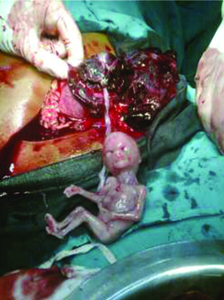
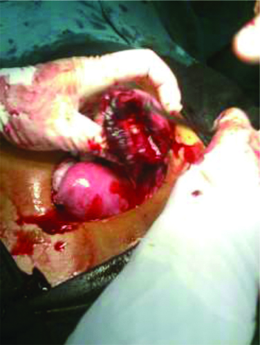
A primigravida at 14 weeks of gestation presented to Emergency Room (ER) in shock. Bedside USG showed a foetus of 13 weeks in the abdomen with haemoperitoneum. Another woman at 16 weeks of gestation also presented in the similar way- in shock and with haemoperitoneum. Both cases underwent emergency laparotomy which revealed a ruptured pregnancy in the rudimentary uterine horn and a free-floating foetus in the abdomen [Table/Fig-1,2]. Hemi-hysterectomy was performed in both cases.
Ruptured rudimentary horn with foetus extruded into the peritoneal cavity.
Ruptured rudimentary horn.
Case 7
A 28-year-old lady, known case of epilepsy, was found unconscious at her home. She was rushed to the ER and was admitted in the Intensive Care Unit (ICU) for further evaluation and management. She became tachycardic with falling Blood Pressure (BP). An ultrasound of her abdomen and pelvis showed an ectopic pregnancy of about 12 weeks in the right adnexa. She underwent laparotomy which revealed the pregnancy in rudimentary uterine horn.
Case 8
Primigravida at 13 weeks of gestation presented with pain in abdomen since a week. Examination revealed a tender mass in the lower abdomen. The USG abdomen and pelvis showed that she had a complex adnexal mass. It was followed-up with an MRI which revealed that the adnexal mass was actually a rudimentary horn with the pregnancy inside. The wall of the horn was thinned out suggesting impending rupture, surgical excision was done.
Discussion
The unicornuate uterus (Class U4) constitutes about 2.4-13% of all Mullerian anomalies [5]. The subtype consisting of a non-communicating rudimentary horn with a functional endometrium can lead to many gynaecological manifestations such as dysmenorrhoea and pelvic pain due to haematometra or endometriosis. Also, pregnancies can occur in these rudimentary horns due to the transperitoneal migration of sperm or zygote and usually ends in uterine rupture [6]. This phenomenon is very rare, occurring in about 1 per 100000 to 140000 pregnancies [3]. The horn rupture in such pregnancies occur usually before 20 weeks [7]. But there are a few case reports of these pregnancies resulting in live births. If the pregnancy continues beyond the second trimester, it might present as malformed foetus, foetal growth restriction, pathological placentation, oligohydramnios and foetal malpresentation [8]. Another manifestation of rudimentary horn pregnancy is failure of termination of pregnancy by medical methods and uterine evacuation.
Over the past ten years, the authors have encountered all these varied manifestations of a unicornuate uterus with canalised rudimentary horn in the tertiary care hospital. Various tertiary care centres have reported similar cases of unicornuate uterus with rudimentary horn presenting in varied ways [Table/Fig-3] [4,9-11].
List of similar cases in literature [4,9-11].
| Year | Study by | Presenting complaints | Diagnosis | Treatment |
|---|
| 2010 | Medeiros LR et al., [9] | Severe dysmenorrhoea | Right unicornuate uterus with left non communicating rudimentary horn with endometrioma; absent left kidney | Laparoscopic removal of left horn with endometrioma excision |
| 2013 | Ambusaidi Q and Jha C [10] | Failed medical methods for termination of pregnancy at 23 weeks with intrauterine foetal demise | Pregnancy in the right rudimentary uterine horn; absent right kidney | Laparotomy with hysterotomy and rudimentary horn excision |
| 2013 | Gonçalves E et al., [4] | 34 weeks pregnancy with hypertensive disorder of pregnancy | Left unicornuate uterus with rudimentary horn pregnancy; absent right kidney | Caesarean section with excision of the rudimentary horn |
| 2018 | Kathpalia S [11] | Primigravida with 5 months gestation with pain abdomen | Ruptured left rudimentary horn of unicornuate uterus with extrusion of foetus and placenta | Excision of the ruptured rudimentary horn |
| 2018 | Kathpalia S [11] | Long standing pain abdomen with acute exacerbation | Right rudimentary non-communicating uterine horn | Laparotomy and rudimentary horn excision |
All the above presentations have been noted in the authors' centre too with management done in similar way, though the authors have not witnessed a case of rudimentary horn pregnancy continuing near term.
The diagnosis of this condition is always challenging and is often not considered, leading to life threatening situations. The clinical suspicion and diagnosis of a rudimentary horn when the patient presents with gynaecological symptoms is difficult as there are no tell-tale signs. However, in cases with pregnancy in the rudimentary horn a few clinical signs might point towards diagnosis. Bimanual palpation in the early gestation of these cases, shows a deviated uterus with a palpable adnexal mass extending outside the uterine angle known as the Baart de la faille’s sign [12]. Even with the advancements in the field of medical imaging, sensitivity of USG in diagnosing a pregnant uterine horn remains a mere 30% [13]. Tsafrir A et al., in their article, suggested ultrasound criteria for early diagnosis of this condition which includes: (a) a pseudo pattern of asymmetrical bicornuate uterus; (b) absent visual continuity between the cervical canal and the lumen of the pregnant horn; and (c) presence of myometrial tissue surrounding the gestational sac [14].
In non obstetric cases MRI plays a major role in the diagnosis of Mullerian anomalies. When ultrasonography becomes inconclusive or does not point towards a diagnosis in cases of refractory dysmenorrhoea or chronic pelvic pain, higher imaging modalities such as MRI should be considered. It is also important for all cases of uterine malformations to undergo evaluation for urinary tract and skeletal abnormalities, as these are commonly associated.
The management of unicornuate uterus with a rudimentary horn is not as challenging as the diagnosis itself. The standard treatment is excision of the rudimentary horn with ipsilateral salpingectomy. This relieves the patient symptoms such as dysmenorrhoea, chronic pelvic pain and also reduces the chances of ectopic pregnancy. It is important to remove the rudimentary horn even when diagnosed incidentally in an asymptomatic patient, due to the high rates of ectopic pregnancy [15]. There are many literatures which support a laparoscopic approach for these surgeries [9,16],
The management of pregnancy in the rudimentary horn remains the same, laparotomy and excision of the rudimentary horn with ipsilateral salpingectomy. Hysterectomy may be necessary in cases with massive haemorrhage. The surgical principles remain the same, but the increased vascularity of the pedicles in pregnancy leading to haemorrhage should be kept in mind. Medical management of rudimentary horn pregnancy with methotrexate is unconventional, though a successful case has been documented [17].
Among the eight cases noted in the present study, three cases presented with gynaecological symptoms whereas five cases had obstetric manifestations. In majority of the cases, the rudimentary horn as the cause of symptoms was only diagnosed intraoperatively as opposed to three cases wherein the diagnosis was established through imaging preoperatively. None of the cases had other associated anomalies.
As the article presents cases over past decade, few of the cases included were done in 2009-2010. It was during the time when there were no cameras in phone and our hospital was not digitalised. Hence, the images of all the cases were not procured.
Conclusion(s)
Mullerian malformations, including rudimentary horn of the uterus can present in a wide spectrum of clinical signs and symptoms. It also poses a diagnostic dilemma as many other gynaecological and obstetric problems can present in the same manner. Appropriate usage of clinical examination and various imaging modalities, as well as diagnostic laparoscopy/hysteroscopy is the key in clinching the diagnosis and averting its complications.
[1]. Iverson R, DeCherney A, Laufer M, Clinical manifestations and diagnosis of congenital anomalies of the uterusUptodate 2012 [Google Scholar]
[2]. Grimbizis GF, Gordts S, Di Spiezio Sardo A, Brucker S, De Angelis C, Gergolet M, The ESHRE/ESGE consensus on the classification of female genital tract congenital anomaliesHum Reprod 2013 28(8):2032-44.10.1093/humrep/det09823771171 [Google Scholar] [CrossRef] [PubMed]
[3]. Chopra S, Keepanasseril A, Rohilla M, Bagga R, Kalra J, Jain V, Obstetric morbidity and the diagnostic dilemma in pregnancy in rudimentary horn: Retrospective analysisArchives of Gynaecology and Obstetrics 2009 280(6):907-10.10.1007/s00404-009-1013-419283398 [Google Scholar] [CrossRef] [PubMed]
[4]. Gonçalves E, Prata JP, Ferreira S, Abreu R, Mesquita J, Carvalho A, An unexpected near-term pregnancy in a rudimentary uterine hornCase Rep Obstet Gynecol 2013 2013:30782810.1155/2013/30782823710390 [Google Scholar] [CrossRef] [PubMed]
[5]. Grimbizis GF, Camus M, Tarlatzis BC, Bontis JN, Devroey P, Clinical implications of uterine malformations and hysteroscopic treatment resultsHum Reprod Update 2001 7(2):161-74.10.1093/humupd/7.2.16111284660 [Google Scholar] [CrossRef] [PubMed]
[6]. Kadir R, Hart J, Nagele F, O’Connor H, Magos A, Laparoscopic excision of a noncommunicating rudimentary uterine hornBJOG Int J Obstet Gynaecol 1996 103(4):371-72.10.1111/j.1471-0528.1996.tb09744.x8605136 [Google Scholar] [CrossRef] [PubMed]
[7]. Kanagal DV, Hanumanalu LC, Ruptured rudimentary horn pregnancy at 25 weeks with previous vaginal delivery: A case reportCase Rep Obstet Gynecol 2012 2012:98507610.1155/2012/98507622720180 [Google Scholar] [CrossRef] [PubMed]
[8]. Shin JW, Kim HJ, Case of live birth in a non-communicating rudimentary horn pregnancyJournal of Obstetrics and Gynaecology Research 2005 31(4):329-31.10.1111/j.1447-0756.2005.00296.x16018780 [Google Scholar] [CrossRef] [PubMed]
[9]. Medeiros LR, Rosa DD, Rosa Silva F, Rosa Silva B, Rosa MI, Laparoscopic approach of a unicornuate uterus with noncommunicating rudimentary hornsISRN Obstet Gynecol 2011 2011:90613810.5402/2011/90613821647229 [Google Scholar] [CrossRef] [PubMed]
[10]. Ambusaidi Q, Jha C, Pregnancy in the rudimentary uterine horn: Case report of an unusual presentationSultan Qaboos University Med J 2014 14(1):e134-38.10.12816/000334924516746 [Google Scholar] [CrossRef] [PubMed]
[11]. Kathpalia S, Rudimentary horn- Different clinical presentationsObstet Gynecol Int J 2018 9(6):440-42. [Google Scholar]
[12]. Anupama H, Madhuri Y, Murthy NLN, Pregnancy in non-communicating rudimentary hornSaudi J Med Pharm Sci 2016 2(9):270-72. [Google Scholar]
[13]. Hassen C, Karim A, Ismail N, Omar M, Case report of a ruptured non-communicating right rudimentary horn pregnancy: An acute emergencyActa Medica 2011 54(3):125-26.10.14712/18059694.2016.3422250483 [Google Scholar] [CrossRef] [PubMed]
[14]. Tsafrir A, Rojansky N, Sela HY, Gomori JM, Nadjari M, Rudimentary horn pregnancy: First-trimester prerupture sonographic diagnosis and confirmation by magnetic resonance imagingJournal of Ultrasound in Medicine 2005 24(2):219-23.10.7863/jum.2005.24.2.21915661954 [Google Scholar] [CrossRef] [PubMed]
[15]. Heinonen PK, Unicornuate uterus and rudimentary hornFertil Steril 1997 68(2):224-30.10.1016/S0015-0282(97)81506-3 [Google Scholar] [CrossRef]
[16]. Dicker D, Nitke S, Shoenfeld A, Fish B, Meizner I, Ben-Rafael Z, Laparoscopic management of rudimentary horn pregnancyHuman Reproduction 1998 13(9):2643-44.10.1093/humrep/13.9.26439806300 [Google Scholar] [CrossRef] [PubMed]
[17]. Rodrigues A, Neves AR, Castro MG, Branco M, Geraldes F, Águas F, Successful management of a rudimentary uterine horn ectopic pregnancy by combining methotrexate and surgery: A case reportCase Rep Womens Health 2019 24:e0015810.1016/j.crwh.2019.e0015831799126 [Google Scholar] [CrossRef] [PubMed]