Boxing Match Gone Awry- A Punch over the Head Leading to Eccentric Proptosis in a Child
Gayathri Nithianandam1, S Prabakaran2, Geeta Anusha Loya3, S Rajasekaran4, Premnath Gnaneswaran5
1 Senior Resident, Department of Ophthalmology, Chettinad Academy of Research and Education, Chennai, Tamil Nadu, India.
2 Assistant Professor, Department of Otorhinolaryngology, Chettinad Academy of Research and Education, Chennai, Tamil Nadu, India.
3 Postgraduate Student, Department of Ophthalmology, Chettinad Academy of Research and Education, Chennai, Tamil Nadu, India.
4 Professor and Head, Department of Otorhinolaryngology, Chettinad Academy of Research and Education, Chennai, Tamil Nadu, India.
5 Professor, Department of Ophthalmology, Chettinad Academy of Research and Education, Chennai, Tamil Nadu, India.
NAME, ADDRESS, E-MAIL ID OF THE CORRESPONDING AUTHOR: Premnath Gnaneswaran, Chettinad Academy of Research and Education, Chennai, Tamil Nadu, India.
E-mail: drpremn18@yahoo.co.in
The most common ocular pathologic conditions in amateur boxing are sub conjunctival haemorrhage, lid injuries, cataract, pupil deformation, angle abnormalities and retinal tear. Proptosis due to frontoethmoidal mucocele in young is one of the least common complications seen in boxers. Here, we discuss a rare case of unilateral eccentric proptosis of right eye in a nine-year-old child after he was punched on the right side of the head during a boxing match. He came with complaint of swelling of upper eyelid. Examination revealed right sided eccentric proptosis with restricted ocular movements and defective vision. Computed Tomography (CT) of orbit showed a well-defined isodense lesion with smooth margins arising from the frontal sinus, extending inferiorly to anterior ethmoidal sinus which caused mass effect over right eye and ocular muscles which resulted in displacement of the eye ball. Otolaryngologist’s opinion was obtained. Functional Endoscopic Sinus Surgery (FESS) was done. Postoperatively vision improved with no proptosis and eyeball returned to its normal position.
Eyeball protrusion, Mucocele, Ocular injuries
Case Report
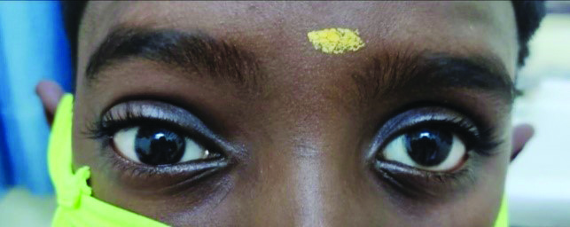
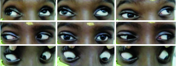
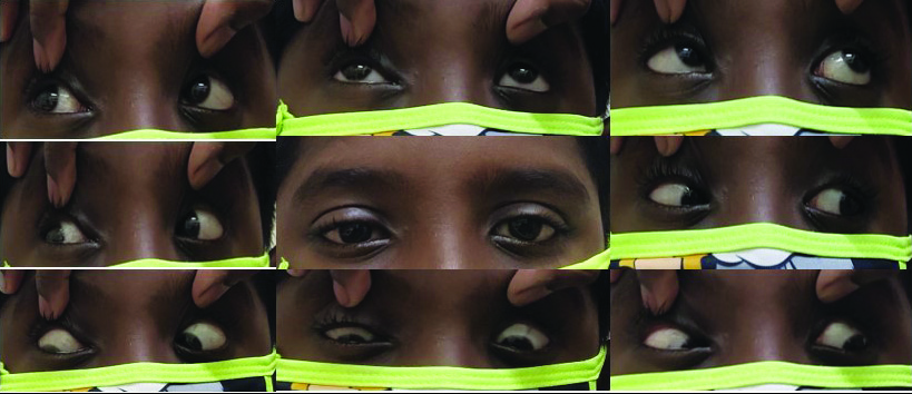
A nine-year-old boy came with complaints of swelling of right upper eyelid since three days. He had a history of punch over the right side of the head during a boxing match. Ophthalmological examination of left eye was normal. Right eye had vision of 6/9 with non-pulsatile eccentric proptosis and upper eyelid oedema [Table/Fig-1]. The globe appeared to be pushed downwards and forwards. There was discontinuity in the superior orbital margin. Extraocular movements like elevation was restricted and depression was exaggerated [Table/Fig-2]. Hirschberg test showed hypotropia. Diplopia was absent in all gazes. Pupillary reaction to light and accommodation was normal. There was no relative afferent pupillary defect. Intraocular pressure was within normal limits. Fundus examination was unremarkable.
Photograph showing proptosis of right eye.
Photograph showing restricted extraocular movements.
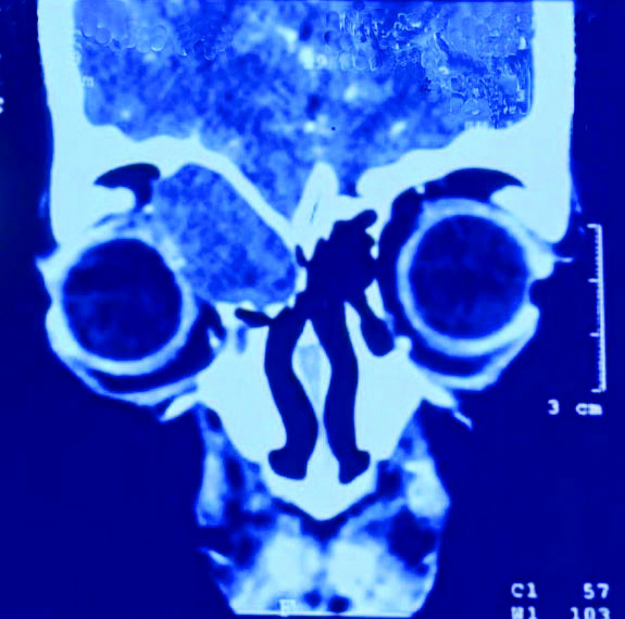
The CT of orbit showed a well-defined isodense lesion with smooth margins arising from the frontal sinus, extending inferiorly to anterior ethmoidal sinus measuring 2.4×2.1 cm suggestive of a mucocele which caused mass effect over right eye and ocular muscles resulting in lateral, downward and forward displacement of the eye ball [Table/Fig-3]. It also indented over superior rectus and medial rectus muscle. Smooth remodelling of adjacent bone with cortical erosion was evident. CT of Paranasal Sinuses (CT-PNS) showed homogenous opacification over right anterior ethmoid extending laterally into right orbit pushing right orbital contents laterally. Evidence of absence of lamina papyracea over right side could be due to pressure effect.
CT orbit showing frontoethmoidal mucocele causing a mass effect over right eye and ocular muscles.
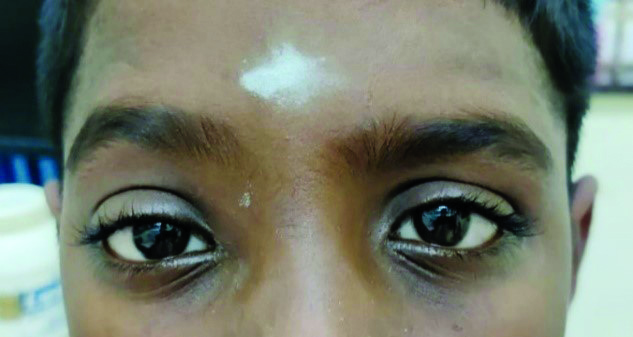
Patient was examined by the otolaryngologist who performed Diagnostic Nasal Endoscopy (DNE) and found lateralised uncinate process and a high septal deviation on right side. Functional Endoscopic Sinus Surgery (FESS) procedure was performed under general anaesthesia. Uncinate process was visualised and right uncinectomy was done. Superior attachment of uncinate was removed to expose the mucocele, a thin bone present over the mucocele was removed and the mucocele sac was incised. Approximately, 10-15 mL of pus was drained out and sent for culture and sensitivity. Frontal recess area was widened. Saline wash was given to the frontal recess and right middle meatal antrostomy was performed. Postoperatively patient was on antibiotics and analgesics. Unaided visual acuity had improved to 6/6, with regression of proptosis, eyeball returned to its normal position and extraocular movements were regained [Table/Fig-4,5].
Photograph showing reduction in proptosis.
Photograph showing restored extraocular movements.
Discussion
Boxing is a well-known sport in which opponent punches the anterolateral part of the face, the chest and the abdomen. Eye traumas in boxing rely on many parameters such as the distance from which the blow occurs, the fist power and placement, and the presence of protective gear [1]. Significantly, there are three mechanisms known to cause eye damage in boxing: direct (coup), indirect (countercoup), and equatorial expansion [2]. Various ocular injuries are reported in boxers. According to a 16-year study done in Australia, 46% of boxing injuries were around eye region, but none had proptosis [3]. Probably there are no reported cases of mucocele leading to proptosis (PubMed search). A mucocele is a sac containing mucus lined by epithelium which completely fills the sinus and is capable of expanding. Mucocele can develop in all paranasal sinuses, but frontal and ethmoidal sinuses are most commonly affected. Tumours, fibrosis, trauma, surgical procedures may result in sinus ostium obstruction which leads to mucoceles [4,5]. Patients with frontoethmoidal mucoceles may come with complaints of frontal headache, facial asymmetry, or swelling, as well as ocular manifestations such as impaired visual acuity, limited ocular mobility and proptosis. Infective organisms are detected by the culture of the aspirate from the mucocele [6]. In case of a mucocele, CT scan is the preferred choice of imaging which shows a homogenous smooth-walled mass expanding the sinus with thinning or loss of translucence. CT scan is an ideal investigation in delineating the extent of the lesion and its relations to other surrounding structures [7]. Mucocele is treated surgically. The access routes to approach may be either external or endonasal [8]. The purpose of the surgery is to eradicate the mucocele with minimal morbidity and to prevent its recurrence. Surgical access is based on the size, location and extent of the mucocele. It could be surgically approached either by an external approach (Lynch Howarth Frontoethmoidectomy) or osteoplastic flap with sinus cavity obliteration. In recent days, endoscopic drainage is the best choice of treatment for frontal mucoceles as it aids in preservation of the frontal sinus mucosa and maintenance of a patent frontal recess for a better clinical outcome [9,10].
Eye injuries mainly occur due to irregular use of protective measures. Even though headgear doesn’t aid in complete protection of the eyes, nose, maxilla, and eyebrows, its use should be made mandatory for all the boxers, irrespective of their age [11].
Conclusion(s)
This case is reported for its rarity as frontal mucocele is one of the least possible complications post a boxing punch. The frontal mucocele is usually associated with ethmoid mucocele together forming a single sac. Its involvement is mostly through the lamina papyracea. CT orbit was the ideal investigation for diagnosing a sino-orbital lesion in this case. A thorough ophthalmological and Ear Nose Throat (ENT) examination is essential in the early diagnosis of any ocular injuries in case of proptosis. The ophthalmologist plays an important role in preventing serious damage to the boxer’s eye. Therefore, it should be made mandatory for all boxers to have a complete ophthalmological examination periodically.
Author Declaration:
Financial or Other Competing Interests: None
Was informed consent obtained from the subjects involved in the study? Yes
For any images presented appropriate consent has been obtained from the subjects. Yes
Plagiarism Checking Methods: [Jain H et al.]
Plagiarism X-checker: Jan 04, 2021
Manual Googling: May 07, 2021
iThenticate Software: May 29, 2021 (19%)
[1]. Smith DJ, Ocular injuries in boxing. In: Vinger PF, editorPrevention of Ocular Sports Injuries 1988 Boston, MALittle, Brown and Co:242-45.10.1097/00004397-198802830-000163403185 [Google Scholar] [CrossRef] [PubMed]
[2]. Wolter JR, Coup-contrecoup mechanism of ocular injuriesAm J Ophthalmol 1963 56(5):785-96.10.1016/0002-9394(63)92943-X [Google Scholar] [CrossRef]
[3]. Zazryn TR, Finch CF, McCrory P, A 16 year study of injuries to professional boxers in the state of Victoria, AustraliaBr J Sports Med 2003 37(4):321-24.10.1136/bjsm.37.4.32112893717 [Google Scholar] [CrossRef] [PubMed]
[4]. Tan CS, Yong VK, Yip LW, Amritj S, An unusual presentation of a giant frontal sinus mucocele manifesting with a subcutaneous forehead massAnn Acad Med Singapore 2005 34(5):397-98. [Google Scholar]
[5]. Galiè M, Mandrioli S, Tieghi R, Clauser L, Giant mucocele of the frontal sinusJ Craniofac Surg 2005 16(5):933-35.10.1097/01.scs.0000168999.20258.ca16192886 [Google Scholar] [CrossRef] [PubMed]
[6]. Brook I, Frazier EH, The microbiology of mucopyoceleLaryngoscope 2001 111(10):1771-73.10.1097/00005537-200110000-0002011801943 [Google Scholar] [CrossRef] [PubMed]
[7]. Bilaniuk LT, Zimmerman RA, Computer-assisted tomography: Sinus lesions with orbital involvementHead Neck Surg 1980 2(4):293-301.10.1002/hed.28900204077364584 [Google Scholar] [CrossRef] [PubMed]
[8]. Khong JJ, Malhotra R, Selva D, Wormald PJ, Efficacy of endoscopic sinus surgery for paranasal sinus mucocele including modified endoscopic Lothrop procedure for frontal sinus mucoceleJournal of Laryngology & Otology 2004 118:352-56.10.1258/00222150432308653415165309 [Google Scholar] [CrossRef] [PubMed]
[9]. Kuhn FA, Javer AR, Primary endoscopic management of the frontal sinusOtolaryngol Clin North Am 2001 34(1):59-75.10.1016/S0030-6665(05)70295-4 [Google Scholar] [CrossRef]
[10]. Dickinson P, Rempel P, Prohibiting headgear for safety in amateur Boxing? Opinion of the Canadian Boxing Community: An Online PollSports Med Open 2016 2(1):1910.1186/s40798-016-0043-226913220 [Google Scholar] [CrossRef] [PubMed]
[11]. Chew YK, Noorizan Y, Khir A, Brito-Mutunayagam S, Prepageran N, Frontal mucocoele secondary to nasal polyposis: An unusual complicationSingapore Med J 2009 50(11):374-75. [Google Scholar]