Oral squamous cell carcinoma is the most common malignancy of the head neck region [1]. Approximately, 90% of oral and oro-pharyngeal malignancies are diagnosed as Squamous Cell Carcinoma (SCC) [2]. The conventional therapeutic schedule consisting of surgery combined with adjuvant radiotherapy or chemotherapy had shown five year survival rate approximately 60% despite all sorts of effort for decades [3]. So, in recent years, targeted therapy has become the primary area of research. There are many therapeutic trials, of which anti-EGFR (Epidermal Growth Factor Receptor) therapy have been found to achieve moderate success in recurrent amd metastatic head-neck SCC [4]. It is pivotal to investigate and identify the predictive bio-markers which can overcome the mechanism of resistance.
Amongst various components of cytoskeleton, MT comprises one of the most important structure that helps in the various cellular functions like cell migration, mitosis, development and gene regulation. It consists of long hollow cylinders made up of polymerised alfa and beta dimers [5]. They always keep a dynamic balance, assembly and disassembly continues at their ends. Stathmin or oncoprotein 18 which is a MT destabilising protein, overexpressed across a broad range of malignancies including leukaemia, lymphoma, OSCC, neuroblastoma, ovarian carcinoma, lung carcinoma, prostate carcinoma and mesothelioma [6]. The aberrant expression of stathmin protein results in the depolymerisation of MT which ultimately leads to uncontrolled cellular proliferation [7]. It also plays role in cancer cell metastasis and migration [8]. Several chemotherapeutic agents like paclitaxel and vinblastine are thought to act by targeting these MTs, disrupting mitotic spindle function, activating spindle assembly check point and inducing cell death [9-11]. Stathmin has been found to be up-regulated in some cancers and correlates well with cell differentiation, proliferation and migration, especially in solid tumour cells. Thus, stathmin may be an attractive target for drug design as targeting this molecule could simultaneously inhibit several aspects of tumour progression [12].
In this study, authors have tried to investigate and explore the association between different clinicopathological factors including histomorphology and stathmin expression in OSCC by immunohistochemical staining.
Materials and Methods
An institutional based descriptive, cross-sectional, observational study was conducted in a tertiary care centre of Kolkata, West Bengal, India from January to March 2020 in the Department of Pathology in RG Kar Medical College and Hospital in collaboration with Department of Oto-Rhino-Laryngology of the same institute. The work was initiated after obtaining ethical clearance from Institutional Ethics Committee (No- RKC/54, dated: 20.01.2020) and informed consent from the study population.
Inclusion criteria: The study included total 28 surgically excised and clinically diagnosed OSCC samples were received during the study period and examined histopathologically.
Exclusion criteria: Patients who had received any chemotherapy or radiation therapy were excluded from this study.
Study Procedure
The sample size was calculated on the basis of statistical formula with the help of Department of Community Medicine of same institute. Data was collected using a pre-designed, pretested semi-structured schedule on dependent variables like stathmin expression and independent variables like clinicopathological profile including age at presentation, sex, history of tobacco chewing, histological type, stage, grade, worst pattern of invasion and other relevant parameters. Data was collected by record review and laboratory techniques including histopathological reports and IHC. Reporting was done by Pathology experts.
Histopathology: All tissue samples that were collected, fixed in 10% neutrally buffered formalin and processing was done for routine histopathological examination. Four micrometers thick sections were cut from formalin fixed paraffin embedded blocks and stained with routine Haematoxylin and Eosin (H&E) stain for histopathological diagnosis of tumour type.
Immunohistochemistry (IHC): For IHC staining procedure, 3 μm thick sections from formalin fixed paraffin embedded tissues were taken and mounted on poly-L-Lysine coated slides. IHC was done manually using rabbit monoclonal Stathmin antibody (Bio SB Inc., California, USA: clone EP 247) and the steps mentioned in the supplied kit were followed. After IHC staining the microscopic examination and reporting was done by two expert histopathologist and blinding was followed as usual. Prostate cancer tissue and Phosphate Buffer Saline (PBS) solution were used as positive and negative control respectively.
Scoring of Immunohistochemistry: The scoring of intensity of stathmin immunoreaction was done on the basis of membranous and cytoplasmic staining as follows: 0=absent staining, 1=weak, 2=moderate, 3=strong. The final score was calculated by multiplying the percentage of positive cell and the staining intensity [13]. Cases with a stathmin score greater than 86.16 (the highest score for normal tissue) were defined as positive [14].
Statistical Analysis
Statistical significance was evaluated using by Fisher’s-exact test and Chi-square test, p<0.05 was considered significant (software SPSS version 25.0).
Results
Total 28 cases of OSCC were studied. The average age of the participants was 56.07±10.78 (mean±SD) years with a range of 50 (28-78) years. There were 18 (64.3%) male patients and 10 (35.7%) female patients, majority of the patients had history of tobacco chewing. Among 28 cases of OSCC, 12 (42.9%) cases were well differentiated, 12 (42.9%) cases were moderately differentiated and 4 (14.2%) cases were poorly differentiated. Baseline patient characteristics are shown in [Table/Fig-1].
Basic demographic and tumour characteristics.
| Characteristics | N (%) |
|---|
| Average age, years (range) | 56.07 (28-78) |
| Sex |
| Male | 18 (64.3) |
| Female | 10 (35.7) |
| Tobacco chewing |
| Present | 22 (78.6) |
| Absent | 06 (21.4) |
| Histological types |
| Well differentiated | 12 (42.9) |
| Moderately differentiated | 12 (42.9) |
| Poorly differentiated | 04 (14.2%) |
The results of IHC expression of stathmin is shown in [Table/Fig-2].
Results of IHC with stathmin antibodies.
| Characteristics | N (%) |
|---|
| Stathmin IHC |
| Positive | 18 (64.3) |
| Negative | 10 (35.7) |
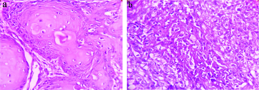
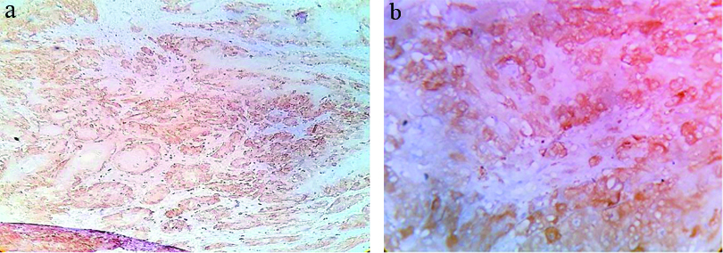
The [Table/Fig-3a,b] show photomicrograph of histopathological images of OSCC with their corresponding IHC results of stathmin expression is shown in [Table/Fig-4a,b].
a) Photomicrograph showing histopathology of well differentiated OSCC H&E (400X); b) Photomicrograph showing histopathology of poorly differentiated OSCC H&E (400X).
a) Photomicrograph showing well differentiated OSCC positive for stathmin IHC (100X); b) Photomicrograph showing poorly differentiated OSCC positive for stathmin IHC (400X).
The stathmin expression and its significance with other clinicopathological parameters like age, sex, tobacco chewing, tumour site, tumour stage and WPOI of the tumour are shown in [Table/Fig-5].
Distribution of stathmin expression in different age, sex, tobacco chewing, histological types, tumour stage, tumour site and WPOI of OSCC cases.
| Variable | Stathmin | p-value |
|---|
| Positive | Negative |
|---|
| Gender | Female | 5 | 5 | 0.234Fisher’s-exact test |
| Male | 13 | 5 |
| Age (years) | <60 | 13 | 3 | 0.030*Fisher’s-exact test |
| >60 | 5 | 7 |
| Tobacco chewing | Positive | 17 | 5 | 0.006*Fisher’s-exact test |
| Negative | 1 | 5 |
| Histological types | Well differentiated | 6 | 6 | 0.172Chi-square test |
| Moderately differentiated | 8 | 4 |
| Poorly differentiated | 4 | 0 |
| T stage | T1 | 0 | 2 | 0.036*chi-square test |
| T2 | 7 | 6 |
| T3 | 6 | 2 |
| T4 | 5 | 0 |
| N stage | N(-) | 5 | 6 | 0.094Fisher’s-exact test |
| N(+) | 13 | 4 |
| pTNM stage | I | 0 | 2 | 0.022*Chi-square test |
| II | 1 | 2 |
| III | 6 | 4 |
| IV | 11 | 2 |
| Worst Pattern Of Invasion (WPOI) | 1-4 | 4 | 6 | 0.045*Fisher’s-exact test |
| 5 | 14 | 4 |
| Tumour site | Buccal mucosa | 4 | 4 | 0.220Chi-square test |
| Tongue | 9 | 2 |
| Lip | 5 | 3 |
| Soft palate | 0 | 1 |
p-values were evaluated by using chi-square test and Fisher’s-exact test; *p-value <0.05 was considered statistically significant
Discussion
In this study, it was observed the overexpression of stathmin in OSCC and the intensity of expression increased with histopathological grading and staging of tumour. In Several studies, it has been observed that in malignant cells there is overexpression of stathmin which leads to uncontrolled proliferation of cell by interfering with the cell cycle and inhibition of stathmin overexpression changes the transformed phenotype [15-17]. So, stathmin has been considered as potential targeted molecule from therapeutic point of view as it arrests the growth of malignant cells. Besides, it has been shown that lung cancer induced by the stathmin transfection has increased sensitivity to vinka alkaloid therapy [18].
Moreover, while inhibiting the stathmin expression in antisense showed a combined synergistic effect of apoptosis along with paclitaxel treatment [19,20]. Ma HL et al., conducted the study on stathmin expression and its correlation with p53 mutation in 48 cases of OSCC. The study explored the relationship between stathmin and p53 expression and a positive correlation was found between stathmin and p53 expression (p=0.008). The immunohistochemical score of stathmin expression was significantly higher in patients with neomorphic mutant p53 (mutp53) expression than in those with wild-type p53 (wtp53) expression [21]. Stathmin expression levels were studied by Kouzu Y et al., on OSCC and found significantly higher in advanced stage of the disease than early stage [15].
Several recent studies show that stathmin not only control the function of MT but also have other roles in various biological functions and a basic level of active stathmin is necessary to maintain the balance between MT stabilising and destabilising factors and thereby normal cellular functions. The stathmin is overexpressed in a number of human cancers including ovarian carcinoma, Wilms’ tumour, breast cancer, adenoid cystic carcinoma of salivary gland, prostate cancer etc., [6]. In high grade breast cancers and ovarian cancers stathmin has been found to be overexpressed, specially those cases that are resistant to the platinum drug therapy [22]. Genomic study in human breast cancer was conducted by Bieche I et al., on the basis of DNA, mRNA and the findings were correlated with the protein analysis of stathmin expression. Total 50 breast tumours were studied. The expression of protein was also assessed by IHC. Out of 50 tumours one third cases i.e, 15 cases showed high mRNA expression and high stathmin protein level detected by the immunoreactivity in the cytoplasm. So, these three methods of DNA, mRNA and stathmin protein expression in the cytoplasm were observed to be highly correlated and could be used as a screening tool for larger studies [23]. Stathmin, the transcribed protein from a higly conserved gene reported to have varieties of function including control of cell cycle, regulation of signal transduction and function of MT. On quantitative analysis of 12 malignant ovarian tumours eight benign ovarian tumours and 10 normal ovarian tissue samples by northern blot analysis showed overexpression of stathmin mRNA in the malignant cancers. Besides, IHC analysis also showed overexpression of stathmin protein in association with proliferating cells [24]. Stathmin expression in context to different malignant tumours including breast carcinoma creates further interest in the study of this protein and its relation with cell signalling, apoptosis, cytoskeletal abnormalities and response to chemotherapy [25]. Studies were done on the expression of stathmin on Wilm’s Tumour by Takahashi M et al., and reported it was highly expressed in high grade tumour in comparison with the low grade tumour. Besides, high chemosensitivity of Wilm’s tumour was related to high expression of stathmin as well as expression of topoisomerase II alpha and MT related genes such as tubulin [26]. However, the status and significance of stathmin expression in OSCC is still under investigation and thus stathmin was selected for further investigation in primary OSCC cases and its association with clinicopathological findings.
In this study, the level of stathmin expression was significantly higher in OSCC. A higher stathmin expression was observed in younger age group patients (<60 years) than older age group (≥60 years) and the result was statistically significant (p=0.030). Higher stathmin staining score was also seen in the patients who had history of tobacco chewing and the finding was statistically significant (p=0.006). Present study showed a significant association between stathmin expression with advanced T stage (p=0.036) and advanced pTNM stage (III and IV) (p=0.022), which indicates the tumour aggressiveness and high histopathological grading of OSCC. This finding was consistent with study of Kouzu Y et al., and Ma HL et al., [15,21]. The stathmin expression levels were significantly associated with WPOI in primary OSCC (p=0.045), suggesting that stathmin expression and its relation with WPOI could be a strong prognostic criteria for OSCC. In this study, no significant association between stathmin expression and gender of patient, site of disease, lymph node status, could be established.
Limitation(s)
The present study had few limitations. Firstly, the study was a hospital based with small sample size and was done in a limited time period. Secondly, genetic and proteomic study which is more informative and advanced could not be carried out in association with IHC. Thirdly, as the patients could not be followed-up, the chemotherapy and prognostic significance of stathmin could not be assessed.
Conclusion(s)
Based on the study and observation from the above data it could be concluded that not only the stathmin is overexpressed in OSCC but highly associated with histopathological grading, pTNM staging and WPOI. Besides, the pattern of expression in different age groups, presence and absence of tobacco chewing, site of tumour, grade of tumour and early and advanced stage of tumour provides a novel insights in the process of development and progress of tumour. Moreover, the association of WPOI of OSCC with higher levels of expression of stathmin can emphasise the use of WPOI as a strong prognostic indicator for OSCC. As the high likelihood of response to targeted therapy against stathmin can improve the overall survival of patients, the importance of detection of stathmin expression in primary OSCC should get more attention in near future.
p-values were evaluated by using chi-square test and Fisher’s-exact test; *p-value <0.05 was considered statistically significant