Aesthetic Repair of Zirconia Supported Porcelain Prosthesis with Ceramic Veneer: A Case Report with Three-Year Follow-up
Tugce Merve Ordueri1, Asena Kaptanoglu2, Mehmet Muzaffer Ates3
1 Dentist, Department of Prosthodontics, Medipol University, Istanbul, Turkey.
2 Dentist, Department of Prosthodontics, Medipol University, Istanbul, Turkey.
3 Professor and Head, Department of Prosthodontics, Medipol University, Istanbul, Turkey.
NAME, ADDRESS, E-MAIL ID OF THE CORRESPONDING AUTHOR: Dr. Tugce Merve Ordueri, Kazımiye Mah, Barbaros 2, Sok. Tellioğlu Plaza. K:4 D:5 Çorlu, Tekirdağ, Turkey.
E-mail: tugceordueri@gmail.com
Chipping is the most common complication in zirconia-supported porcelain prosthesis. If the prosthesis has ideal adaptation and there is no problem other than chipping, intraoral repair is the most practical solution for such failures. Composite resins are often preferred for intraoral porcelain repair. However, the wear and unstable colour of composite resins negatively affect aesthetics. This complication could be restored intraorally and aesthetically with ceramic veneer. This case report presents the intraoral repair of a zirconia supported Fixed Partial Denture (FPD) consisting of four units. The cohesive fracture of the ceramic material in the incisal part of maxillary right central incisor was restored with ceramic veneers. Preparation was done with a tapered, round end diamond bur under water-cooling. The impression was taken with elastomeric impression material. Ceramic veneer was manufactured with a leucite-reinforced glass-ceramic and cemented with light cure resin cement. Based on the three-year follow-up of the performed intraoral repair, ceramic veneers have shown an alternative treatment for fractured FPD.
Chipping, Fracture, Intraoral repair, Porcelain veneer
Case Report
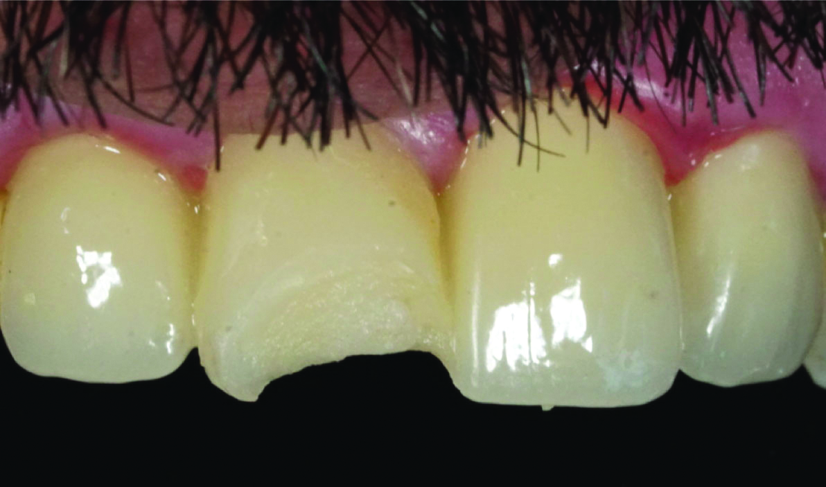
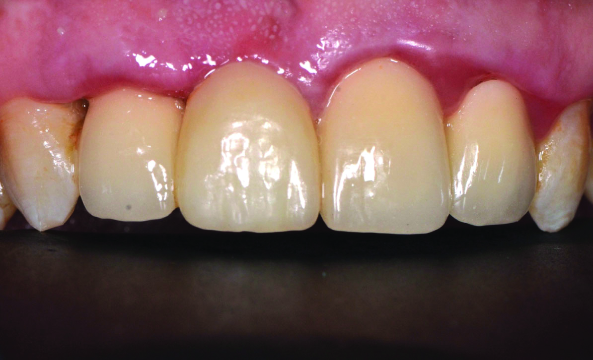
A 38-year-old male patient visited a private dental office with complaints of fractured tooth which occurred two days back, in a four unit porcelain veneered zirconia FPD, which was delivered one year ago. The FPD was located between upper right and left second incisors. After the oral examination, it was observed that the fracture was in the ceramic fragment of the right central incisor [Table/Fig-1]. All treatment options regarding the porcelain chipping (porcelain fracture without zirconia framework exposure) were explained to the patient. The patient refused to renew his FPD due to clinical time and cost. Patient approved of ceramic veneer, as it would be more aesthetic than to repair with composite resin.
Patient presents chipping at the incisal area of veneered zirconia fused to multiunit zirconia bridge at tooth.
The labial surface of the right central incisor was prepared with a tapered, round end diamond bur (Meisinger, Hager and Meisinger GmbH, Dusseldorf, Germany) under water coolant until a flat surface was obtained on the ceramic. All sharp edges and corners were smoothened. Chamfer marginal finish line was prepared in order to avoid stress build-up [Table/Fig-2a,b].
Close-up view of preparation.
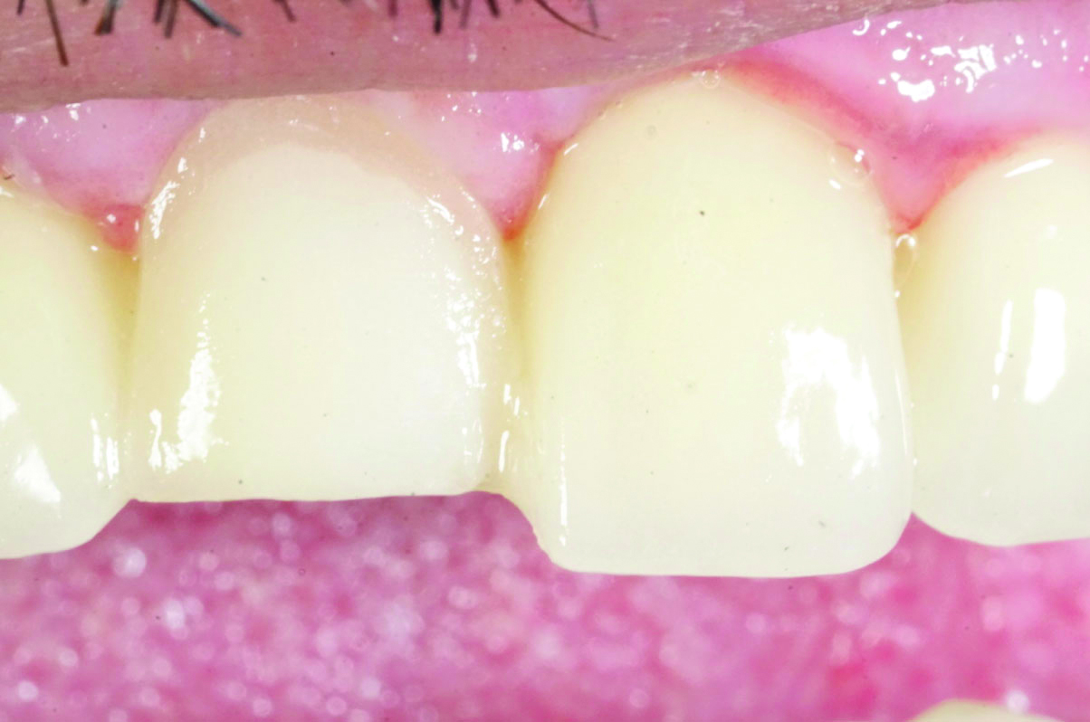
Prepared porcelain in tooth 11.

After the completion of the preparation, the impression was taken via additional type elastomeric impression material (Elite H-D, Zhermack, Germany). The porcelain veneers were fabricated using a leucite-reinforced glass-ceramic (IPS Empress E.max, IvoclarVivadent, Schaan, Liechtenstein).
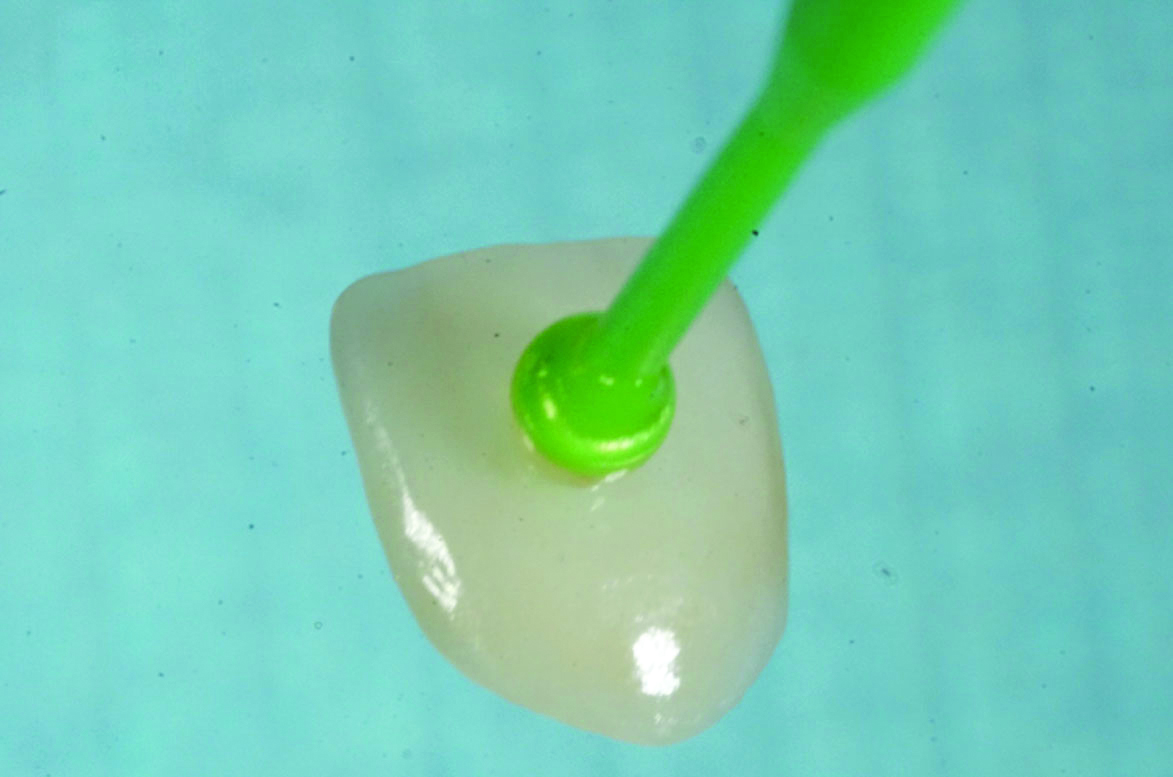
A light curing gingival barrier (OpalDam, Ultradent) was applied to protect the periodontal tissues. The porcelain surface and inner surface of the laminate veneer were etched with 9.5% Hydrofluoric (HF) acid (Ultradent) for one minute, washed, and dried [Table/Fig-3]. Silane (Monobond N, IvoclarVivadent, Liechtenstein) was applied to both surfaces with microbrush and waited for one minute. A thin layer of bonding (Heliobond, IvoclarVivadent, Liechtenstein) was applied and light cured for 20 seconds. It was cemented with Variolink N LC (IvoclarVivadent, Liechtenstein). Residual cement was removed. The polymerisation was completed with the application of additional 40 seconds of light curing [Table/Fig-4]. As the patient had tooth wear in lower anterior incisal surfaces and posterior occlusal surfaces (bruxism), a stabilisation splint was made and the patient was recommended to use it during sleep.
Ceramic veneer etched with Hydrofluoric (HF) acide, washed and dried.
Final view of repaired porcelain.
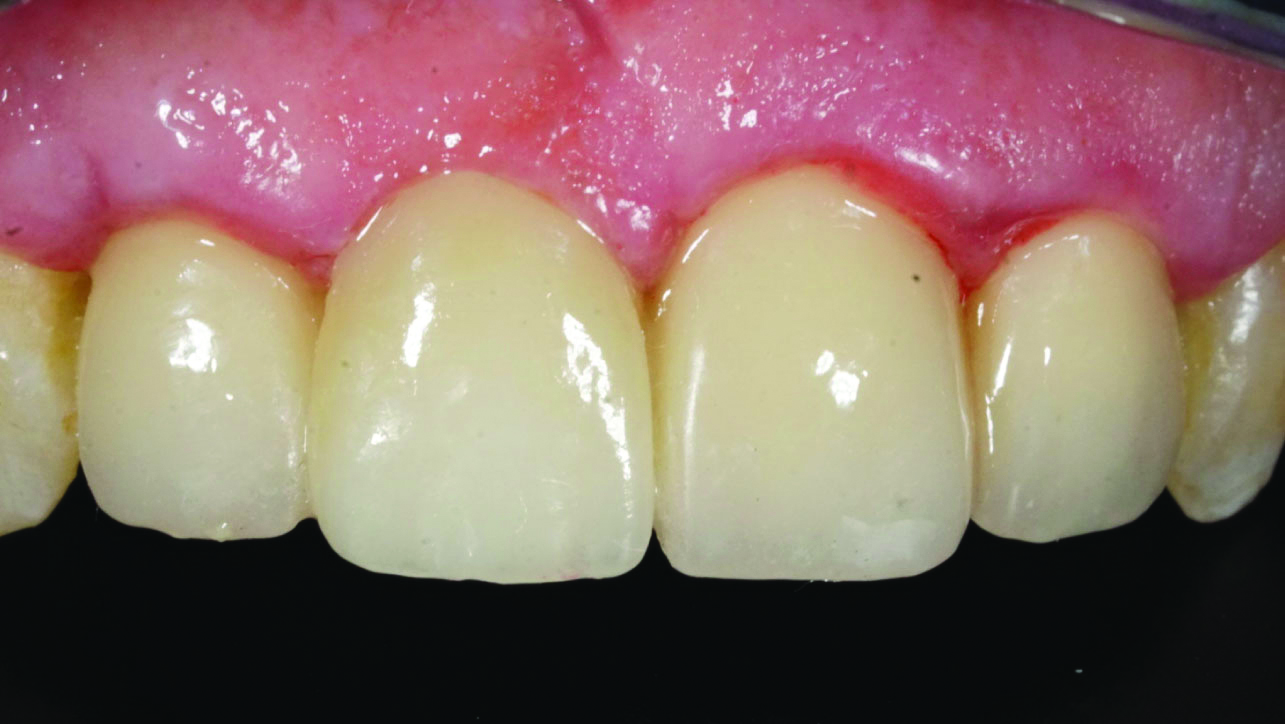
The FPD and the ceramic veneer intraoral repair survived for three years to this date [Table/Fig-5]. Marginal integrity, marginal discolouration, anatomical form, restoration colour stability and periodontal health were evaluated after three years. All of the criteria were acceptable except periodontal health. The patient was motivated about tooth brushing and dental floss use.
A three-year follow-up of repaired FPD.
Discussion
Zirconia-supported fixed prostheses are preferred as they have been used successfully for the past two to three decades due to their colour, excellent biocompatibility, and mechanical properties [1]. Also, they are more aesthetic than metal-supported prostheses [2]. Nevertheless, the disadvantage of zirconia-supported prosthesis is the inadequate bonding between Zirconia core and the veneering ceramic layer due to failure of adhesion between two chemically different materials [3]. Separation of porcelain fragment from the framework as plates (delamination) or by chipping are most common failures in zirconia-supported restorations [4,5].
Due to the increase in application frequency of zirconia-based restorations and the higher fracture rates in long-term follow-up studies compared to metal-based restorations [6,7], the need of repair has become more important.
In literature, veneer fractures are classified in 3 grades. Grade-1 refers to chipping of a small amount of veneer fragment that can be repaired with re-shaping and polishing. Grade-2 contains moderate chipping that can be repaired intraorally with composite. Grade-3 refers to severe chipping in need replacement of the FPD [8]. Considering the potential risks of removing the broken FPD, financial limitations and the time spent for renewal of restoration, this treatment option is not a practical solution. If Grade-3 fracture is seen, replacement of FPD is inevitable [9-11].
If the restoration can continue its function in the mouth after fracture and there is no other reason for renewal of prosthetic restoration, the intraoral repair provides convenience for both the patient and the physician [12].
Composite resins are often applied to porcelains that have failure and there are many systems developed for intraoral repair with composite resins [13-15]. In the meantime, the prognosis of the intraoral repair with composite is controversial due to the wearing of the composite over the time and its lack of colour stability compared to ceramic used in direct intraoral repair methods [16]. Amongst the intraoral repair systems, ceramic veneers have a superior success rate compared to using composite resins especially in cases where the ceramic contains large fractures [17]. The aesthetic properties of full ceramics are more successful than composite resins. They have colour stability and high resistance to wear, and their bond strength to the substructures during repair is found to be sufficient in the studies. These properties has led to the idea that the use of ceramics for porcelain repair can be increased in clinically [18].
The recent studies performed with zirconia supported restorations have concluded that the shear bond strength of the repair made with ceramics is higher than composite, meaning that the intraoral repair made with ceramics is better alternative [17,19]. In intraoral repair methods, diamond burs are used to smoothen the chipped surface of the ceramic. Rising of heat can cause possible formation and propagation of cracks. In literature, abundant cooling was performed in order to avoid ceramic heating at this stage [20]. The most common methods for the surface treatment of ceramics after chipping are acid etching, sandblasting with aluminum oxide or sandblasting with silica-coated particles [21]. Etching with acid increases the surface area and energy of the ceramic, and the bonding ratio of resin to ceramic increases with the surface energy [18,22].
Although it is a subject of discussion to use HF acid in etching due to the harmful effects of HF acid on tissues, there has been no reports of complications related to HF acid in the dental literature [23], While applying HF acid intraorally for porcelain repair, the isolation of gingival area with rubberdam will protect patients from possible hazardous effects [21]. Traditionally, porcelain-chipped restorations are repaired with composite resin technique. More well-designed intraoral studies are necessary to justify this ceramic veneer repair technique.
Conclusion(s)
Direct repair techniques are good alternative treatments in fracture or chipping of the veneering ceramic, without the need for replacement of the prosthesis. In case of fracture in porcelain veneered zirconia framework restorations, ceramic veneers can be used in intraoral repair technique, as a better alternative than composite resins. The fact that no complications were observed during the three years follow-up of the patient showed that porcelain ceramic veneers can be considered as a long-term repair option.
Author Declaration:
Financial or Other Competing Interests: None
Was informed consent obtained from the subjects involved in the study? Yes
For any images presented appropriate consent has been obtained from the subjects. Yes
Plagiarism Checking Methods: [Jain H et al.]
Plagiarism X-checker: Mar 30, 2021
Manual Googling: Apr 12, 2021
iThenticate Software: Apr 28, 2021 (11%)
[1]. Daou EE, The Zirconia ceramic: Strengths and weaknessesOpen Dent J 2014 8(1):33-42.10.2174/187421060140801003324851138 [Google Scholar] [CrossRef] [PubMed]
[2]. Meirelles PD, da Rocha LS, Pecho OE, Della Bona A, Benetti P, Intraoral repair of a chipped porcelain-zirconia restorationJ Esthet Restor Dent 2020 32(5):444-50.10.1111/jerd.1259232442351 [Google Scholar] [CrossRef] [PubMed]
[3]. Urapepon S, Taenguthai P, The effect of zirconia framework design on the failure of all-ceramic crown under static loadingJ Adv Prosthodont 2015 7(2):146-50.10.4047/jap.2015.7.2.14625932313 [Google Scholar] [CrossRef] [PubMed]
[4]. Raigrodski AJ, Hillstead MB, Meng GK, Chung KH, Survival and complications of zirconia-based fixed dental prostheses: A systematic reviewJ Prosthet Dent 2012 107(3):170-77.10.1016/S0022-3913(12)60051-1 [Google Scholar] [CrossRef]
[5]. Pihlaja J, Näpänkangas R, Raustia A, Early complications and short-term failures of zirconia single crowns and partial fixed dental prosthesesJ Prosthet Dent 2014 112(4):778-83.10.1016/j.prosdent.2014.03.00824840907 [Google Scholar] [CrossRef] [PubMed]
[6]. Sailer I, Fehér A, Filser F, Lüthy H, Gauckler LJ, Schärer P, Prospective clinical study of zirconia posterior fixed partial dentures: 3-year follow-upQuintessence Int 2006 37(9):685-93. [Google Scholar]
[7]. Raigrodski AJ, Chiche GJ, Potiket N, Hochstedler LJ, Mohamed SE, Billiot S, The efficacy of posterior three-unit zirconium-oxide-based ceramic fixed partial dental prostheses: A prospective clinical pilot studyJ Prosthet Dent 2006 96(4):237-44.10.1016/j.prosdent.2006.08.01017052467 [Google Scholar] [CrossRef] [PubMed]
[8]. Özcan M, Volpato CAM, Surface conditioning protocol for the adhesion of resin-based materials to glassy matrix ceramics: How to condition and why?J Adhes Dent 2015 17(3):292-93. [Google Scholar]
[9]. Demirekin ZB, Çavdarlı K, Türkaslan SS, Ceramic veneers: A conservative alternative for intraoral repairSDU J Heal Sci Inst 2016 7(11):64-68. [Google Scholar]
[10]. Agingu C, yuan ZC, wu JN, Cheng H, Özcan M, Yu H, Intraoral repair of chipped or fractured veneered zirconia crowns and fixed dental prosthesis: Clinical guidelines based on literature reviewJ Adhes Sci Technol 2018 32(15):1711-23.10.1080/01694243.2018.1443639 [Google Scholar] [CrossRef]
[11]. Lee SJ, Cheong CW, Wright RF, Chang BM, Bond strength of the porcelain repair system to all-ceramic copings and porcelainJ Prosthodont 2014 23(2):112-16.10.1111/jopr.1206423725343 [Google Scholar] [CrossRef] [PubMed]
[12]. Özcan M, Valandro LF, Amaral R, Leite F, Bottino MA, Bond strength durability of a resin composite on a reinforced ceramic using various repair systemsDent Mater 2009 25(12):1477-83.10.1016/j.dental.2009.06.02019671476 [Google Scholar] [CrossRef] [PubMed]
[13]. Han IH, Kang DW, Chung CH, Choe HC, Son MK, Effect of various intraoral repair systems on the shear bond strength of composite resin to zirconiaJ Adv Prosthodont 2013 5(3):248-55.10.4047/jap.2013.5.3.24824049565 [Google Scholar] [CrossRef] [PubMed]
[14]. Özcan M, Van Der Sleen JM, Kurunmäki H, Vallittu PK, Comparison of repair methods for ceramic-fused-to-metal crownsJ Prosthodont 2006 15(5):283-88.10.1111/j.1532-849X.2006.00124.x16958728 [Google Scholar] [CrossRef] [PubMed]
[15]. Kocaağaoğlu H, Manav T, Albayrak H, In Vitro Comparison of the Bond Strength between Ceramic Repair Systems and Ceramic Materials and Evaluation of the WettabilityJ Prosthodont 2017 26(3):238-43.10.1111/jopr.1238126524614 [Google Scholar] [CrossRef] [PubMed]
[16]. Cardoso AC, Spinelli Filho P, Clinical and laboratory techniques for repair of fractured porcelain in fixed prostheses: A case reportQuintessence Int 1994 25(12):835-38. [Google Scholar]
[17]. Kumchai H, Juntavee P, Sun AF, Nathanson D, Comparing the repair of veneered zirconia crowns with ceramic or composite resin: An in vitro studyDent J 2020 8(2):01-12.10.3390/dj802003732349281 [Google Scholar] [CrossRef] [PubMed]
[18]. Aladağ A, Çömlekoğlu EM, Indirect ceramic repair with a metal-ceramic laminate veneer: case reportEÜ Diş hek Fak Derg 2009 30:47-51. [Google Scholar]
[19]. Habib SR, Bajunaid S, Almansour A, AbuHaimed A, Almuqrin MN, Alhadlaq A, Shear bond strength of veneered zirconia repaired using various methods and adhesive systems: A comparative studyPolymers (Basel) 2021 13(6):91010.3390/polym1306091033809539 [Google Scholar] [CrossRef] [PubMed]
[20]. Özcan M, Volpato M, Intra-oral repair technique for ceramic fracture using direct resin compositeItal J Dent Med 2016 (8):67-70. [Google Scholar]
[21]. Kimmich M, Stappert CFJ, Intraoral treatment of veneering porcelain chipping of fixed dental restorationsJ Am Dent Assoc 2014 144(1):31-44.10.14219/jada.archive.2013.001123283924 [Google Scholar] [CrossRef] [PubMed]
[22]. Carrabba M, Vichi A, Louca C, Ferrari M, Comparison of traditional and simplified methods for repairing CAD/CAM feldspathic ceramicsJ Adv Prosthodont 2017 9(4):257-64.10.4047/jap.2017.9.4.25728874992 [Google Scholar] [CrossRef] [PubMed]
[23]. Özcan M, Allahbeickaraghi A, Dündar M, Possible hazardous effects of hydrofluoric acid and recommendations for treatment approach: A reviewClin Oral Investig 2012 16(1):15-23.10.1007/s00784-011-0636-622065247 [Google Scholar] [CrossRef] [PubMed]