Composite resins are currently the most popular restorative materials due to their superior esthetics, strong mechanical properties and high resistance to dissolution. However, in spite of the improvements in resin composite formulations over the years, polymerisation shrinkage which necessitates incremental placement techniques still presents clinical challenges [1].
Bulk fill composite resins have reportedly shown to have lower polymerisation shrinkage and greater depth of cure [2]. The stress decreasing resin technology associated with bulk fill composites eliminates the need for incremental placement and at the same time exhibit reduced shrinkage. These are available both as flowable and regular consistency [2,3].
The success of resin composites is directly linked to an intimate bond with tooth tissues and a leak proof marginal seal [3]. Dental adhesives are a requisite to form optimum sealed cavity walls to avoid leakage as poor adhesion between the enamel/dentin and restorative material causes gap formation [4]. This in turn may be accountable for rise in postoperative sensitivity, pulpal inflammation, marginal discoloration or staining and recurrent caries [4,5].
An innovative approach in advanced dental adhesive technology is the development of universal adhesives that can be applied in either etch and rinse, self-etch, or selective-etch protocols [4]. However, etching should be limited to enamel only, as etching dentin can lead to the formation of a low-quality hybrid layer which is prone to nanoleakage and to reduced bond strengths to dentin. It is now being recommended that a preliminary phosphoric acid etching (selective etch) be used to enhance the adhesion to enamel of all-in-one adhesives [4,5]. It is hence necessary to study the relative efficacy of both these techniques in achieving a fluid tight seal [5].
No previous studies have been conducted to evaluate cavosurface marginal integrity of bulk fill and conventional composites using universal adhesive. Thus, the aim of the study is to evaluate cavosurface marginal integrity in class I restorations using Surefil Bulk fill, Tetric N- Flow Bulk fill and Filtek Z350 XT composites using selective etch and self etch mode of a universal adhesive.
Materials and Methods
The in vitro research study was performed out in the Department of Conservative Dentistry and Endodontics, Bapuji Dental College and Hospital, Davangere, Karnataka, India for one month duration from April 2018 to May 2018. The study was commenced after obtaining the ethical clearance (BDC/Exam/283/2016-17).
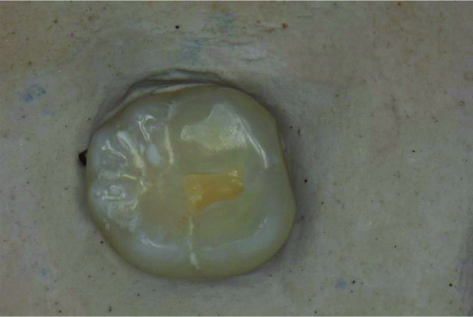
Sixty extracted non carious, intact human mandibular molar teeth were selected for this study. Standardised Class I cavities of 4 mm depth were prepared by a single operator (restorative dentist) with a high-speed handpiece using carbide fissure #245 (SS White Inc) bur under air-water coolant [Table/Fig-1]. The burs were discarded after five cavity preparations and dimensions were measured with a periodontal probe to maintain consistency. Samples were divided into two main groups in which Single Bond Universal (3M ESPE) was used.
Class I cavity preparation.
Experimental Groups
Group I: Selective Etch Mode (n=30)
Group II: Self Etch Mode (n=30)
These two main groups were further divided into three subgroups each
Subgroup FC: Filtek Z350 XT (3M ESPE) (n=10)
Subgroup TF: Tetric N- Flow Bulk fill (Ivoclar Vivadent) (n=10)
Subgroup SB: Surefil Bulk fill (Dentsply) (n=10)
Bonding Protocol
Group I: Selective Enamel Etching was done. Scotchbond Universal Etchant (3M ESPE) was applied to the prepared tooth enamel and allowed to react for 15 seconds. It was then rinsed thoroughly with water followed by drying. According to manufacturer’s instructions, thin layer of Single Bond Universal (3M ESPE) was applied onto the prepared cavity using applicator tip and rubbed for 20 seconds followed by mild air drying for 5 seconds to evaporate the solvent. Subsequently, adhesive was cured for 10 seconds with Light Emitting Diode (LED) curing unit.
Group II: According to manufacturer’s instructions a thin layer of Self-Etch adhesive Single Bond Universal (3M ESPE) was applied onto the prepared cavity using applicator tip and rubbed for 20 seconds followed by mild air drying for 5 seconds to evaporate the solvent. Subsequently adhesive was cured for 10 seconds with LED curing unit.
Subgroup FC: Filtek Z350 XT (Conventional) (3M ESPE) was condensed into a Class I cavity and two increments of 2 mm each was cured for 20 seconds using LED curing light with the tip of the light source held as close as possible to the surface.
Subgroup TF: A 4 mm of Tetric N- Flow Bulk fill (Ivoclar Vivadent) was placed and cured for 20 seconds using LED curing light.
Subgroup SB: A 4 mm of Surefil Bulk fill (Dentsply) was placed and cured for 20 seconds using LED curing light.
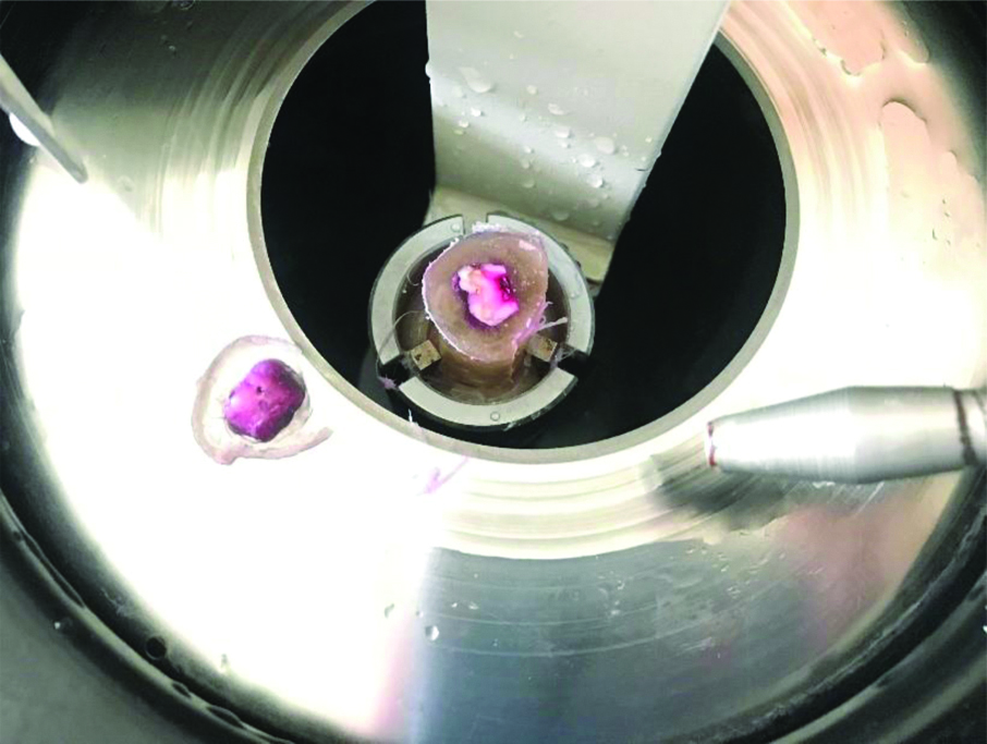
After the restoration, the samples were stored for 24 hours in distilled water at 37°C. Apical openings of the teeth were occluded with sticky wax and then the entire tooth surface was painted with two coats of varnish (nail polish) to within 1 mm of the restoration margins. The specimens were subjected to thermocycling for 500 cycles (5°C±2°C and 55°C±2°) with a dwell time of one minute in each temperature bath. The teeth were immersed in 0.5% basic fuschin dye for 24 hours. Following this, teeth were rinsed in distilled water. All the specimens were then sectioned longitudinally in a mesio-distal direction towards the center of the restorations using a hard tissue microtome [Table/Fig-2] [6].
Dye Penetration Evaluation
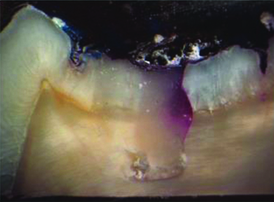
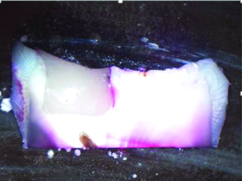
Dye penetration was evaluated according to the scoring criteria [7] [Table/Fig-3] and each specimen was examined with a stereomicroscope (Leica, Germany) [Table/Fig-4,5].
Grading/scoring of Dye infiltration in the specimens.
| Score | Tooth-restoration interface [7] |
|---|
| 0 | No dye infiltration. |
| 1 | Dye infiltration up to the first third of the prepared cavity wall |
| 2 | Dye infiltration up to the second third of the prepared cavity wall |
| 3 | Dye infiltration onto the entire prepared cavity wall |
| 4 | Dye infiltration onto the entire prepared cavity wall and the pulpal floor |
Stereomicroscopic image I.
Stereomicroscopic image II.
Statistical Analysis
The data of microleakage scores were entered into computer database for statistical analysis and responses were analysed by Statistical Package for Social Sciences (SPSS) Version 22.0. The descriptive statistics, mean, medians range, minimum and maximum values were calculated for each group tested. Kruskal-Wallis test was used to compare means of more than two groups, Mann-Whitney U-test were used to compare means of two groups and the p-value <0.05 was considered as statistically significant. Since, microleakage was expressed in terms of scores, non parametric methods were used for analysis.
Results
Tetric N flow Bulk fill flowable composite (subgroup TF) (1.15±1.268) yielded the lowest microleakage score followed by SureFil bulk fill (subgroup SB) (1.65±0.875) and the highest microleakage score was recorded in the Filtek Z350 XT group (subgroup FC) (2.85±1.137) [Table/Fig-6].
Microleakage scores in two different modes of universal adhesive.
| Composite | Mode | N | Min | Max | Median | Mean | SD |
|---|
| Subgroup TF | Group I | 10 | 0 | 2 | 1.00 | 0.70 | 0.675 |
| Group II | 10 | 0 | 4 | 1.50 | 1.60 | 1.578 |
| Total | 20 | 0 | 4 | 1.00 | 1.15 | 1.268 |
| Subgroup SB | Group I | 10 | 0 | 3 | 1.00 | 1.20 | 0.919 |
| Group II | 10 | 1 | 3 | 2.00 | 2.10 | 0.568 |
| Total | 20 | 0 | 3 | 2.00 | 1.65 | 0.875 |
| Subgroup FC | Group I | 10 | 0 | 4 | 3.00 | 2.50 | 1.354 |
| Group II | 10 | 2 | 4 | 3.00 | 3.20 | 0.789 |
| Total | 20 | 0 | 4 | 3.00 | 2.85 | 1.137 |
FC: Filtek Z350 XT (3M ESPE); TF: Tetric N- Flow bulk fill (Ivoclar Vivadent); SB: Surefil bulk fill (Dentsply); Group I: Selective- etch mode; Group II: Self-etch mode
[Table/Fig-7] shows microleakage scores of group I and II (modes of universal adhesive) with the three different composites. There was a statistically highly significant difference (p<0.001) between the mean microleakage scores of the three groups.
Microleakage scores in various subgroups.
| Ranks |
|---|
| Composite | N | Mean rank |
|---|
| Microleakage | Subgroup TF | 20 | 20.90 |
| Subgroup SB | 20 | 27.45 |
| Subgroup FC | 20 | 43.15 |
| Test statisticsa,b |
| Analytical test | Microleakage |
| Chi-square | 17.973 |
| df | 2 |
| Asymp. Sig. | <0.001 |
a. Kruskal wallis test; b. Grouping variable: Composite
Group I subgroup TF yielded lower microleakage score 0.70±0.675 as compared to group II subgroup TF which had a higher score 1.60±1.578 [Table/Fig-6]. This difference was not statistically significant (p-value=0.266) [Table/Fig-8].
Microleakage scores between Group I and Group II in subgroup TF.
| Ranksa |
|---|
| Mode | N | Mean rank | Sum of ranks |
|---|
| Microleakage | Group I | 10 | 9.10 | 91.00 |
| Group II | 10 | 11.90 | 119.00 |
| Test statisticsb,c |
| Analytical test | Microleakage |
| Mann-Whitney U | 36.000 |
| Wilcoxon W | 91.000 |
| Z | -1.111 |
| Asymp. Sig. (2-tailed) | 0.266 |
| Exact Sig. [2*(1-tailed Sig.)] | 0.315a |
a. Not corrected for ties; b. Composite=1; c. Grouping variable: Mode
Group I subgroup SB yielded lower microleakage score 1.20±0.919 as compared to group II subgroup SB which had a higher score 2.10±0.568 [Table/Fig-6]. This difference was statistically significant. (p-value=0.017) [Table/Fig-9].
Microleakage Scores between group I and group II in subgroup SB.
| Ranksa |
|---|
| Mode | N | Mean rank | Sum of ranks |
|---|
| Microleakage | Group I | 10 | 7.55 | 75.50 |
| Group II | 10 | 13.45 | 134.50 |
| Test statisticsb,c |
| Analytical test | Microleakage |
| Mann-Whitney U | 20.500 |
| Wilcoxon W | 75.500 |
| Z | -2.378 |
| Asymp. Sig. (2-tailed) | 0.017 |
| Exact Sig. {2*(1-tailed Sig.)} | 0.023a |
a. Not corrected for ties; b. Composite=2; c. Grouping Variable: Mode
Group I subgroup FC yielded lower microleakage score 2.50±1.354 as compared to group II subgroup FC which had a higher score 3.20±0.789 [Table/Fig-6]. This difference was not statistically significant (p-value=0.260) [Table/Fig-10].
Microleakage scores between group i and group ii in subgroup FC.
| Ranksa |
|---|
| Mode | N | Mean rank | Sum of ranks |
|---|
| Microleakage | Group I | 10 | 9.10 | 91.00 |
| Group II | 10 | 11.90 | 119.00 |
| Total | 20 | | |
| Test statisticsb,c |
| Analytical test | Microleakage |
| Mann-Whitney U | 36.000 |
| Wilcoxon W | 91.000 |
| Z | -1.127 |
| Asymp. Sig. (2-tailed) | 0.260 |
| Exact Sig. [2*(1-tailed Sig.)] | 0.315a |
a. Not corrected for ties; b. Composite=3; c. Grouping variable: Mod
Discussion
Polymerization composite resins is associated with volumetric shrinkage, resulting from the conversion of the monomer molecules into a cross-linked polymer network, which induces stress and strain in the tooth-restoration interface [8]. With recent advances, composites have desired physical and aesthetic properties, but their polymerisation shrinkage and related stress is one of the most important complications [8,9]. The marginal gap caused by the polymerisation shrinkage may lead to failure of adhesion. In addition, cavity-wall gap formation may cause marginal staining, postoperative sensitivity and secondary caries [9].
The incremental layering technique associated with composite resins is time consuming, may include voids and increase the risk of failure [9]. The major concern behind applying thicker increments is whether the resin composite cures enough in the deeper parts of the cavity to obtain acceptable mechanical and physical properties [8]. In the current study, subgroup TF (Tetric N Flow) showed least microleakage when compared to subgroup SB (SureFil Bulk fill) and subgroup FC (Filtek Z350 XT) irrespective of the adhesive mode.
Arslan S et al., in their study attributed the reduction in microleakage of flowable composites due to lower stress exhibited by them as a result of low elastic modulus and wettability as compared to that of conventional composites [10]. Furthermore, flowable composites can be placed into small cavities and have a tendency to adapt better to the internal cavity wall as compared to conventional composites, which are more viscous [4]. These features of flowable composites can be the reason for our findings of their superior behaviour. This is in agreement to the study done by Sadeghi M and Lynch CD and Scotti N et al., [11,12].
Ilie N and Hickel R, studied the dynamics of SureFil in which a photoactive group was incorporated in a urethane-based methacrylate resin (SureFil) that showed a 60-70% reduction in shrinkage stress in the unfilled resin as compared compared to conventional methacrylate-based resins [13]. The activated resin demonstrated slow radical polymerisation rate, therefore indicating that the radical polymerisation was affected due to the photoinitiator incorporated into the resin. This lower curing stress was also shown to be retained in filled compositions [9]. According to the results obtained in the present study, group I subgroup SB (SureFil Bulk fill in selective etch mode) showed less microleakage as compared to group II subgroup SB (SureFil bulk fill in self-etch mode). This difference was statistically significant (p-value <0.05).
The success of resin composites is directly linked to an intimate bond with tooth tissues. Dental adhesives are a requisite to form optimum sealed cavity walls to avoid leakage as poor adhesion between the enamel/dentin and restorative material causes gap formation. From a biological viewpoint, mechanisms of adhesion are quite different for all adhesive systems [14].
Phosphoric acid etching is the most successful approach in bonding to enamel. Phosphoric acid selectively dissolves enamel rods, yielding a porous surface where the adhesive can penetrate [14,15]. The thus established micromechanical interlocking provides satisfactory bond strength and interfacial seal [16,17]. Phosphoric acid etching of the enamel margin has also been recommended prior to the use of mild self-etch adhesives, whose limited demineralising potential is adequate to dentin, yet defective on more highly mineralised substrates [18-20].
Selective enamel etching prior to the application of a mild self-etch adhesive is recommended to achieve an optimal level of bonding to both enamel and dentin [17]. The results of this study confirmed previous investigations showing that etching using phosphoric acid remains the most definitive mode of pre-treatment to achieve fatigue resistant enamel bonds [21,22]. Although the self-etch adhesives in the study performed well with cut enamel preceding the functional and thermal stresses, this particular adhesive approach was notably less effective after fatigue testing. It is known that self-etching systems provide a network of intercrystallite retention leading to a large surface for bonding. It was shown that this type of enamel bond was initially able to compensate polymerisation shrinkage stress, but after fatigue testing marginal quality of these interfaces exhibited more gaps [22].
Limitation(s)
Further studies evaluating the longevity of bulk fill composite resins and universal adhesives conducted in-vivo would provide more reliable results. The SEM method evaluates qualitative and quantitative margins. Moreover, it can be used for in-vitro and in-vivo screening of restorations and have shown to be more precise in assessing the marginal integrity of restorations as compared to stereomicroscope.
Conclusion(s)
As shown by microleakage scores analysis, the Tetric N flow bulk fill flowable composite resin can be considered as a better choice of resin composite material when compared to SureFil bulk fill and Filtek Z350XT composite resins. Selective enamel etching with SureFil bulk fill should be considered as the better strategy as compared to self-etch for providing adequate seal in mild universal adhesives in Class I cavities.
FC: Filtek Z350 XT (3M ESPE); TF: Tetric N- Flow bulk fill (Ivoclar Vivadent); SB: Surefil bulk fill (Dentsply); Group I: Selective- etch mode; Group II: Self-etch mode
a. Kruskal wallis test; b. Grouping variable: Composite
a. Not corrected for ties; b. Composite=1; c. Grouping variable: Mode
a. Not corrected for ties; b. Composite=2; c. Grouping Variable: Mode
a. Not corrected for ties; b. Composite=3; c. Grouping variable: Mod