There’s More to a Bergmeister’s Papilla Than Which Meets the Eye
Shruthy Vaishali Ramesh1, Prajnya Ray2, Prasanna Venkatesh Ramesh3, Meena Kumari Ramesh4, Ramesh Rajasekaran5
1 Medical Officer, Department of Cataract and Refractive Surgery, Mahathma Eye Hospital Private Limited, Trichy, Tamil Nadu, India.
2 Consultant, Department of Optometry, Mahathma Eye Hospital Private Limited, Trichy, Tamil Nadu, India.
3 Medical Officer, Department of Glaucoma and Research, Mahathma Eye Hospital Private Limited, Trichy, Tamil Nadu, India.
4 Head, Department of Cataract and Refractive Surgery, Mahathma Eye Hospital Private Limited, Trichy, Tamil Nadu, India.
5 Head, Department of Paediatric Ophthalmology and Strabismus, Mahathma Eye Hospital Private Limited, Trichy, Tamil Nadu, India.
NAME, ADDRESS, E-MAIL ID OF THE CORRESPONDING AUTHOR: Prasanna Venkatesh Ramesh, Medical Officer, Department of Glaucoma and Research, Mahathma Eye Hospital Private Limited, No. 6, Tennur, Seshapuram, Trichy-620017, Tamil Nadu, India.
E-mail: a2prajann@gmail.com
Hyaloid artery remnant, Optical coherence tomography, Vitreo-retinal traction
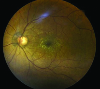
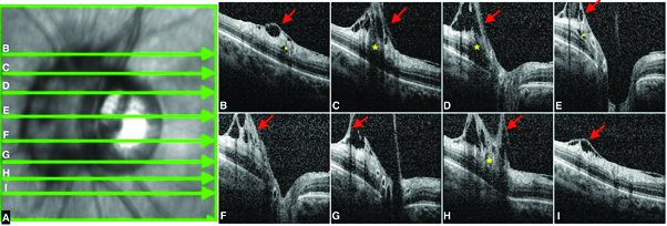
A 60-year-old female presented with bilateral immature cataract with best-corrected visual acuity of 20/200 in both eyes Oculus Uterque (OU). Fundus examination of left eye Oculus Sinister (OS) [Table/Fig-1] revealed Bergmeister’s papilla. The remainder of OS examination, including the right eye Oculus Dextrus (OD), was unremarkable. Optical Coherence Tomography (OCT) revealed that the Bergmeister’s papilla was creating peripapillary traction on the adjacent nasal retina with disruption of inner retinal layers [Table/Fig-2]. Vigilant monitoring of traction complex following cataract surgery was advised.
Fundus photograph Oculus Sinister (OS) revealing Bergmeister’s papilla and epiretinal membrane over the posterior pole with surface wrinkling of the macula which was also incidentally noted.
A. OCT infrared image of the same Optic Nerve Head (ONH) with green arrows depicting the sections shown in the forth coming images. B,C,D,E,F,G,H and I, shows multiple sections of the OCT infrared image of the ONH revealing the Bergmeister’s papilla causing peripapillary overlying traction (red arrows) with disorganisation of the adjacent inner retinal layers (yellow asterisks).
Bergmeister’s papillae is an uncommon anomaly that arises from the centre of the optic disc consisting of fibrous sheath, and represents the remnant of the hyaloid artery which is frequently observed as an incidental clinical finding [1]. The papillae are usually asymptomatic. The Bergmeister’s papillae is a developmental congenital anomaly of the embryogenic hyaloid artery, which appears at the third week of gestation and reaches the lens by the fourth or the fifth week of gestation, to form tunica vasculosa lentis, eventually undergoing complete atrophy and disappearing at birth [1]. The remnant of anterior portion of the hyaloid artery is called the Mittendorf’s dot, which is commonly located on the infero-nasal aspect of the posterior lens capsule and the remnant of the posterior portion of the hyaloid artery is called the Bergmeister’s papillae which is present at the Optic Nerve Head (ONH) usually composed of glial tissue [2]. In most cases, the Bergmeister’s papillae is an occasional finding with no clinical impact. But in most severe cases it can be associated with, pigmentary changes, amblyopia, posterior polar cataract, microphthalmia, persistence of primitive vitreous, vitreo-macular traction, vitreous haemorrhages and sometimes tractional retinal detachment due to contraction of the residual fibrovascular tissue [3-5]. In this case scenario [Table/Fig-2], the condition is prone to contraction of the fibrovascular tissue, which may exert traction on the retina causing peripapillary traction on the adjacent retina with disruption of inner retinal layers. For diagnosis, slit-lamp examination, fundus examination and OCT have been used. Anticipating a sequential peripapillary area vitreous thickening, vigilant monitoring needs to be done for the development of vitreo-macular traction, tractional retinal detachment or retinoschisis as they may become a possible cause of future visual deterioration [5].
Though Bergmeister’s papillae is most often inconsequential, the need for OCT evaluation in view of a rare vitreoretinal adhesion proves to be a useful non invasive test, to monitor its clinical sequelae.
Author Declaration:
Financial or Other Competing Interests: None
Was informed consent obtained from the subjects involved in the study? Yes
For any images presented appropriate consent has been obtained from the subjects. Yes
Plagiarism Checking Methods: [Jain H et al.]
Plagiarism X-checker: Feb 16, 2021
Manual Googling: Mar 26, 2021
iThenticate Software: Apr 17, 2021 (20%)
[1]. Mann I, The development of the human eye 1964 New YorkGrune & Stratton [Google Scholar]
[2]. Jones HE, Hyaloid remnants in the eye of premature babiesBr J Ophthalmol 1963 47(1):39-44.10.1136/bjo.47.1.3914186858 [Google Scholar] [CrossRef] [PubMed]
[3]. Kaur R, Khan B, Syal E, Sidhu H, Kaur M, Persistent hyaloid artery with posterior polar cataract in a young male: A rare presentationKerala J Ophthalmol 2018 30(3):203-05.10.4103/kjo.kjo_64_18 [Google Scholar] [CrossRef]
[4]. Azrak C, Campos-Mollo E, Lledó-Riquelme M, Ibañez FA, Toldos JJM, Vitreous haemorrhage associated with persistent hyaloid arteryArch Soc Espanola Oftalmol 2011 86(10):331-34.10.1016/j.oftal.2011.03.00622004579 [Google Scholar] [CrossRef] [PubMed]
[5]. Gandorfer A, Rohleder M, Charteris D, Kampik A, Luthert P, Ultrastructure of vitreomacular traction syndrome associated with persistent hyaloid arteryEye 2005 19(3):333-36.10.1038/sj.eye.670145915258604 [Google Scholar] [CrossRef] [PubMed]