Among all PNS, the maxillary sinuses are the first sinuses that begin to grow and the largest of the PNS [1]. It is contained within maxillary bones, filled with air and padded with respiratory epithelium. It reduces skull weight, increasing acoustic resonance, absorbing direct blows to the facial bones and insulating the upper teeth and eyes from heat, humidifying the inhaled air and supporting the development of the upper jaw [2]. Its location in the midfacial region makes them vulnerable to complications of tumours and inflammation in the neighboring structures [3].
Measurement and precise assessment of tissue size in reconstructive surgery led to significant advances in volume calculation [4]. Furthermore, measurement of the volume is useful in several medical applications such as determination of the relationship between gum bone loss and mucus thickness in the maxillary sinuses [5]. The normal shape of the facial bones is an indicator of the normal development of the PNS [6]. Therefore, it can be claimed that the normal topography of the face might indicate the normal development of the maxillary sinuses.
Computed Tomography (CT) scans imaging occupies a prominent position in the recent studies to measure the size of the PNS compared to conventional X-rays in living and human cadavers [7]. The advantage of examining the size of maxillary sinuses in a three-dimensional CT scan can be used for determination of gender for both criminal investigations and forensic anthropology [8,9]. In addition, CT imaging plays a major role in the diagnosis and management of PNS diseases, such as nasal polyps, sinus tumours and complications of sinusitis [7]. In this context, the CT of 2 Dimensional (2D) scans has been applied in various studies to estimate the volume of the sinuses with the application of several formulae [10,11]. Other studies used 3 Dimensional (3D) CT scans in order to obtain data for volumetric assessment of the maxillary sinuses [12-14]. There are differences in the PNS between the right and the left sides in terms of the shape and size. The anatomical structures are intricate and differ from person to another [10]. Several studies have been conducted, but consensus about the effect of age and gender on the PNS is not clear, reduction in the volume of the PNS in relation to age was reported after examination of the sphenoid and maxillary sinuses [15,16].
Previous studies have shown inconsistent results concerning the effect of gender on the volume of the sinuses. This was evident in the results of the maxillary sinus examination [17,18]. Pneumatisation of the PNS in the human, primarily, was evaluated by injection of various substances into the cadaver and measurement of the anatomical borders of the sinus and by taking a traditional X-ray. At present, trial of studies using of imaging techniques, such as Magnetic Resonance Imaging (MRI) that provided numerous sections of coronal, sagittal and axial images which enable an evaluation of the structure of the paranasal cavity. Moreover, the availability of many imaging modalities, presented a new perspective for using the morphometric analysis of these images [19]. The large volume of the maxillary sinuses, their anatomical position in the upper jaw and their relationship to the eye, nose and dental roots make them an interesting area for endoscopic sinus surgery, maxillofacial surgery and dental implant surgery [20]. To our knowledge, no information is currently available on the volume of the maxillary sinuses for Sudanese population. For this reason, present study was intended to be the first of its type to measure the volume of the maxillary sinuses on the CT images of normal Sudanese subjects using ImageJ 1.52p software program and to discover the correlation of the maxillary sinuses volume to the age and gender.
Materials and Methods
A retrospective observational study was conducted in the Department of Radiology, Royal Care Hospital, Khartoum, Sudan, during the period from August 2018 to July 2019, after approval from Human Research Ethical Committee (HREC0013/RCIH.07/19).
Inclusion criteria: CT images of adults aged more than 17 years from both genders of Sudanese subjects, who underwent a CT scan of the Paranasal sinuses (PNS) showed positive clinical signs and symptoms and proved to be normal, were included in the study.
Exclusion criteria: The CT images with congenital anomalies, sinus diseases or sinus fractures were excluded, as well as the CT images of individuals below the age of 17 were also excluded from the study. These healthy Sudanese CT images of the maxillary sinuses had been used in the final assessment. Multi slice CT scanner (Toshiba Aquilion 64 CT scanner) was used to obtain an axial CT image of the PNS. The projection was parallel to the orbito-meatal line, the slice reconstruction interval was 0.600 mm and its thickness was 1 mm. Coronal and sagittal planes were constructed using the Multiplanar reconstruction (MPR) processing by the workstation. Advance technical factors were applied to receive high quality data, 120 kVp; 210 mAs; rotation time=1.0 sec; collimation=64×0.625; FOV=220 mm and bone window (1500 HU window width, and 200HU window level). The patients were provided with perfect technical information about the CT scan imaging process.
Maxillary sinus volume was calculated using Cavalieri principle which is one of the stereological methods with ImageJ program (ImageJ 1.52p Wayne Rasband, National Institutes of Health, USA). The Cavalieri principle is an effective method in volumetric measurements of life structures. It is used to estimate the volume of the irregular shaped constituents from a set of two-dimensional cuts through it, provided that they are analogous, separated by an identified distance and start haphazardly inside the object. These parameters are encountered by standard CT and MRI techniques [21].
Coronal planes that were constructed using the MPR were used for measurement by ImageJ. Reconstructed consecutive parallel images with thicknesses of 1 mm and 6 mm interval (maxillary sinuses are visible every 10 slices) were used for ImageJ application. These cross-sectional image series were constant due to the stepwise movement of the computerised tomography machine. All consecutive images of CT were transferred to the ImageJ software for calculation. ImageJ software was matched using the reduction scale of the images. Using the program, the number of pixels enclosed by the traced maxillary sinus contours on each section was automatically calculated and it provided us with the cross-sectional area of the maxillary sinus of interest on a slice-by-slice basis.
The sum of the areas was multiplied by the section thickness to provide the volume of the maxillary sinus, as shown in the formula Vmax=t×∑A. Where Vmax is Volume of maxillary sinus; t is the section thickness of consecutive sections; and ∑A is the total sectional area of the consecutive sections. The maxillary volume was measured three times. All measurements were done using the same stereological software. The volume of the maxillary sinuses was compared in both sides. Relation of the maxillary sinuses volume to the gender was examined.
Statistical Analysis
Data was statistically analysed by SPSS version 21.0. The normality test was done before analysis and the results of the skewness and kurtosis tests confirmed the normality of the data. A paired-sample t-test was applied to compare between the volume of the right and left maxillary sinuses. An independent t-test was used to evaluate the difference between the gender and the volume of the maxillary sinuses. The p-value less than 0.05 was considered significant. Pearson’s r test was applied to investigate the correlation of age to the maxillary sinus volume.
Results
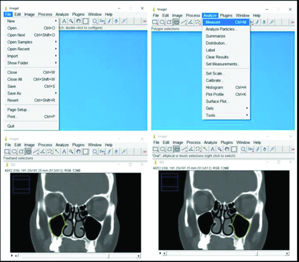
The study sample consisted of 81 CT images (46 males and 35 females). Their age ranged between 17 and 78 years, with the mean of 43.46 and 42.06 years for male and female groups, respectively. Sectional cut surface areas of the maxillary sinus were estimated using the planimetry method. For this aim, the polygonal selection tool of the ImageJ was used and the borders of the maxillary sinus were delineated [Table/Fig-1]. The mean total volume (cm3±SD) of both sinuses in the male and female groups was 38.70±12.12, and 38.34±11.50, respectively.
Computed Tomography (CT) images opened in ImageJ program shows the delineation of the maxillary sinuses’ border, thus enabled its volume measurement.
The different mean volumes of maxillary sinus are shown in [Table/Fig-2].
Mean and total of the maxillary sinuses volume (cm3±SD).
| Maxillary sinus | Males | Females |
|---|
| Volume (cm3) |
|---|
| min.-max. | mean±SD | min.-max. | mean±SD |
|---|
| Right | 7.17-30.76 | 19.22±6.05 | 7.59-33.44 | 18.84±5.85 |
| Left | 7.85-32.78 | 19.48±6.11 | 6.84-31.25 | 19.50±5.89 |
| Total | 15.02-63.54 | 38.70±12.12 | 16.76-62.30 | 38.34±11.50 |
| Age (Year) |
| 17-71 | 43.46±16.27 | 19-78 | 42.06±15.68 |
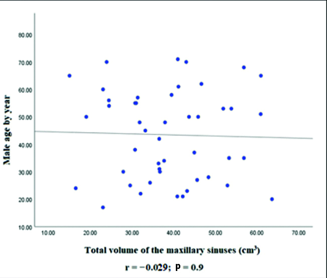
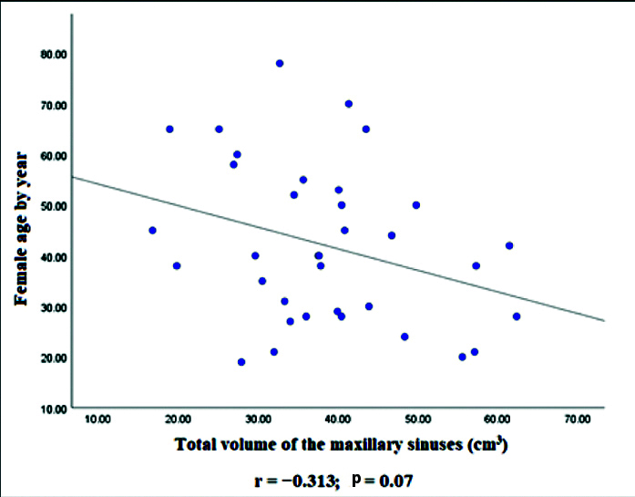
No significant differences were observed between the volume of the left and right maxillary sinuses in either male or female groups p=0.07 and p=0.11, respectively. There was a minor negative correlation between the total volume of maxillary sinuses and age in the male group which did not reach the level of statistical significance (r=0.029; p=0.9) [Table/Fig-3]. A negative correlation was also found between the total volume of the maxillary sinuses and age in the female group; however, the p-value did not reach the level of statistical significance (r=0.313; p=0.07) [Table/Fig-4]. This means, the size of the maxillary sinuses was inversely related to age. The effect of increasing age on the size of the maxillary sinuses was more pronounced in female than in male [Table/Fig-3,4]. Using independent t-test, no significant difference was determined between the gender in relation to the total volume of the maxillary sinuses p=0.9.
The graph shows the correlation between male age and the total volume of the maxillary sinuses.
The graph shows the correlation between female age and the total volume of the maxillary sinuses.
During the study, one male patient was found with right side nasal septal deviation, in spite, this observation did not significantly affect the ipsilateral maxillary sinus volume.
Discussion
After birth, there are momentous changes in the morphology, size and dimensions of the maxillary sinuses. Therefore, it is necessary to consider the amount of changes that may occur in the volume at the required surgical intervention. As maxillary hypoplasia is greatly affecting the size of the maxillary sinus [22]. Studies had reported that the size of the maxillary sinuses decreases in both sexes after reaching the maximum level of development, this could be due to mineral loss in the entire bone matrix enclosing the maxillary sinuses as a result the bony wall of the maxillary sinus shrinks to reduce the size of the sinuses [23-25].
It was found that the size of the maxillary sinuses begins to increase from birth to continue during childhood and adolescence and then ceases at the end of the age of 18 [26]. The volume of the maxillary sinuses continues to increase until the third molar has completely erupted this had been highlighted in the report by Thomas A and Raman R [27], whereas Degermenci M et al., concluded that the maxillary sinus reaches its maximum size at the age of 16 [28]. In spite of that Ariji Y et al., and Baweja S et al., had been pointed that the maxillary sinuses increased up to 20 and 25 years respectively, then after gradual reduction was observed [29,30]. The volume of the maxillary sinus was evaluated in CT Scan by Ariji Y et al., found that the volume of the maxillary sinus appears to decrease when the age is more than 20 years [29]. For these reasons, the age of more than 17 years was adopted in the current series.
Review of English literature revealed that there are several studies and efforts have been done to analyse the maxillary sinuses volume and their relation to gender and age. Rani SU et al., in India, Jasim HH and Al-Taei JA in Iraq and Kim J et al., in Korea had reported the volume of the maxillary sinuses are larger in male than in female [31-33].
These observations are in contrary with findings of this study where the gender does not affect the size of the maxillary sinus. Based on the current obtained results, it is not possible to identify the gender based on the size of the maxillary sinus. Depending to these, it can be saying that the maxillary bones in both sexes for some Sudanese subjects may have the same pattern of sinuses aeration development. On the other hand, the current study came in support to previous studies where there was no statistically significant difference in the maxillary sinuses volume between the sexes and sides [6,28].
The unique study of Fernandes CL had stated that the size of the maxillary sinus varies according to ethnic and gender, and male sinuses were significantly larger than those of females (p=0.002) [17].
Uchida Y et al., reported that no statistically significant differences were observed with respect to various factors such as age, sex and side when measuring the total volume of the maxillary sinus [34]. The data regarding the volume of the maxillary sinus on CT scan shows negative correlation r=-0.43 with regard to maxillary sinus volume and age, as for sex and side there was no significant difference in relation to the volume of the maxillary sinus [29]. As well, negative correlation r=-0.238 was also seen between the total volume of the maxillary, frontal, ethmoid and frontal sinuses to age, which means the total volume of these sinuses reduced with age [10].
The aforementioned results to large extent correspond to the findings of this study where negative correlation was observed when compared the age with total maxillary sinuses volume in male and female subjects, as it was r=-0.029, and r=-0.313, respectively. This means that the age reduces the size of the maxillary sinus after reaching its maximum size. In this context, since the volume of the maxillary sinuses ceases at a certain age and there is continuous increase in age, it could be suggested that the significant differences observed between the sinus volume and age are due to the actual reduction in the size of the maxillary sinuses with age. Hence, the highly statistical significance might be seen in the maxillary sinus volume of elderly when compared with their age.
Barghouth G et al., reported that no difference was observed in volume indexes between the right and left sides in the maxillary, sphenoid, and frontal sinuses. A difference between the sides in maxillary sinuses length was observed, when the age was more than 8 years [35]. Similarly, difference in the volume of the maxillary sinuses between both sides among gender was statistically insignificant. As, it was suggested in the above mentioned studies that the volume of the adult maxillary sinuses changes with age, this may be associated with the presence or loss of maxillary molars and premolars. Previously, had been reported that the absence of posterior maxillary teeth causes a decrease in maxillary sinus volume [36]. Therefore, it is essential to be aware about the anatomical approximate between the floor of the maxillary sinus and the maxillary third molar in the preoperative strategic planning when dealing with the maxillary third molar [37]. As well as, Velasco-Torres M et al., had declared that the total size of the maxillary sinuses is much smaller in individuals with total or partial tooth loss than in individuals with teeth, this result was affected by age, since the elderly showed a smaller size, regardless of dental condition and gender [38]. Şentürk M et al., reported that no significant difference was detected between the maxillary sinuses volume in children with and without nasal septal deviation and concluded that the presence of severe septal deviation did not affect the maxillary sinus volume [39]. This conclusion supported the observation of the current study, where analysed CT image found that the deviation of the nasal septum had no effect on the maxillary sinus volume.
The strength of this study is that, it is the first study to be performed among Sudanese individuals in this manner. The current study would be a database for the size of the maxillary sinuses in Sudanese population. Additional large prospective cross-sectional studies might be required to support the findings of the current study. As well as, future large studies including children are needed to ascertain the developmental overlap and beginning of the maxillary sinuses in both sexes in our local population. Lastly, but not the least large cohort study in abnormal maxillary sinuses as in case of absence of posterior maxillary teeth, or change in clinical conditions, like sinusitis, tumour, fracture of Maxilla may be included.
Limitation(s)
The limitation of the current study is being retrospective in nature, as well as, it included patients only from Khartoum, the capital of Sudan, and did not included patients from the other different regions of the country who are arising from different ethnicity.
Conclusion(s)
The volume of maxillary sinuses was negatively correlated with individual age, not affected by gender and no difference between sides. So, it cannot be used to determine the sex in Sudanese population for medicolegal purposes. An elaborated flag of the anatomy of the sinuses and its volumetric comparison of both sides might be essential in performing therapeutic interventional maxillary sinuses procedures. Maxillary sinus volumetry using CT scan is feasible under different circumstances, and it might contribute to a better clinical evaluation.