The temporomandibular joint and the condylar inclinations is the utmost important factor in prosthodontic rehabilitation, and the maintenance of the stomatognathic system. Precise recording of the maxillomandibular relationship and establishing the condylar guidance will aid in improved efficiency of the complete denture prosthesis. Fabrication of the balanced complete denture prostheses is often carried out indirectly in the absence of the patient on a semi-adjustable articulator, hence to re-establish the lost maxillomandibular relationship in a completely edentulous patient is the most difficult task. However, a historical review in dentistry reveals that there are numerous techniques, philosophies, and materials that can be manipulated and used to record the maxillomandibular relationship, in centric and all the eccentric movements (left lateral, right lateral and protrusive movement) of the maxillary and mandibular jaws. The conventional and the gold standard technique for recording the condylar guidance is Gothic arch tracing technique, the only disadvantage being time consuming and manipulation errors during the transfer to the articulator [1,2]. Thus, there is a need to exercise the newer and easy techniques to record the condylar guidance that is less time consuming technique. Recent development in a semi-adjustable articulator has introduced the use of interocclusal bite records and axiographic techniques to record static, as well as the dynamic movement of the mandible, and also record the condylar guidance while programming the articulator. Axiographic technique is less cumbersome as compared to the gothic arch tracing. The reason for determining the condylar guidance differences on right and left side was that the right and left eminences seldom have exactly the same contours, slants and declivities [3-6]. The present study was conducted to evaluate and compare the axiographic technique and the Panoramic radiographic technique that can be used as an alternative method to record condylar guidance values over the conventional Gothic arch tracing technique.
Materials and Methods
This observational study under clinical set up was conducted over a period of 6 months between Oct 2018-March 2019. Based on the observations made in the pilot study, the following sample size of 20 edentulous subjects was calculated using the G*Power 3.1 software.
Inclusion criteria: Patients with completely edentulous maxillary and mandibular arches and with good neuromuscular function, visiting Department of Prosthodontics, AB Shetty Memorial Institute of Dental Science, Deralakatte, Mangalore were selected to participate in the study after obtaining the informed consent.
Exclusion criteria: The patients with restricted and deviated mouth opening, and patients with the history of disorders of temporomandibular joint and muscles of mastication were excluded from the study.
The ethical clearance was obtained from the ethical committee- Nitte (Deemed to be university) Mangalore (approval no. NU/CEC/2016-2017/0080).
The subjects fulfilling the inclusion criteria were selected for the study and the treatment was planned to fabricate the balanced complete denture prosthesis. The treatment was initiated with making of primary impression using the conventional techniques following which border molding and secondary impressions were made. The master casts were prepared using dental stone, the denture bases and occlusal rims were fabricated on the master casts. The occlusal rims were adjusted in the patient’s mouth to register the maxillomandibular relationship; the tentative centric relation was registered at the conventionally established vertical dimension. The face bow transfer was done following which the maxillary cast was mounted on the semi adjustable articulator (Artex®, GIRRBACH DENTAL). Later the mandibular cast was mounted in the articulator using the tentatively registered jaw relation; the mandibular occlusal rims were reduced by 3 mm as a space for the attachment of the Extraoral gothic arch tracers and for the interocclusal recording medium.
Extraoral Tracing with Interocclusal Bite Recording Technique
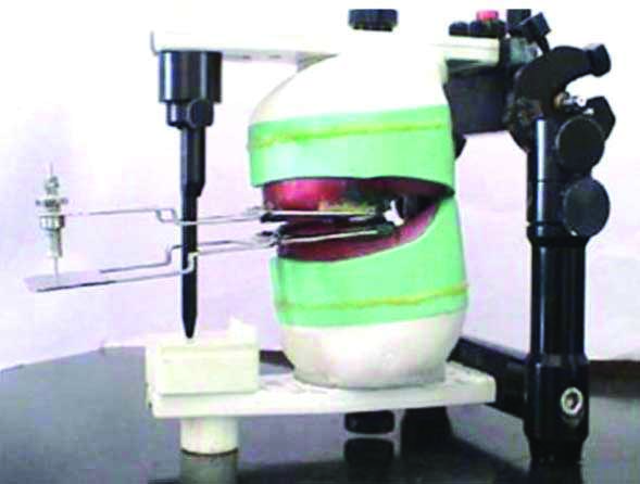
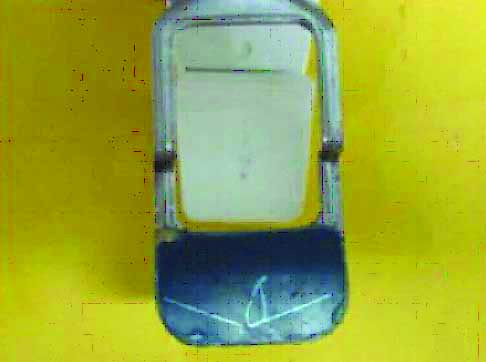
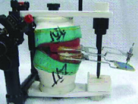
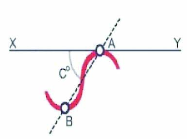
The extraoral tracing assembly (Hight extraoral tracer assembly) was attached to the mounted bases on the articulator [Table/Fig-1]. Patient was asked to do required movements. When the patient was proficient in executing all the movements, the tracing plate was dried and thin layer of soot was applied evenly so that no resistance was offered to the movement of the stylus that was attached to the maxillary occlusal rims, following which the patient was asked to move the mandible in centric relation and all the eccentric relations, the clearly visible tracing were made by the stylus on the tracing plate. The definitive arrow point tracing with sharp apex was obtained, the pointer of the stylus was adjusted to be at the point of the apex of the arrow point tracing. Quick setting dental plaster was injected between the occlusal rims and the plaster was allowed to set. The tracing plate was removed and conformed for sharp apex indicating centric relation [Table/Fig-2]. The record bases were placed back into the articulator; the mark made by the pin was confirmed to coincide with the apex of the tracing following which the protrusive interocclusal record was obtained. A hole was made 6 mm from centric hole in secured tracing plate with blank radiographic film, and the bases were replaced in patients mouth and the lower jaw was guided in a protrusive movement till the stylus enters the notch made on the radiographic film. The protrusive record was obtained using quick setting impression plaster; tracing assembly with recording bases was transferred to the articulator. The condylar posts were set at 0 degrees, the incisal pin was raised of the incisal table and the locknuts were loosened. The recording bases were returned to the articulator (Artex®. GIRRBACH DENTAL) and maxillary rim was aligned so that it coincides with the imprints on mandibular rim. The locknuts were moved back and forth one side at a time until the maxillary rim was firmly and securely seated against the lower index, and the stylus pointer coincided with the hole made i.e., 6 mm from the centric relation point. Protrusive relation record was adjusted three times and results were averaged before the horizontal condylar guidance was set and locknut was tightened with hand pressure [Table/Fig-3]. In all the cases articulator was programmed by single operator.
Record bases with tracing devices attached.
Extraoral Gothic arch tracing.
Interocclusal record is positioned in the articulator. (Images clockwise)
Panoramic Radiography
A panoramic radiograph was taken using orthopantomogram (PM2002CCPROLINE). All radiographs were made by the same operator and with the same radiographic unit; the image was acquired at 74 kvp, and 10 mA. Tracings were made of the images of each radiograph on tracing paper. Images were traced for Frankfort horizontal plane which passes through the lowest point in the margin of the orbit and highest point in the margin of auditory meatus. Outline for most superior point on the articular eminence and inferior point on the articular tubercle just adjacent to zygomatic arch were traced [Table/Fig-4]. A line was drawn connecting most superior and inferior points of curvature representing the mean curvature line [Table/Fig-5]. The angle was measured between mean curvature line and the reference line, each measurement was repeated twice, twenty panoramic radiographic images were traced independently before making interpretation, and the data were subjected to the statistical analysis [Table/Fig-5].
Panoramic radiograph showing landmark used.
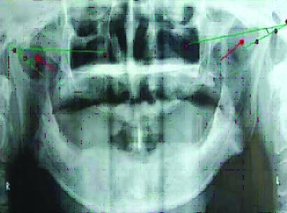
Tracing of line AB joining height of superior curvature A and inferior curvature B, representing inclination of articular eminence is angle made by intersection of mean curvature line and horizontal reference line.
Axiographic Technique
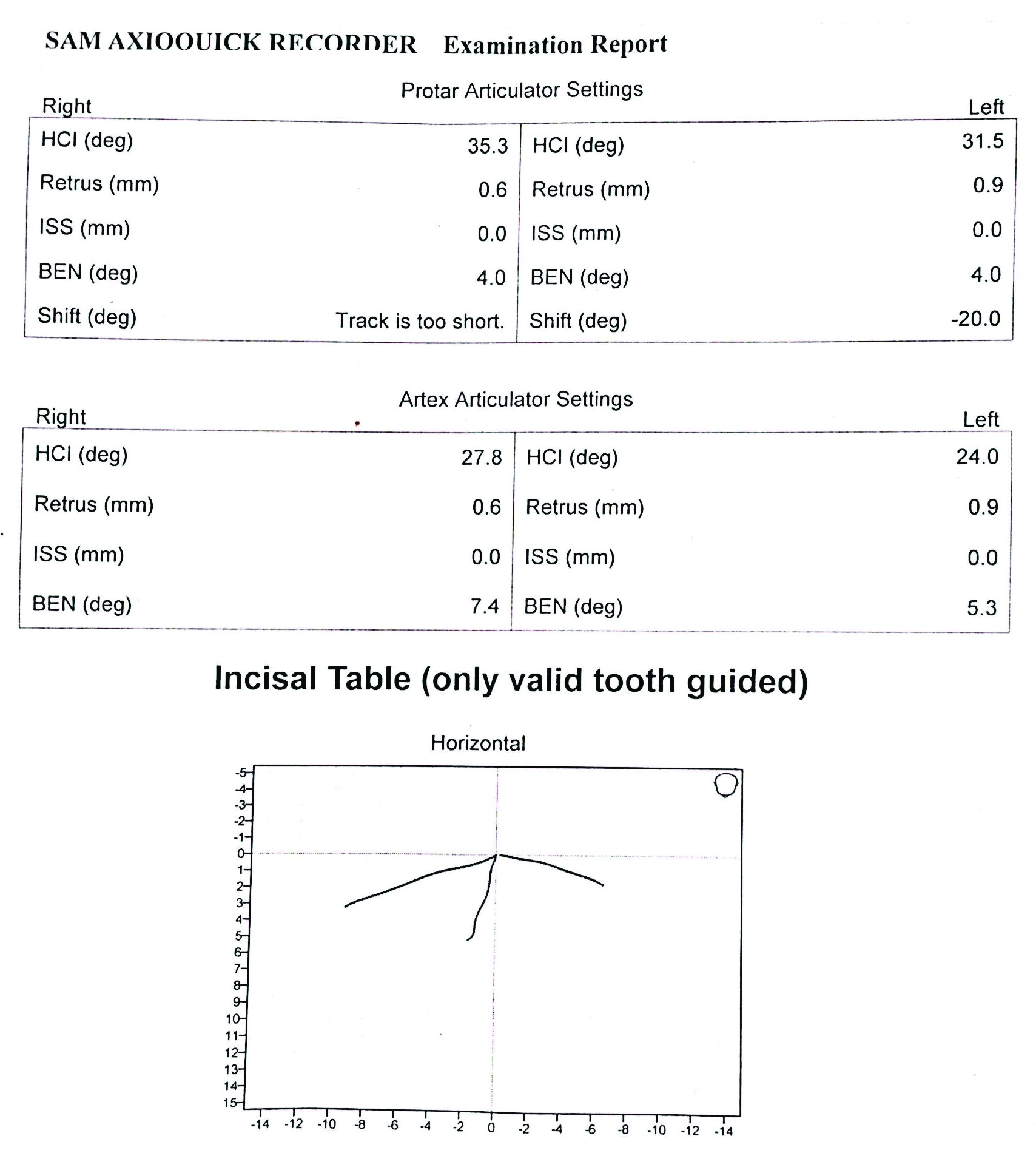
The patients were asked to visit the Temporomandibular joints (TMJ) Clinic where the computerised recording of mandibular movement was made by using axiographic jaw tracking device, the axio-quick recorder, ultrasonic Axiograph, was used to record mandibular movement. The combination of Axioquick® with a contactless ultrasonic recording device allows the 3D acquisition of all mandibular movements [Table/Fig-6]. The patients were seated in an upright position following which the jaw tracking device was attached to the waxed up complete denture, the device was attached to the monitor that works on software version 1.0, SAM 3 to record the jaw movement. The recording of the jaw movements was received while patients was asked to open the jaw in rotational movement and translational movement of the jaw, later the patient was asked to make left lateral and right lateral movement and protrusive movement. The values obtained from the axiograph were utilised to program the semi-adjustable articulator [Table/Fig-6,7]. These values obtained from axiographic tracing were compared with the values obtained from Gothic arch tracers, and the orthopantomographic values. Following which the obtained data was statistical analysed.
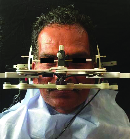
Condylar guidance angle on axiograph.
HCI: Horizontal chondylar inclination; Retrus: Retrusion; ISS: Immediate side shift; BEN: Biomechanical evaluation
Statistical Analysis
The correlation between the Axiographic tracing technique, Gothic arch tracing technique and the Panoramic radiographic technique was assessed by using Karl Pearson correlation test. Paired t-test was used to compare right and left side. The comparison between the Axiographic tracing technique, Gothic arch tracing technique and the Panoramic radiographic technique on left and right sides was assessed by using Anova test and Bonferroni Post-Hoc Test. A p-value less than 0.05 were considered as statistically significant.
Results
Data collected was tabulated and statistically analysed. The correlation between the right and left side of the extraoral Gothic arch tracing, orthopantomogram and axiographic tracing values. A statistically significant positive correlation was found for horizontal condylar guidance between both the sides of panoramic radiograph technique and axiographic technique (r=0.46, p=0.04, r=0.81, p=0.001) [Table/Fig-8].
Correlation between right and left side Extraoral gothic arch tracing, orthopantomogram and axiographic tracing values.
| Techniques studied | Left |
|---|
| Extraoral gothic arch tracing | Orthopantomogram | Axiographic tracing |
|---|
| Right | Extraoral gothic arch tracing | r | 0.302 | | |
| p-value | 0.20 | | |
| Orthopantomogram | r | | 0.46 | |
| p-value | | 0.04* | |
| Axiographic tracing | r | | | 0.81 |
| p-value | | | <0.001* |
Pearson’s Correlation Test; *p<0.05 Statistically Significant
Correlation between extraoral Gothic arch tracing, orthopantomogram, and the axiographic tracing values of right and left side of the subjects [Table/Fig-9]. A positive correlation was found between the right Extraoral gothic arch tracing and right and left side of OPG. Also, a positive correlation was observed between the left Extraoral gothic arch tracing and left OPG.
Correlation between extra oral gothic arch tracing, orthopantomogram, and axiographic tracing values of right and left side of the subjects.
| Techniques studied | Orthopantomogram | Axiographic tracing |
|---|
| Right | Left | Right | Left |
|---|
| Extraoral gothic arch tracing | Right | r | 0.47 | 0.57 | 0.003 | -0.03 |
| p-value | 0.04* | 0.008* | 0.99 | 0.91 |
| Left | r | 0.37 | 0.55 | 0.27 | 0.33 |
| p-value | 0.10 | 0.01* | 0.25 | 0.15 |
| Orthopantomogram | Right | r | | | 0.19 | 0.07 |
| p-value | | | 0.43 | 0.78 |
| Left | r | | | -0.10 | -0.11 |
| p-value | | | 0.68 | 0.64 |
Pearson’s Correlation Test; *p<0.05 Statistically Significant; p>0.05 Non Significant, NS
The comparison between right and left side of the Extraoral gothic arch tracing, Orthopantomogram, and Axiographic tracing values [Table/Fig-10].
Comparison between right and left side Extraoral gothic arch tracing, orthopantomogram, and axiographic tracing values.
| Techniques studied | Sides studied | N | Mean (°) | Std. Deviation (°) | Mean Difference | 95% Confidence interval of the difference | t | df | p-value |
|---|
| Lower | Upper |
|---|
| Extraoral gothic arch tracing | Right | 20 | 33.00 | 7.50 | 5.25 | 0.48 | 10.02 | 2.30 | 19 | 0.03* |
| Left | 20 | 27.75 | 9.52 |
| Orthopantomogram | Right | 20 | 36.50 | 7.72 | 4.15 | -0.08 | 8.38 | 2.05 | 19 | 0.05 |
| Left | 20 | 32.35 | 9.45 |
| Axiographic tracing | Right | 20 | 25.65 | 2.03 | 0.10 | -0.49 | 0.69 | 0.36 | 19 | 0.73 |
| Left | 20 | 25.55 | 2.06 |
Paired t-test; *p<0.05 Statistically Significant
The average horizontal condylar guidance angles and the standard deviations between the right side and the left side of the Extraoral Gothic arch tracing was 33±7.50° on the right side and on the left side was 27.92±9.52°, respectively. The average horizontal condylar guidance angles and the standard deviations between the right side and the left side of the orthopantomography was 36.5±7.72° and 32.35±9.45°, respectively. The average horizontal condylar guidance angles and the standard deviations between the right side and the left side of the axiographic tracing were 25.65±2.03° and 25.55±2.06°, respectively. Statistically significant differences were observed between right and left sides in Extraoral gothic arch Tracing. The other two techniques showed nonsignificant differences between both the sides.
[Table/Fig-11a] shows the comparison between Extraoral gothic arch tracing, orthopantomogram, and axiographic tracing values on the right and left sides. Significant differences were obtained between the techniques on right and left sides.
Comparison between Extraoral gothic arch tracing, Orthopantomogram, and Axiographic tracing values on the right and left sides.
| Sides studied | Techniques studied | N | Mean (°) | SD | Repeated measures ANOVA |
|---|
| F | p-value |
|---|
| Right | Extraoral gothic arch tracing | 20 | 33.00 | 7.50 | 20.47 | <0.001* |
| Orthopantomogram | 20 | 36.50 | 7.72 |
| Axiographic tracing | 20 | 25.65 | 2.03 |
| Left | Extraoral gothic arch tracing | 20 | 27.75 | 9.52 | 5.55 | 0.008* |
| Orthopantomogram | 20 | 32.35 | 9.45 |
| Axiographic tracing | 20 | 25.55 | 2.06 |
*p<0.05 Statistically Significant; p>0.05 Non Significant, NS
Statistically nonsignificant values were obtained with pairwise comparison on right side where the mean difference between extraoral tracing and orthopantomography was -3.50 [Table/Fig-11b]. On pairwise comparison on the left side where the mean difference between extraoral tracing and orthopantomography, and between the extraoral tracing and the axiographic tracing was -4.60 and 2.20, respectively. Statistically nonsignificant values were obtained between the extraoral tracing and orthopantomography; and the extraoral tracing and axiographic tracing on left side [Table/Fig-11b].
Pairwise comparison between extraoral Gothic arch tracing, Orthopantomogram, and Axiographic tracing values according to right and left side.
| Sides studied | (I) Group | (J) Group | Mean Difference (I-J) | Std. Error | p-value | 95% Confidence interval for difference |
|---|
| Lower bound | Upper bound |
|---|
| Right | Extraoral gothic arch tracing | Orthopantomogram | -3.50 | 1.76 | 0.18 | -8.11 | 1.11 |
| Axiographic tracing | 7.35 | 1.74 | 0.001* | 2.79 | 11.91 |
| Orthopantomogram | Axiographic tracing | 10.85 | 1.70 | <0.001* | 6.39 | 15.31 |
| Left | Extraoral gothic arch tracing | Orthopantomogram | -4.60 | 2.01 | 0.10 | -9.87 | 0.67 |
| Axiographic tracing | 2.20 | 2.02 | 0.87 | -3.11 | 7.51 |
| Orthopantomogram | Axiographic tracing | 6.80 | 2.21 | 0.02* | 0.99 | 12.61 |
Bonferroni Post-Hoc Test; *p<0.05 Statistically Significant; p>0.05 Non Significant, NS
Discussion
The successful treatment of complete denture prosthesis is based on the long-term preservation of alveolar bone along with functional balance of Stomatognathic system [3,4]. Posselt U and Nevstadt P concluded in their study that individual registration of condylar path inclination should be used instead of mean value adjustment [5]. Payne JA stressed the importance of determining and obtaining the values of the condylar guidance rather than relying on average values. Author also reviewed the literature of various studies of recording protrusive condylar angle and found it ranged from 22-65°, with the average of 38° [7]. Arstad T compared the articulator setting from various techniques like Gysi, Stansbery, Hanau, and it was concluded that none of the methods using situational interocclusal records can be considered reliable when a thermoplastic material is used and that functional engraving or grinding in method can register the movements of the mandible and those of the bases on the soft tissues [8]. The prominent outline of the articular eminence can be viewed on the panoramic radiograph and can be utilised to program the articulator. However, the tracings obtained from axiograph may also be used to program the semi-adjustable articulator [9]. All the values were evaluated, compared and statistically analysed. Based on the results obtained, the Gothic arch tracing method is the ideal technique of recording the mandibular movement in centric and eccentric motion, which helps to obtain accurate condylar guidance values, with the help of traced arrowhead and plaster records of centric and protrusive movements. The low error index was obtained between the two radiographs; the angular values are acceptable since the variation found were less than 5 degrees. Variations of horizontal condylar guidance greater than 5 degrees were noted after repeated registrations in patients with TMJ disturbances as well as in patients treated with complete denture. This could be explained by the fact that horizontal condylar angulations vary greatly on either sides and from one individual to another and the variations can be determined with Gothic arch tracing techniques [10]. There are some inherent inaccuracies and limitations in this application regarding panoramic distortion, head and reference plane orientation, and difficulty in distinguishing the articular eminence outline from the zygomatic arch. Parallax errors may arise, if the beam becomes directed from either above or below due to positioning errors and the positions of these two lines relative to each other may vary. The articular eminence inclination in the radiographic image was traced from the most superior to the most inferior points of curvature, representing mean inclination of the curve. This may be different from the guiding inclination with approximately 4mm to 6mm of protrusion, which is the clinically significant range of protrusion and condylar guidance. Considering the inaccuracies of the interocclusal record technique within the range of errors of up to 30 degrees, the radiographic articular eminence image may have clinical relevance. Based on the literature survey, the average condyle guide settings values were considered to be accurate by the clinicians to program the articulator [11]. However, the mean setting value of condylar guidance on a semi adjustable articulator is 30 to 40 degrees is used. When the true incline is flat (0-10 degrees) may be used or if the true incline is steep (70- 80 degrees) may be used, the discrepancy while obtaining the condylar guidance values will have an influence on the outcome of prosthetic restorations, as these will be used to programme the semi-adjustable articulator following which the complete denture prosthesis will be fabricated, the inaccurate condylar guidance settings may lead to failure of the prosthesis, the control over the protrusive and lateral eccentric guidance and posterior disocclusion or the attainment of a completely balanced articulation may be adversely affected [12]. Based on the observations made on overall results the panoramic radiographic images can be used to program the semi adjustable articulator and to check the reliability of using the Axiographic tracing techniques to programme the semi adjustable articulator further studies have to be carried out.
The present study demonstrated three techniques Gothic arch tracing technique, Panoromic radiographic technique, and axiographic technique. The Panoramic radiographic technique had considerably reliable readings. Thus, it can be used as an adjunct to the traditional Gothic arch tracing in programming articulator and thus providing effective treatment.
Limitation(s)
The limitation of the study was the small sample size. The distortion of the panoramic radiographs may result in blurring of the morphology of the anatomical landmark. Radiation exposure and positioning of the patients head may alter the readings. The Axiographic tracing technique may cause distortion of the mandibular arch if highly resorbed. Also, the tracings can be done only in the patients with good interarch space.
Conclusion(s)
The practicability of the three techniques and the consistency of the results considered that the radiographic method can be used as an alternative to the gold standard Gothic arch tracing technique to record condylar guidance values that seems to be most suitable for clinical use which is less time consuming and easy to manipulate. However, the axiographic technique also can be used as an alternative technique to panoramic radiographic imaging technique and the Gothic arch tracing technique, as all these three techniques were used in individual patients and values obtained were compared, further the articulator was programmed and complete denture prosthesis were fabricated and inserted in patients mouth, the follow-up report showed no clinical issues while the complete denture prosthesis was in function. Further studies with large sample size are recommended in future.
Pearson’s Correlation Test; *p<0.05 Statistically Significant
Pearson’s Correlation Test; *p<0.05 Statistically Significant; p>0.05 Non Significant, NS
Paired t-test; *p<0.05 Statistically Significant
*p<0.05 Statistically Significant; p>0.05 Non Significant, NS
Bonferroni Post-Hoc Test; *p<0.05 Statistically Significant; p>0.05 Non Significant, NS