A dye refers to a coloured substance that has affinity to a substrate on which it is being applied. Eosin is the most commonly used synthetic dye belonging to xanthene group. Although, these synthetic dyes are efficient, they are hazardous to human and animal health. Turmeric (Curcuma longa) has been in-use-in the subcontinent as a spice and flavoring agent in most of the food preparation. It is a natural antibiotic and has anti-inflammatory property [1]. Turmeric is used as a dye for various purposes since centuries [2]. The turmeric extract has been used as a potential alternative for eosin on routine histopathology sections [1]. Extracts of wing fruit have been used as a promising histological stain [3]. Henna (Lowsonia inermis) extract has been used as an effective alternative to eosin in histological sections [4]. Kumkum is a red coloured powder which is mainly used for religious purposes in India. It is made from saffron flowers of Crocus sativus L. with mild use of turmeric. The use of kumkum was considered as safer than other chemicals for staining pectinolytic actinomycetes [5].
To the best of the knowledge of the authors, kumkum solution has not been used in histopathology to stain the tissues. The present study highlights the novel use of kumkum solution in the field of histopathology for the purpose of staining cervix tissue. The present study was undertaken to evaluate the staining characteristics of kumkum solution as a counterstain and to compare the staining characteristics of kumkum solution and turmeric solution in histopathology sections of cervix tissue.
Materials and Methods
The present study was a prospective observational study conducted from May 2019 to July 2019 for a period of three months, at a rural tertiary care referral institute. The study was conducted in the histopathology section of Department of Pathology, PES Institute of Medical Sciences and Research, Kuppam, Andhra Pradesh, India. The study was approved by Institutional Ethics Committee bearing number PESIMSR/IHEC/46.
Inclusion and Exclusion criteria: All cases in which cervix tissue specimens were received for histopathological evaluation were included in the study. Those cases in which cervix tissue specimens were unsatisfactory for evaluation were excluded from the study.
Sample size calculation: The sample size was calculated using following formula:
“n” is the sample size
“Z2(1-α/2)” is the level of significance at 5% that is 95% confidence interval
“p” is the expected proportion of histopathology samples
“d” is the desired error of margin
A total of 57 cases were analysed. Each case was routinely processed and H&E stained cervix tissue sections were screened. Representative tissue section was selected for the study. For each case, three unstained representative tissue sections were taken. One control section was stained by H&E stain using eosin as the counterstain. The control section was stained by H&E staining technique according to the standard operating procedure. One tissue section was stained by H&T stain by using turmeric solution as the counterstain. Other tissue section was stained by H&K using kumkum solution as the counterstain. Turmeric solution and kumkum solutions were prepared by maceration technique [2]. Turmeric solution was prepared by dissolving 15 gm of commercially available turmeric powder (branded company) in 100 mL of isopropyl alcohol. Kumkum solution was prepared by dissolving 15 gm of commercially available kumkum powder (branded company) in 100 mL isopropyl alcohol. The turmeric solution and kumkum solution were left undisturbed for 48 hours. The solutions were filtered and the filtrates were stored in labeled bottles and were utilised for staining.
For the purpose of standardisation and uniformity, single brand of commercially available kumkum was used to stain the slides throughout the study. Similarly, single brand of commercially available turmeric was used to stain the slides throughout the study. To standardise each batch of the kumkum solution, the staining was performed on quality control slide of the cervix. The staining was performed on the slides only when the staining characteristics were found to be satisfactory on quality control slides. Batch to batch verification was performed for each batch before staining the slides to be evaluated in the study. Similar procedure was followed for turmeric solution. The pH of the solutions was determined by pH meter. The pH of turmeric solution was 5.54-5.64 and the pH of kumkum solution was 5.45-5.52.
After treating the sections with nuclear stains and blueing, the sections were treated with filtered counterstains viz., turmeric and kumkum for eight minutes for each of the new staining techniques. The timing was standardised by trial and error method to achieve optimal staining. The histomorphological details were documented in H&T sections and H&K sections. The staining characteristics of H&T sections and H&K sections were evaluated by a single pathologist using a scoring system by comparing with that of conventional H&E sections. The scoring system was indigenously designed for evaluating the stained sections based on the parameters documented by Raju L et al., [4].
Scoring system: Each section was compared with H&E section and scored 1 to 4 according to the following scoring system. Contrast at low power (X100) was scored as score 1 (very poor- unsuitable for interpretation), score 2 (sub-optimal- just acceptable for interpretation), score 3 (optimal contrast at low power), score 4 (excellent contrast at low power). Morphological details of the cytoplasm was scored as 1 (very poor- unsuitable for interpretation), score 2 (sub-optimal- just acceptable for interpretation), score 3 (optimal cytoplasmic features) and score 4 (excellent preservation and sharp cytoplasmic features). Morphological details of the nucleus was scored as 1 (very poor- nuclear features overtly obscured by counterstain), score 2 (sub-optimal- nuclear features moderately obscured by counterstain), score 3 (optimal nuclear features- nuclear features mildly obscured by counterstain) and score 4 (excellent- nuclear features not obscured by counterstain). Morphological details of the acellular structures were scored as 1 (very poor- unsuitable for interpretation), score 2 (sub-optimal- just acceptable for interpretation), score 3 (optimal morphological features) and score 4 (excellent preservation and sharp features).
Statistical Analysis
The socio-demographic variables and descriptive statistics were represented using frequencies and percentages. Two sample t-test was used for continuous variables and Chi-square test or Fischer-Exact test was used for categorical variables was used. All results were analysed by considering statistical significance at a level of p-value <0.05. All statistical calculations were done through statistical software STATA version 14.1.
Results
In the present study, 57 cases of cervix tissue was examined and analysed. The lesions were seen in females in the range of 36-71 years (mean=46.58). Clustering of cases was seen in fifth decade. Most common histopathological finding in the cervix tissue was squamous metaplasia {40 cases (70.18%)}. The other findings included non-specific chronic inflammation {36 cases (63.16%)}, chronic polypoidal endocervicitis {35 cases (61.4%)}, Low grade squamous intraepithelial lesion {34 cases (59.65%)}, Nabothian follicle cyst {27 cases (47.37%)}, microglandular hyperplasia {1 case (1.75%)}, High grade squamous intraepithelial lesion {1 case (1.75%)} and squamous cell carcinoma {1 case (1.75%)} [Table/Fig-1,2].
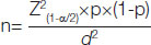
a-f: Microphotographs of cervix lesions: Chronic polypoidal endocervicitis with squamous metaplasia in (a) H&T, (b) H&E and (c) H&K in HPF (X400). Low grade squamous intraepithelial lesion in (d) H&T, (e) H&E and (f) H&K in HPF (X400).
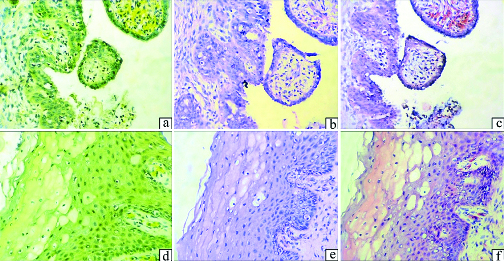
a-f: Microphotographs of cervix lesions: Microglandular hyperplasia in (a) H&T, (b) H&E and (c) H&K in HPF (X400); Squamous cell carcinoma in (d) H&T, (e) H&E and (f) H&K in HPF (X400).
The staining characteristics of H&T sections and H&K sections were evaluated by using a scoring system as described earlier and compared with that of conventional H&E sections. The staining characteristics included contrast at low power, morphological details of the cytoplasm, morphological details of the nucleus and morphological details of the acellular structures. The contrast was evaluated at low power for the ability of a stain to appreciate and differentiate various morphological structures (nerve bundles, muscle tissue, erythrocytes, etc.,). The morphological details of cytoplasm included cytoplasmic characteristics like cytoplasmic borders, vacuolations, granules and other features. The morphological details of nucleus included nuclear characteristics like nuclear borders, chromatin pattern, nucleoli and other features. Whether any stain obscures the nuclear morphology was also documented. Morphological details of the acellular structures included characteristics of elastic fibers, collagen fibers, proteinaceous material and other features.
Contrast at low power was better appreciated in H&K sections in comparison with H&T sections. H&K showed better average score than H&T with respect to contrast at low power. The p-values were statistically highly significant. Morphological details of cytoplasm were better appreciated in H&K sections in comparison with H&T sections. H&K showed better average score than H&T with respect to morphological details of cytoplasm. The p-values were statistically highly significant. Morphological details of nucleus were better appreciated in H&K sections in comparison with H&T sections. The nucleus showed light yellowish tinge in H&T stained sections. H&K showed better average score than H&T with respect to morphological details of nucleus. The p-values were statistically highly significant. Morphological details of acellular structures were better appreciated in H&K sections in comparison with H&T sections. H&K showed better average score than H&T with respect to morphological details of acellular structures. H&K showed better overall performance than H&T. The p-values were statistically highly significant [Table/Fig-3,4].
Comparison of staining characteristics between H&T and H&K.
| Parameters | Score 2 | Score 3 | Score 4 | Total | p-value |
|---|
| Contrast at low power |
| H&T | 9 (15.79%) | 31 (54.39%) | 17 (29.82%) | 57 | <0.001* |
| H&K | 0 | 1 (1.75%) | 56 (98.25%) |
| Morphological details of cytoplasm |
| H&T | 2 (3.51%) | 28 (49.12%) | 27 (47.37%) | 57 | <0.001* |
| H&K | 0 | 3 (5.26%) | 54 (94.74%) |
| Morphological details of nucleus |
| H&T | 12 (21.05%) | 37 (64.91%) | 8 (14.04%) | 57 | <0.001* |
| H&K | 0 | 1 (1.75%) | 56 (98.25%) |
| Morphological details of acellular structures |
| H&T | 5 (8.77%) | 40 (70.18%) | 12 (21.05%) | 57 | <0.001* |
| H&K | 0 | 10 (17.54%) | 47 (82.46%) |
*Chi-square test was the statistical tool used to perform statistical analysis; p-value <0.05 considered significant; *No subjects were present in Score 1
Comparison of average scores and overall performance between H&T and H&K.
| Stain | Contrast at low power | Morphological details of cytoplasm | Morphological details of nucleus | Morphological details of acellular structures | Total | p-value |
|---|
| H&T | 3.14 | 3.44 | 2.93 | 3.12 | 12.63 | <0.001* |
| H&K | 3.98 | 3.95 | 3.98 | 3.82 | 15.73 |
*Two sample t-test was the statistical tool used to perform statistical analysis; p-value <0.05 considered significant
Morphological appearance of various tissues in H&T and H&K stained sections [Table/Fig-5,6].
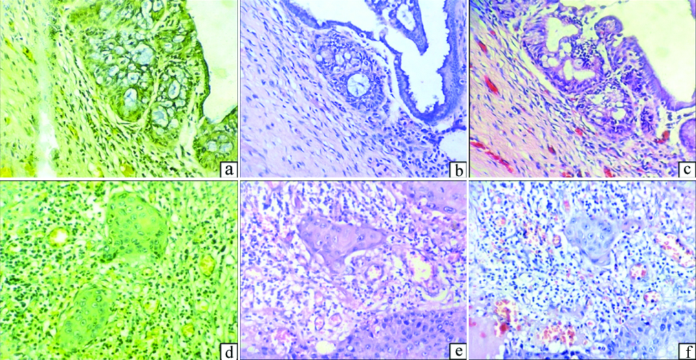
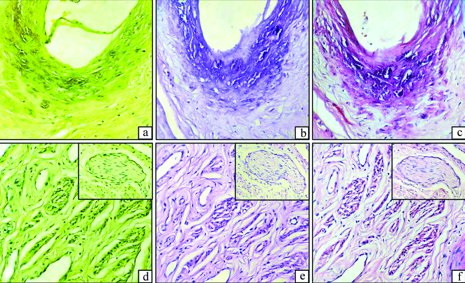
a-f: Microphotographs of various tissues: Blood vessels with erythrocytes in (a) H&T, (b) H&E and (c) H&K in HPF (X400). Elastic lamina in (d) H&T, (e) H&E and (f) H&K in HPF (X400).
a-f: Microphotographs of various tissues: Calcification in (a) H&T, (b) H&E and (c) H&K in HPF (X400); Smooth muscle tissue. Inset: nerve bundle in (d) H&T, (e) H&E and (f) H&K in HPF (X400).
Ectocervical Squamous Epithelium
H&T sections: Squamous epithelium appeared as polygonal shaped cells having moderate to abundant cytoplasm and central oval nucleus. Cytoplasm showed yellow to green hue of varying intensity depending on the degree of maturation of the cell. Basal cells showed greenish yellow cytoplasm. Intermediate cells showed light yellow cytoplasm and mature superficial cells showed yellowish green cytoplasm. Nucleus appeared greenish-black coloured. But nuclei of many cells showed yellowish tinge.
H&K sections: Squamous epithelium appeared as polygonal shaped cells having moderate to abundant cytoplasm and central oval nucleus. Cytoplasm showed pink to red hue of varying intensity depending on the degree of maturation of the cell. Basal cells showed reddish pink cytoplasm. Intermediate cells showed light pink cytoplasm and mature superficial cells showed pinkish-red cytoplasm. Nucleus appeared blue coloured.
Endocervical Glandular Epithelium
H&T sections: Glandular epithelium appeared as columnar shaped cells having moderate to abundant cytoplasm and basal oval nucleus. Cytoplasm showed yellowish blue hue. Nucleus appeared greenish-black coloured. But nuclei of many cells showed yellowish tinge.
H&K sections: Glandular epithelium appeared as columnar shaped cells having moderate to abundant cytoplasm and basal oval nucleus. Cytoplasm showed pinkish bluish hue. Nucleus appeared blue coloured.
Erythrocytes
H&T sections: Erythrocytes appeared as bright yellow coloured round to oval structures. But, it was not showing good contrast in comparison with the surrounding tissue.
H&K sections: Erythrocytes appeared as very bright red coloured round to oval structures. It was showing excellent contrast in comparison with the surrounding tissue. In fact, in many cases, it appeared better than the corresponding H&E stained sections.
Elastic Fibers
H&T sections: Elastic fibers were seen as refractile yellow coloured fibers in the connective tissue stroma and in the blood vessels (arteries) as elastic lamina. But the fibers were not easily discernible in all the cases.
H&K sections: Elastic fibers were seen as refractile bright reddish pink coloured fibers in the connective tissue stroma and in the blood vessels (arteries) as elastic lamina. The fibers were clearly discernible in most of the cases. In many cases, the elastic fibers were better appreciated in than the corresponding H&E stained sections.
Collagen Fibers
H&T sections: Collagen fibers were seen as light yellowish green coloured fibers in the connective tissue stroma and basement membrane.
H&K sections: Collagen fibers were seen as pink coloured fibers in the connective tissue stroma and basement membrane.
Smooth Muscle Tissue
H&T sections: Smooth muscle tissue was composed of dark yellowish green coloured spindle shaped cells arranged in bundles or fascicles.
H&K sections: Smooth muscle tissue was composed of dark pinkish red coloured spindle shaped cells arranged in bundles or fascicles.
Nerve Tissue
H&T sections: Nerve tissue was composed of light yellowish green coloured spindle shaped cells arranged in well delineated bundles. The nerve cells were identified by wavy nucleus.
H&K sections: Nerve tissue was composed of light pink coloured spindle shaped cells arranged in well delineated bundles. The nerve cells were identified by wavy nucleus.
Calcification
Calcification was due to Monckeberg medial sclerosis involving the wall of the muscular arteries.
H&T sections: Calcifications were seen as blackish brown coloured amorphous material.
H&K sections: Calcifications were seen as bluish black coloured amorphous material.
Mucin Material
H&T sections: Mucin material was seen as blue coloured material.
H&K sections: Mucin material was seen as bluish pink coloured material.
a-f: Microphotographs of artifacts: Muck (Arrow) in (a) H&T and (b) H&K in HPF (X400). Stain precipitate (Arrow) in (c) H&T and (d) H&K in HPF (X400). Patchy unstained area (Arrow) in (e) H&T and (f) H&K in HPF (X400).
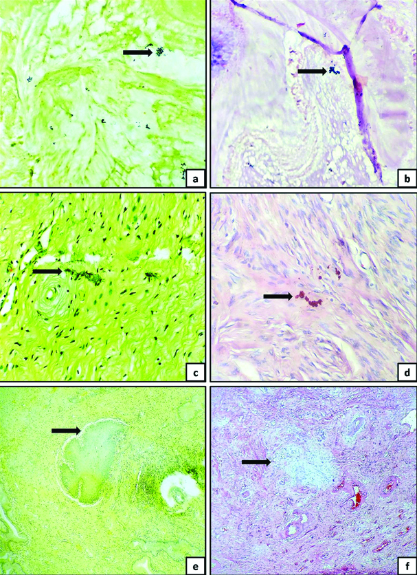
H&T sections: Artifacts were seen in 14 cases (24.56%). Various artifacts included mucks {4 cases (7.02%)}, stain precipitates {3 cases (5.26%)} and patchy unstained areas {7 cases (12.28%)}. Muck appeared as black to brown coloured material. Stain precipitate appeared as dark yellow coloured material. Patchy unstained areas appeared as irregular patches of areas devoid of stain.
H&K sections: Artifacts were seen in 6 cases (10.53%). Various artifacts included mucks {3 cases (5.26%)}, stain precipitates {1 cases (1.75%)} and patchy unstained areas {2 cases (3.51%)}. Muck appeared as black to brown coloured material. Stain precipitate appeared as dark pinkish-red coloured material. Patchy unstained areas appeared as irregular patches of areas devoid of stain.
Discussion
Dyes are used to stain a variety of substrates. Biological stains are often used to add colour to tissues, microbes or spores to make them optically distinct [1]. Stains form an integral part of laboratory technique. Most of the stains used in histology are synthetic dyes. Synthetic dyes are often efficient but may pose hazards to humans and animals [6]. Plants are a rich source of natural dyes which can be extracted from different parts like leaves, fruits, seeds, flowers, barks and roots. The hazardous effect of chemicals and synthetic dyes such as eosin has led to quest for alternative organic dyes from the natural sources [7]. Many natural dyes have been used in diagnostic fields of histology, histochemistry and histopathology [2]. Turmeric has been considered as safer alternative to eosin. But eosin was proved to be undoubtedly better over turmeric [1]. Extracts of Lowsonia inermis (henna), Carcade (Hibiscus sabdariffa) and wing fruit (Pterocarpus osun) have been used as substitutes for eosin. Henna needs oxidation with potassium permanganate. Alkaline and acid extracts of wing fruit are hazardous [3,4].
Kumkum is a red coloured powder used for religious and spiritual purposes in India [5,8]. Conventionally, it is prepared from flowers of Crocus sativus L. with mild use of turmeric [5]. Kumkum was employed to stain Escherichia coli (E.coli) in a study conducted by Gupta A et al., [8]. Suneetha V and Praveen Kumar G used kumkum to demonstrate pectinolytic actinomycetes and suggested that it was safer than other chemicals for staining purpose [5]. The current study emphasises the utility of kumkum as a counterstain in histopathology sections of cervix tissue.
The total number of cases was highest in the present study. In contrast, other studies had less number of cases. Suryawanshi H et al., and Kumar S et al., compared turmeric stain with conventional H&E [1,9]. Abraham M et al., compared turmeric stain extracted by maceration method and soxhlet extraction method with conventional H&E stain [2]. Sudhakaran A et al., compared ginger stain and turmeric stain with conventional H&E stain [6]. In contrast to the other studies, the present study compared kumkum and turmeric stains with conventional H&E stain. Abraham M et al., used both maceration method and soxhlet method to prepare the staining solution [2]. In contrast, the studies including the present study employed only maceration method to prepare the staining solution. Suryawanshi H et al., had used 50% alcohol in their study [1]. Sudhakaran A et al., employed 90% alcohol to prepare ginger stain and 70% alcohol to prepare turmeric stain [6]. Abraham M et al., and Kumar S et al., employed 70% alcohol as solvent to prepare the staining solution [2,9]. Seventy percent isopropyl alcohol was used as a solvent to prepare staining solutions (both turmeric and kumkum) in the present study. However, the other studies had not mentioned the nature of alcohol used. Suryawanshi H et al., and Kumar S et al., performed staining on a variety of tissues [1,9]. Sudhakaran A et al., employed buccal tissue in their study [6]. Abraham M et al., performed staining on 20 normal oral mucosa and 20 pathological oral tissue [2].
In the present study, staining was performed on cervix tissue. Cervix tissue was considered for the study because it was the most common tissue specimen received in the laboratory. It is composed of variety of structures such as ectocervix tissue (squamous epithelium), endocervix tissue (mucinous epithelium), blood vessels, smooth muscle tissue, nerve bundles, mucin, collagen fibers and elastic fibers. Furthermore, a variety of lesions may be seen in cervix ranging from inflammatory lesions to premalignant and malignant lesions [10,11].
Hence, the staining characteristics of variety of structures could be examined. Abraham M et al., found that maceration method of extraction of turmeric was better than soxhlet method [2]. Sudhakaran A et al., found that ginger staining was better than turmeric [6]. But the statistical significance varied between the observers. The p-value was 0.05 with one observer and it was 0.07 with other observer. Suryawanshi H et al., found that staining ability of turmeric was as good as eosin with respect to epithelium, keratin, muscles, adipocytes, blood vessels and erythrocytes [1]. But staining ability of turmeric was not as good as eosin with respect to collagen fibers, cartilage and bone. Kumar S et al., found that staining ability of turmeric was comparable to eosin with respect to collagen and skeletal muscle fibers [9]. But, Suryawanshi H et al., and Kumar S et al., found that overall performance of H&E was better than turmeric [1,9].
In the present study, it was observed that staining characteristics of kumkum was superior to both H&E and H&T with respect to various structures. But statistical significance was tested by comparing only kumkum and turmeric considering H&E as the gold standard. In H&T stained sections, nucleus of the cells had a yellowish tinge. This indicates that turmeric may have non-selective staining properties. It may stain both nucleus and cytoplasm. In contrast, nucleus of the cells appeared blue and was not obscured in H&K stained section. In comparison with H&E stained sections, H&K stained sections showed minor variation in hue of the cells, but was not interfering with identification of the cells or the tissue. It was found that the overall performance of kumkum was better than turmeric and was statistically highly significant. [Table/Fig-8] shows comparison of staining parameters in various studies as compared to present study.
Comparison of staining parameters in various studies [1,2,6,9].
| Parameters | Present study | Sudhakaran A et al., [6] (India, 2018) | Abraham M et al., [2] (India, 2017) | Suryawanshi H et al., [1] (India, 2017) | Kumar S et al., [9] (India, 2014) |
|---|
| Total number of cases | 57 | 25 | 40 | 10 | 10 |
| Stains employed | Kumkum, Turmeric, H&E | Ginger, Turmeric, H&E | Turmeric, H&E | Turmeric, H&E | Turmeric, H&E |
| Tissues employed | Cervix tissue | Buccal tissue | Normal oral mucosa, Oral squamous cell carcinoma | Epithelium, keratin, collagen fibers, muscles, adipocytes, blood vessels and RBCs, cartilage, bone | Epithelium, collagen, muscles, blood vessels, nerves, bone, adipose tissue, keratin |
| Method of extraction | Maceration | Maceration | Maceration, Soxhlet | Maceration | Maceration |
| Solvent employed | Isopropyl alcohol (70%) | Alcohol (90% and 70%) | Alcohol (70%) | Alcohol (50%) | Alcohol (70%) |
| Inference | Kumkum staining was better than turmeric | Ginger staining was better than turmeric | Maceration method was better than Soxhlet method | H&E was better than turmeric | H&E was better than turmeric |
| p-value | p<0.001(Highly significant) | p=0.05 (Just significant) | p=0.04 (Significant) | p<0.001(Highly significant) | p<0.001(Highly significant) |
The principles of staining suggest that staining is due to an ionic bond between the dye and the tissue components. This is associated with electrostatic attraction between dissimilar ions. The pattern of binding of tissue with the stain depends on the bond between the dye and the tissue components. The factors associated with selective staining are dye concentration, its aqueous or alcoholic nature, time of action on the solvent and pH [2].
Even though the staining solutions were prepared from commercially available kumkum and commercially available turmeric powder from a standard company, the quality of kumkum and turmeric may vary in different places. So, the individual laboratories have to standardise the concentration of the staining solution in order to achieve optimal results. Nath AK and Thappa DM reported occurrence of contact dermatitis due to commercially available kumkum [10]. Lal MBS and Srinivas CR reported occurrence of allergic contact dermatitis due to turmeric in kumkum [12]. So, the only contraindication for kumkum or turmeric may be allergy to the products. Health care professionals who are allergic to kumkum or turmeric are advised to refrain themselves from performing the procedure. It may be suggested to wear personal protective equipments (gloves and face mask) during preparation. However, no such untoward incidence was encountered in the present study.
The turmeric is an ecological compound which is readily available and is non-toxic than synthetic stains. The important staining component of turmeric is curcumin. It has affinity for collagen, cytoplasm, erythrocytes and muscles. The composition of turmeric includes tannins, saponins, alkaloids and flavonoids-polyphenolic compounds. Tannins and flavonoids impart colour and flavonoids present in the curcumin provide a brightened tinge. Saponins lower the surface tension and contribute to the staining efficacy [1,2].
Kumkum is prepared from saffron flowers of Crocus sativus L. with mild use of turmeric [5]. Crocus sativus L. contains 150 volatile compounds and many non-volatile compounds. The characteristic components of saffron include crocin (responsible for colour), picrocin (responsible for bitter taste) and safranal (responsible for odour and aroma). It has been found that Crocus sativus L. has antioxidant, antidepressant, anticonvulsant and anticancer properties. Saffron has been used as a histological colourant to stain connective tissue [13].
The exact composition of commercially available kumkum is not known. But, the components of commercially available kumkum include starch or chalk powder, azodyes, coal tar dyes, toluidine red, erythrosine, lithol red, calcium salt, fragrances, groundnut oil, canaga oil, tragranth gum, turmeric powder and parabens [14]. In the present study, commercially available kumkum proved to be an efficient counterstain for demonstrating various structures in histopathology sections of cervix tissue. Therefore, the use of kumkum stain may be extrapolated to study the histopathology of other tissues and organ systems in the body.
Limitation(s)
The evaluation of stained slide was performed by a single pathologist. The evaluation of stained slides by doubled blinded technique would have been ideal, but evaluation of inter-observer variability was not in the scope of the study. The durability of archival slides was not studied because the duration of the study was done for a period of three months and it was not in the scope of the study. But the staining characteristics appeared to be well preserved even after one year and six months. Artifacts like mucks, stain precipitates and patchy unstained areas were seen in both H&K and H&T stained sections. The mucks due to mucin material may be difficult to avoid because mucin may attract mucks. Stain precipitate may be avoided by proper filtration of staining solutions using qualitative Whatman filterpaper No.1. Patchy unstained areas may be due to residual wax. It is possible to overcome the problem by proper dewaxing of the tissue sections.
Conclusion(s)
Kumkum solution showed better staining characteristics and overall performance than the turmeric solution. Kumkum appears to be an efficient counterstain for demonstrating various structures in histopathology sections of cervix tissue. It may be considered as an unexplored archaic behooveful colourant. The utility of kumkum may be extrapolated to study the histopathology of other tissues and organ systems in the body.
*Chi-square test was the statistical tool used to perform statistical analysis; p-value <0.05 considered significant; *No subjects were present in Score 1
*Two sample t-test was the statistical tool used to perform statistical analysis; p-value <0.05 considered significant