Neonatal Umbilical Myiasis- A Rare Case Report
Sonu Kumar1, Poonam Dalal2, Neha3, Ankit Singla4
1 Junior Resident, Department of Paediatrics, Pt. BD Sharma PGIMS, Rohtak, Haryana, India.
2 Professor, Department of Paediatrics, Pt. BD Sharma PGIMS, Rohtak, Haryana, India.
3 Senior Resident, Department of Paediatrics, Pt. BD Sharma PGIMS, Rohtak, Haryana, India.
4 Junior Resident, Department of Paediatrics, Pt. BD Sharma PGIMS, Rohtak, Haryana, India.
NAME, ADDRESS, E-MAIL ID OF THE CORRESPONDING AUTHOR: Neha, Senior Resident, Department of Paediatrics, Pt. BD Sharma PGIMS, Rohtak, Haryana, India.
E-mail: nehadoc1991@gmail.com
Myiasis is an infestation of live vertebrates (humans and/or animals) by larvae of dipterous fly. Although, it usually infects domestic and wild animals but humans may be rarely affected if they are reared in unhygienic condition. The index case is a seven-day-old male neonate born by normal vaginal delivery, who presented to the Emergency Department with complaint of passage of worms from umbilicus which do not extend to deeper tissues. On examination, periumbilical erythema was also visible along with white glistening worms. After application of turpentine oil to the umbilical stump, the worms were mechanically removed with the help of forceps. Umbilical myiasis is a very rare presentation indicating poor hygiene and a preventable condition.
Hygiene, Infestation, Neonate, Worms
Case Report
A seven-day-old male neonate, born by normal vaginal delivery with birth weight 2.5 kg, presented to Paediatric Emergency with complaint of passage of worms from umbilicus noticed by parents two days back. The parents are resident of rural area and belong to lower socioeconomic group. They were less conscious about maintenance of hygiene. The baby was kept in a small dark room with his mother, since birth. The neonate was delivered in a Government facility. The cord was cut and tied with a cord clamp and was discharged next day after observation period of 24 hours. The umbilical cord had fallen off on day 4 of life and there was no history of any external agent application. There was no history of fever, lethargy, poor feeding or any other sign of systemic illness.
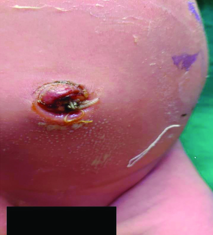
On examination, there was periumbilical erythema and multiple white glistening worms were coming out of umbilicus as shown in [Table/Fig-1]. Baby was afebrile and systemic examination revealed no abnormality.
White glistening worms coming out umbiflicus of neonate.
On investigations, sepsis screen was negative Total Leucocyte Count (TLC)-9600/mL, Polymorphonuclear (PMN)-38/ Leucocyte (L) 50/Monocytes (M) 8/Eosinophils (E) 4/basophils(B) 0, C-reactive Protein (CRP)-4.27 mg/L, Absolute Neutrophil Count (ANC)- 3640/mm3, Procalcitonin-0.3 ng/mL. Ultrasonography showed few echogenic foci in skin and subcutaneous tissue of umbilicus. However, there was no further extension of such foci in deeper tissues and no sinus/tract formation was seen.
Intravenous broad spectrum antibiotics were started after taking sample for blood culture. The umbilical stump was irrigated with normal saline followed by local application of turpentine oil. The worms were then mechanically removed with the help of forceps which vary in size of 7-8 mm. Topical application with turpentine oil, irrigation with normal saline and mechanical removal of larvae was done for next 3 days. Approximately, 30-35 larvae were extracted with the help of forceps during first three days of admission.
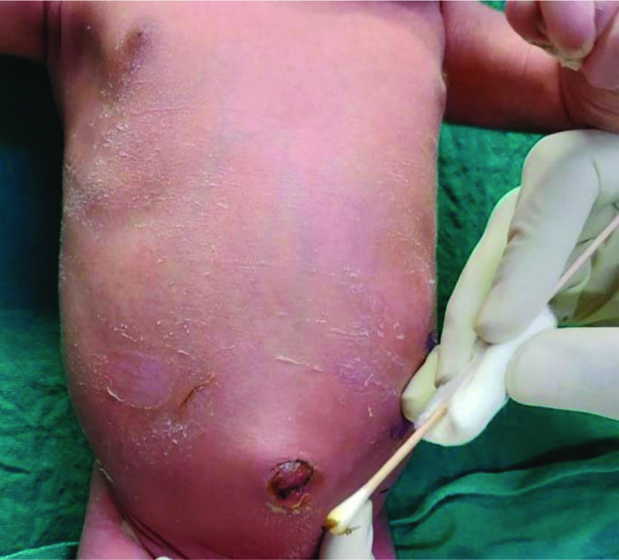
The localised erythema got resolved and there were no larvae visible in the umbilical stump after third day of admission, as depicted in [Table/Fig-2]. Repeat ultrasonography showed no residual larvae or any other abnormality. Blood culture was sterile and antibiotics were discontinued after five days and baby was observed for another 48 hours in hospital. He accepted breast feed well throughout the hospital stay and was discharged in stable condition on day seven of hospitalisation. The parents were counselled about hygiene measures and the need for follow-up. The baby is growing well with no further complaints.
No larvae, No erythema observed on third day.
Discussion
The term myiasis was coined by Reverend Frederick William in 1840. The commonly affected sites by myiasis are ears, nose, mouth, eyes, vagina, intestine and skin. Umbilical myiasis is a type of wound myiasis usually caused by larvae of dipterous fly. The fly lay eggs on umbilicus and dry skin. The eggs then transform into larvae. These larvae subsequently invade the wound and grow rapidly and reach to maturity in 4-8 days [1]. Thus, eggs can be either directly laid by the fly or may transfer from eggs laid on human faeces by contaminated fly legs. The third stage instar larvae is best for species identification [2].
Treatment consists of removal of larvae, cleaning of wound and use of local antiseptics and systemic antibiotic to control any possible associated infection [3]. Local application of irritant substances like turpentine oil, ether, mineral oil, chloroform and phenol etc., causes larvae asphyxia and helps in complete removal [4]. Local irrigation, manipulation and in rare cases surgery may be required. Surgery is used for removal of dead and decayed larvae (not alive) from affected site to prevent secondary infection [4]. Local application of 1% ivermectin has some role in facilitating the larvae [5]. Ivermectin is used against many parasitic infections [6]. It acts by causing the parasite’s cell membrane to increase in permeability resulting in paralysis and death [6]. It has been widely used in cases of animal myiasis successfully [7].
There are occasional case reports of umbilical myiasis [8,9]. In a case report, there were signs of systemic sepsis and umbilical discharge, also revealed growth of Staphylococcus aureus in an eight-day-old neonate [8]. Unlike in the index case, where antibiotics were given on basis of clinical sepsis and blood culture showed no growth. There is another case report of umbilical myiasis where an eight-day-old neonate, akin to index case, delivered in Government Facility developed similar condition due to poor hygiene [10].
Myiasis is generally seen in immunocompromised adult patients, comatose patients and patients on chemotherapeutic drugs [11]. There are other sites also where myiasis is found in children. Aural myiasis has been reported, where worms were found in ears of six paediatric patients. The maggots were removed under light microscopy with a combination of suctioning and forceps. One patient required general anaesthesia for removal of the maggots [12]. There is another case report where a 10-year-old comatose boy that had maggots in his nostrils. The initial presence of blood/mucus around the wounds, the hot/humid climate, and the severe co-morbidities facilitate such condition [11].
Conclusion(s)
Umbilical myiasis is an indicator of poor hygienic condition. Measures should be taken to prevent transmission and breeding of flies. Health education in community and personal hygiene plays a crucial role in prevention of myiasis.
Author Declaration:
Financial or Other Competing Interests: None
Was informed consent obtained from the subjects involved in the study? Yes (from parents)
For any images presented appropriate consent has been obtained from the subjects. Yes (from parents)
Plagiarism Checking Methods: [Jain H et al.]
Plagiarism X-checker: Jun 13, 2020
Manual Googling: Jan 09, 2021
iThenticate Software: Feb 06, 2021 (18%)
[1]. Singh I, Gathwala G, Yadav SP, Wig U, Jakhar KK, Myiasis in children: The Indian perspectiveInt J Pediatr Otorhinolaryngol 1993 25(1-3):127-31.10.1016/0165-5876(93)90045-5 [Google Scholar] [CrossRef]
[2]. Cook GC, Zumla A, Manson’s tropical disease. 21st ed. Medical Acarology and Entomology 2003 PhiladelphiaSaunders (ELST):1727-32. [Google Scholar]
[3]. Gordon PM, Hepburn NC, Williams AE, Bunny MH, Cutaneous myiasis due to Dermatobio hominis: A case report of six casesBr J Dermatol 1995 132(5):811-14.10.1111/j.1365-2133.1995.tb00732.x7772491 [Google Scholar] [CrossRef] [PubMed]
[4]. Kumar SL, Manuel S, John TV, Sivan MP, Extensive gingival myiasis- Diagnosis, treatment, and preventionJ Oral Maxillofac Pathol 2011 15(3):340-43.10.4103/0973-029X.8671522144842 [Google Scholar] [CrossRef] [PubMed]
[5]. Francesconi F, Lupi O, MyiasisClin Microbiol Rev 2012 25(1):79-105.10.1128/CMR.00010-1122232372 [Google Scholar] [CrossRef] [PubMed]
[6]. Juarez M, Cabrera AS, Gonzalez AD, The multitargeted drug ivermectin: From an antiparasitic agent to a repositioned cancer drugAm J Cancer Res 2018 8(2):317-31. [Google Scholar]
[7]. Avni-Magen N, Eshar D, Friedman M, Kirmayer D, Letschert L, Gati I, Retrospective evaluation of a novel sustained-release ivermectin varnish for treatment of wound myiasis in zoo-housed animalsJ Zoo Wildl Med 2018 49(1):201-05.10.1638/2016-0299R2.129517452 [Google Scholar] [CrossRef] [PubMed]
[8]. Singh AK, Nag SS, Mitra P, Roy A, Neonatal umbilical myiasisIndian Dermatol Online J 2015 6(4):312-13.10.4103/2229-5178.16030626225352 [Google Scholar] [CrossRef] [PubMed]
[9]. Kumar V, Gupta SM, Umbilical myiasis in a neonatePaediatr Int Child Health 2012 32(1):58-59.10.1179/1465328111Y.000000002222525451 [Google Scholar] [CrossRef] [PubMed]
[10]. Dey PK, Bhattacharya T, Pal SN, Das S, Pal S, Umbilical myiasis in a newborn: A case reportJournal of College of Medical Sciences-Nepal 2012 8(4):42-45.10.3126/jcmsn.v8i4.8700 [Google Scholar] [CrossRef]
[11]. Hira PR, Assad RM, Okasha G, Ali FM, Iqbal J, Mutawali KE, Myiasis In Kuwait: Nosocomial Infections Caused By Lucilia Sericata and Megaselia ScalarisAm J Trop Med Hyg 2004 70(4):386-89.10.4269/ajtmh.2004.70.38615100451 [Google Scholar] [CrossRef] [PubMed]
[12]. Yuca K, Caksen H, Sakin YF, Yuca SA, Kiris M, Yilmaz H, Aural myiasis in children and literature reviewTohoku J Exp Med 2005 206(2):125-30.10.1620/tjem.206.12515888968 [Google Scholar] [CrossRef] [PubMed]