Genital Wart during Pregnancy: A Rare Case
Vandana Verma1, Pragya Shree2, Shweta Kumar3
1 Assistant Professor, Department of Obstetrics and Gynaecology, UPUMS, Saifai, Etawah, Uttar Pradesh, India.
2 Assistant Professor, Department of Obstetrics and Gynaecology, K.D. Medical College, Mathura, Uttar Pradesh, India.
3 Assistant Professor, Department of Skin and V.D., UPUMS, Saifai, Etawah, Uttar Pradesh, India.
NAME, ADDRESS, E-MAIL ID OF THE CORRESPONDING AUTHOR: Vandana Verma, Flat No. 202, Type 4, Block C, New Campus, UPUMS, Saifai, Etawah, Uttar Pradesh, India.
E-mail: drvandana19@gmail.com
Condyloma Acuminata (CA) or wart is a benign lesion which is caused by Human Papillomavirus (HPV) type-6 or type-11 infection. During pregnancy, condyloma has a tendency to proliferate and may have recurrence. This is because during pregnancy physiological changes takes place to the external genitalia and immunological effects during pregnancy promote HPV replication, and increased vaginal secretions contacting the skin and mucous membranes in pregnancy also lead to proliferation of CA in pregnancy. A 25-year-old primigravida presented to our hospital at 36 weeks pregnancy with extensive genital warts. These lesions regressed itself one month postpartum and remained only on vulva. Podophyllum resin application was planned for remaining lesions two months postpartum. The few treatments that have been tested and recommended for use in pregnancy are Bi and Tri Chloro Acetic Acid (BCA/TCA) application, cryotherapy, electrocautery and surgical excision, including laser treatment.
Condyloma acuminata, Human papillomavirus, Podophyllum resin, Warts in pregnancy
Case Report
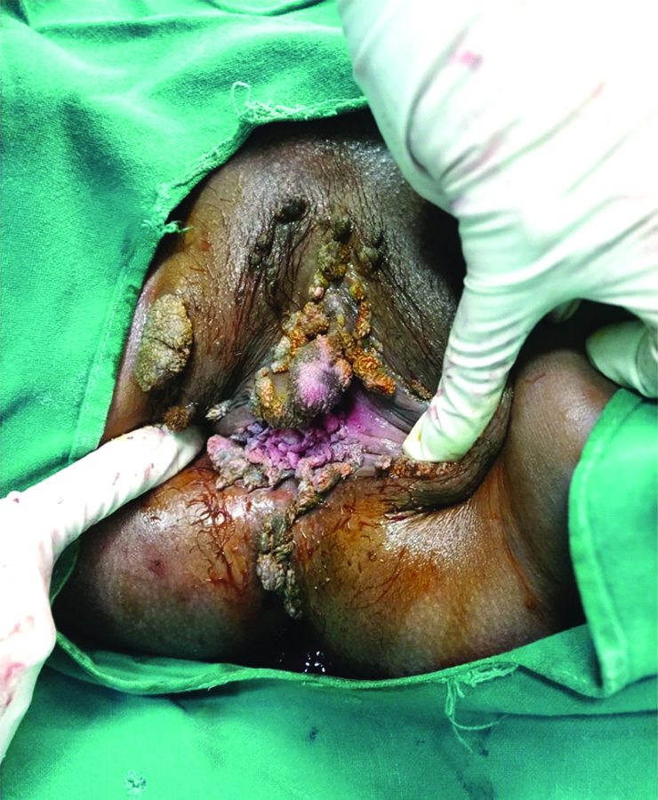
A 25-years-old primigravida presented to the hospital at 36 weeks pregnancy with extensive cauliflower like growth on genitalia. This patient also had history of epilepsy and was on multiple antiepileptic drugs since 13 years of age. Patient’s married life was two years and she had very small pea size lesion around labia majora since one year and after seven months of pregnancy, this lesion gradually became extensive and involved whole vagina and vulva. There was no history of pain itching or bleeding in these lesions. Husband was not having any history of same lesions and he does not have history of sexual contact other than spouse before or after marriage and there was no such history in patient also. There was no history of trauma, diabetes mellitus, oral ulcers, urinary symptoms, vaginal itching or discharge per vaginum, weight loss or loss of appetite. General and systemic examinations were within normal limits. Obstetrical examination findings were corresponding to period of gestation. On genital examination irregular warts like growth was seen involving vulva and vagina (multiple lesions on vulva and vagina largest was 4×5 cm), firm in consistency without signs of inflammation on vulva and multiple wart like growth in vagina (around 0.5 cm in size) [Table/Fig-1].
Warts one month postpartum.
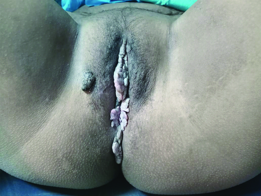
Patient did not have any antenatal investigations so all investigation done and reports were in normal limits. Venereal Disease Research Laboratory (VDRL) and HIV status were non-reactive, HPV IgG was positive and patient was planned for elective caesarean section at 39 weeks because of extensive lesion in vagina. Neonate was alive and healthy. Baby was not having any similar lesion on body. These genital lesions were confusing with carcinoma due to their rapid growth during pregnancy. So, biopsy was needed to confirm the diagnosis. After delivery biopsy of genital lesion was done which showed stratified squamous epithelium lined tissue, hyperkeratosis, parakeratosis, acanthosis, papillomatosis with prominent granular layer, parabasal hyperplasia which confirmed the diagnosis of condyloma accuminata. As the warts regresses on its own after delivery and patient had no complaints other than visible abnormal growth in vulva so no treatment was given during pregnancy. These lesions regressed itself one month postpartum and remained only on vulva [Table/Fig-2]. Patient was reassured and counselled for follow-up. Podophyllum resin application was planned for remaining lesions after one month because patient has not given consent for electrodessication.
Discussion
Condyloma Acuminata (CA) or wart is a benign lesion which is caused by infection with HPV type-6 or type-11. The lesions of CA are most commonly present on genital skin, vaginal mucosa, perianal area or anal mucosa [1]. The lesion of CA has a tendency of proliferation and recurrence during pregnancy. This is because of physiological changes to the external genitalia and immunological effects that promote HPV replication during pregnancy. Increased vaginal secretions coming in contact of genital skin and mucous membranes in pregnancy also causes proliferation of genital lesion during pregnancy [2]. HPV infections are manifested within two years of infection in 90% cases, but long latent period of years may be possible for CA so first lesion of CA or its recurrence may occur after years of infection. Vertical transmission of HPV can occur periconceptional, during pregnancy or during delivery when foetus passes through birth canal by infected secretions [3]. In pregnant women caesarean delivery can be adopted for protection of neonate against the transmission of neonatal herpes who present in late pregnancy with genital warts [4].
Genital warts are characterised by flat, popular, irregular or pedunculated growths on the genital or anal skin and/or mucosa and are mostly asymptomatic, but they may present with itching, friable painful growth. Genital warts are diagnosed by visual inspection but to confirm the diagnosis, histopathological examination of tissue should be done. HPV nucleic acid tests are not recommended for routine diagnosis or management of visible genital warts [4].
In present case report, it was difficult to diagnose condyloma by appearance due to its extensive nature and rapid growth during pregnancy. Similar to this case Devi LT and Pathania K also reported in case report that diagnosis of CA may be difficult during pregnancy [4].
In this case report, multiple warty lesions were present in vagina and vulva so podophyllum resin application was planned for treatment whereas case reported by Nigam A and Mishra A, and Grimaldi L et al., also showed the atypical presentation of CA during pregnancy in which large growth was present involving vulva so excision of growth was done [5,6]. In present case, there were multiple lesions whereas Yavuzcan A et al., reported a case of giant periurethral condyloma with 28 weeks pregnancy and in this case excision of lesion and histopathology was done [7].
Cases of anogenital warts present as a fast growing lesion during pregnancy due to hormonal changes and immunosuppression during pregnancy and have decreased tolerance and response to treatment [8]. BCA/TCA application cryotherapy, electrocautery and surgical excision, including laser treatment are recommended during pregnancy but they have reduced response and there are chances of miscarriage and preterm labour during treatment [8]. It is uncertain that the treatment reduces transmission of HPV and there is also possibility of spontaneous resolution, so there is another acceptable alternative for some persons is to wait for spontaneous resolution before any treatment [4].
Conclusion(s)
Human Papillomavirus (HPV) infection presentation can range from asymptomatic to cervical cancer. Small genital wart lesion may become extensive and cumbersome during pregnancy and again regress after delivery in due course of time. There may be diagnostic dilemma during pregnancy due to its extensiveness and atypical presentation as in this case. Treatment does not reduce the transmission rate and there are chances of spontaneous regression so treatment can only be given if patient has discomfort, burning or itching in genital area or any other complaint.
Author Declaration:
Financial or Other Competing Interests: None
Was informed consent obtained from the subjects involved in the study? Yes
For any images presented appropriate consent has been obtained from the subjects. Yes
Plagiarism Checking Methods: [Jain H et al.]
Plagiarism X-checker: Oct 31, 2020
Manual Googling: Feb 11, 2021
iThenticate Software: Mar 08, 2021 (12%)
[1]. Song G, Zhou X, Wu Y, A pregnant woman with condyloma acuminatum on the vaginal orifice, areola, groin, and umbilicusIndian Journal of Pathology and Microbiology 2019 62(2):310-12.10.4103/IJPM.IJPM_518_1830971564 [Google Scholar] [CrossRef] [PubMed]
[2]. Cohen E, Levy A, Holcberg G, Wiznitzer A, Mazor M, Sheiner E, Perinatal outcomes in condyloma acuminata pregnanciesArch Gynecol Obstet 2011 283(6):1269-73.Epub 2010 Jun 1710.1007/s00404-010-1558-220556405 [Google Scholar] [CrossRef] [PubMed]
[3]. Jach R, Galarowicz B, Huras H, Pawlik D, Basta T, Streb J, Vertical transmission of HPV in pregnancy. A prospective clinical study of HPV-positive pregnant womenGinekol Pol 2014 85(9):672-76.10.17772/gp/179025322538 [Google Scholar] [CrossRef] [PubMed]
[4]. Devi LT, Pathania K, Pregnancy with HPV associated Viral WartsMed J Armed Forces India 2011 65(3):272-73.10.1016/S0377-1237(09)80024-5 [Google Scholar] [CrossRef]
[5]. Nigam A, Mishra A, Condyloma acuminatum: Atypical presentation during pregnancyInternational Journal of STD & AIDS 2011 22(9):534-35.10.1258/ijsa.2009.00911421890557 [Google Scholar] [CrossRef] [PubMed]
[6]. Grimaldi L, Cuomo R, Bilenchi R, Roviello F, Nisi G, Brandi C, A case report of giant genital wartsJ Siena Acad Sci [Internet] 2014[cited 2021Jan.29] 6(1)Available from: https://pagepressjournals.org/index.php/jsas/article/view/jsas.2014.484410.4081/jsas.2014.4844 [Google Scholar] [CrossRef]
[7]. Yavuzcan A, Çağlar M, Turan H, Tekin A, Topuz S, Yavuzcan G, The treatment of giant periurethral condyloma in pregnancy using ultrasonic thermal scalpel: A case report and a new single session treatment optionCase Reports in Obstetrics and Gynaecology 2015 2015:79241210.1155/2015/79241225648983 [Google Scholar] [CrossRef] [PubMed]
[8]. Yang LJ, Zhu DN, Dang YL, Zhao X, Treatment of condyloma acuminata in pregnant women with cryotherapy combined with proanthocyanidins: Outcome and safetyExp Ther Med 2016 11(6):2391-94.10.3892/etm.2016.320727284325 [Google Scholar] [CrossRef] [PubMed]