Lyme disease is a zoonotic disease spread by the bite of Ixodes ticks. These ticks are known to be found in wooded or grassy areas. The disease manifestations can be divided into early Lyme disease and late Lyme disease. The manifestations of late Lyme disease may include arthritis, cranial nerve palsy, short-term memory deficits and Lyme carditis. The disease is diagnosed by a two-step process of Enzyme immunoassay followed by Western blot Test. Disseminated disease is treated with Intravenous (IV) ceftriaxone, cefotaxime or penicillin G, or a combination of oral and IV regimens or in some cases, only oral drugs for up to 28 days. Lyme disease is endemic in temperate regions, especially in America. However, over the years the disease has been reported from various countries of Asia including in India, where there have been sporadic cases. Hereby, the author presents three paediatric cases with varied presentations. The neurological symptoms ranged from Lower Motor Neuron (LMN) facial nerve palsy to acute encephalitis. One patient also had non-erosive arthritis.
Introduction
Lyme disease is a multisystem disease that is caused by the spirochete Borrelia burgdorferi [1]. It was first recognised in children with arthritis in Lyme county of Connecticut, in the United States of America, in 1975. The three common species of Borrelia causing Lyme disease are B. burgdorferi, B. garinii and B. afzelli. Deers serve as a reservoir of this tick, and it is spread by the bite of hard ticks of the Ixodes genus with Ixodes persulcatus being the most common vector in Asia. Though, cases are more often reported from temperate regions, the disease has also been reported from India [2-15]. A case series of Neuroborreliosis has been presented below.
Traditional teaching has considered Lyme disease to be a disease of western nations. However, in the past few years, there have been an increasing number of cases reported from India too. This is a disease that we need to be aware of as an emerging zoonotic disease.
Case Series
Case 1
A 10-year-old male child, presented with fever for six days, followed by acute-onset right LMN type of facial nerve palsy along with cerebellar signs in the form of scanning speech, ataxia, intention tremor, dysdiadochokinesia, past pointing, headache and vomiting for one day without seizures, altered sensorium and other focal deficit or meningeal signs. The child was investigated for rhombencephalitis (brainstem encephalitis) in view of fever with LMN cranial nerve and cerebellar signs. Systemic examination was normal, with no rashes. The clinical possibilities of infectious disease particularly those treatable like Herpes, Listeria, Salmonella and Lyme’s disease were considered. Demyelinating and neoplastic aetiologies were also taken under consideration.
The haemogram showed haemoglobin value of 10.2 g/dL, total leucocyte count of 10,800/mm3 with the differential of neutrophil 87%, lymphocyte 10%, eosinophil 2%, monocyte 1%, platelet count of 3,54,000/mm3, erythrocyte sedimentation rate of 8 mm/1st hour with microcytic hypochromic anaemia on peripheral smear. Blood culture had no growth; serum widal test was negative.
Lumbar puncture was done after ruling out papilloedema on fundus examination. Cerebrospinal Fluid (CSF) analysis showed eight mononuclear cells, sugar of 40 mg/dL against blood sugar of 60 mg%, protein 20 mg/dL. The gram staining showed no pus cells and no organism was seen. Cerebrospinal Fluid (CSF) culture had no growth. Herpes Simplex Virus (HSV)- Polymerase Chain Reaction (PCR) was negative.
Magnetic Resonance Imaging (MRI) brain showed multiple T2/FLAIR hyperintense lesions without perilesional oedema in right parietal, left occipital and pons area suggestive of acute demyelinating encephalomyelitis versus brainstem encephalitis. Serum IgM for Lyme’s disease was positive thus, clinching the diagnosis of Lyme’s Rhombencephalitis.
The child was empirically, initially started on a combination of ceftriaxone, ampicillin and acyclovir. Ampicillin was stopped after five days when culture was negative and acyclovir after seven days when HSV-PCR came out to be negative. Ceftriaxone was continued for 14 days followed by two weeks of oral doxycycline in view of positive Lyme serology. The child improved after seven days of treatment and is well after eight months of follow-up.
Case 2
An eight-year-old girl was referred from a private hospital in view of fever for five days, followed five days later by altered sensorium, status epilepticus and extrapyramidal movements in the form of orofacial dyskinesia and choreoathetosis. A clinical possibility of acute encephalitis syndrome versus autoimmune encephalitis versus acute disseminated encephalomyelitis was presumed. MRI brain showed bilateral hippocampal oedema. Complete haemogram, blood culture, serum widal, CSF examination including PCR for HSV and Anti-N-methyl-d-aspartate (NMDA) antibodies, liver and kidney function tests were unremarkable. She was managed with ceftriaxone, acyclovir for 10 days along with antiepileptic and antidystonia measures and IV immunoglobulin 2 g/Kg with recovery. Four weeks later, she presented with the left LMN facial nerve palsy which was treated with steroids. One month later, she developed non-erosive seronegative pauciarticular arthritis. This prompted serology for Lyme’s disease that came out to be positive. She was admitted and treated with ceftriaxone for 14 days at a dose of 100 mg/kg/day followed by doxycycline at a dose of 5 mg/kg/day for 14 days. The patient is now well after three months of follow-up.
Case 3
A nine-year-old boy came to the Outpatient Department with inability to close the right, eye and with facial deviation. On examination, he was found to have right LMN facial nerve palsy. He was empirically treated with prednisolone at 1 mg/kg/day for one week followed by tapering and oral acyclovir at 80 mg/kg/day. His anti-Lyme’s IgM antibodies were positive. He was also treated with ceftriaxone for 14 days followed by doxycycline for 14 days. His facial nerve weakness had resolved gradually after one month, and does not have any new concerns after two months of follow-up. The details of all three cases are summarised in [Table/Fig-1].
| Cases | Age/gender | Clinical features | Investigations | Outcome |
|---|
| Case 1 | 10 years/Male | Fever, acute-onset right Lower Motor Neuron (LMN) type of facial nerve palsy, cerebellar signs, headache and vomiting. | Cerebrospinal Fluid (CSF) analysis: eight mononuclear cells, Sugar 40 mg/dL against blood sugar of 60 mg%, protein 20 mg/dL. Gram staining: no pus cells, no organism seen. CSF culture: no growth.Herpes Simplex Virus (HSV)- PCR negative.Magnetic Resonance Imaging (MRI) Brain: multiple T2/FLAIR hyperintense lesions without perilesional oedema in right parietal, left occipital and pons areaSerum IgM for Lyme’s disease was positive | Ceftriaxone for 14 days followed by two weeks of oral doxycycline, patient is well after eight months of follow-up. |
| Case 2 | Eight years/Female | Fever, altered sensorium, status epilepticus and extrapyramidal movements. Four weeks later: left Lower Motor Neuron (LMN) facial nerve palsy. One month later: nonerosive seronegative pauciarticular arthritis. | MRI brain: bilateral hippocampal oedema.One month later: serology for Lyme’s disease positive. | Managed with ceftriaxone, acyclovir, antiepileptic and antidystonia measures and Intravenous (IV) immunoglobulin 2 g/kg with recovery.Four weeks later: left Lower Motor Neuron (LMN) facial nerve palsy treated with steroids.One month later: Arthritis treated with ceftriaxone for 14 days followed by doxycycline for 14 days. Well after three months of follow-up. |
| Case 3 | Nine years/Male | Right Lower Motor Neuron (LMN) facial nerve palsy. | Anti-Lyme’s IgM antibodies positive. | Initially empirically treated with prednisolone and acyclovir. Treated with ceftriaxone for 14 days followed by doxycycline for 14 days. His facial nerve weakness had resolved, and was normal on two months of follow-up. |
Discussion
For transmission of Borrelia, the tick needs to be attached to the host for atleast 36-48 hours. This is possible because the transmission is by the bite of the nymphal stage of the tick which is less conspicuous than an adult tick [1]. After the tick separates from the host, there is a small nodule at the site of its attachment which is seen in all tick bite, and is not indicative of the disease [1]. Three to 30 days after the bite, the characteristic skin lesion of the disease occurs. This is called Erythema migrans, which is a bull’s eye-shaped erythematous rash at the site of tick bite. This occurs in 70-80% of infected individuals and may not be well-visualised in dark skinned individuals. The spirochete can be isolated from this skin lesion, though this is not a diagnostic method commonly employed. This stage of the disease is called Early Lyme Disease [1]. Following this the organism spreads and may manifest with arthritis, cranial nerve palsies (the most common being LMN facial nerve palsy), neuroretinitis, meningeal irritation, pain and paraesthesias, short-term memory deficits and Lyme carditis. These symptoms occur days to months after the tick bite. This stage is called Late Lyme Disease [1].
Patial RK et al., reported a 15-year-old male from Shimla, Himachal Pradesh, in whom they isolated the spirochete from blood [2]. Praharaj AK et al., serologically tested 500 individuals from north eastern region of India for prevalence of antibodies against Borrelia burgdorferi [3]. These individuals included service personnel and their families. They found 13% prevalence of antibodies in females and individuals in the age group 16-30 years and were more frequently positive. People hailing from Arunachal Pradesh had the highest seroprevalence. They suggested that this was because females worked more and were more frequently exposed to forest environments and the young active population was also seen to be more involved.
Babu K and Murthy PR, reported the case of a 45-year-old female from Nagarhole forest in South India who presented with neuroretinitis as the sole disease manifestation of Lyme disease [4]. Jairath V et al., described five patients from Haryana, who had skin manifestations of Lyme disease and had a serological diagnosis [5]. Shenthar J et al., from Bengaluru treated a 43-year-old male with Lyme carditis presenting with complete heart block and ventricular tachycardia [6]. The heart block was completely reversed with antibiotic therapy.
There was an outbreak of Lyme disease in the Wayanad district of Kerala, in the vicinity of the Wayanad Wildlife Sanctuary in early 2013. Five patients were affected and there was one fatality. The deer population in the nearby forest was thought to have contributed [7]. Authors had also reported two siblings who had presented with altered mental status and multiple cranial nerve palsies who were found to be serologically positive and who responded to appropriate antibiotic treatment [8].
Sharma A et al., reported a 10-year-old boy from Himachal Pradesh who presented erythema chronicum migrans [9]. There was history of visit to a nearby forest a few days prior to the rash. Serological testing for Borrelia burgdorferi revealed an elevated level of IgM antibody. Patient responded to treatment with oral doxycycline.
Sinha P et al., described a 26-year-old male from Maharashtra who presented with erythema marginatum and fever with headache [15]. There was no history of insect bite or history of travel. The serology for Borrelia showed positive Borrelia IgM. The CSF analysis was suggestive of aseptic meningitis. He responded to IV ceftriaxone given for fourteen days.
The other two common tick-borne diseases in India are Kyasanur Forest Disease and Crimean Congo Haemorrhagic Fever. Of these Kyasanur forest disease had a recent outbreak (2014-2015) in the same Wayanad district of Kerala and most of the patients of this outbreak were based just 1-2 km away from the region of the Lyme disease outbreak [10].
Guliani BP et al., treated a 25-year-old female who visited hilly areas in the Himalayas in North India [11]. She presented with sudden unilateral painless blurring of vision due to neuroretinitis and had positive serology for Lyme disease. Tevatia P et al., reported a young man from Dehradun who had multisystem involvement of Lyme disease [12]. He presented with progressive decrease in vision, headache, quadriparesis, seizures, skin and pulmonary lesions, lymphadenopathy and hypocellular marrow.
Baveja S et al., described a paediatric patient with erythema chronicum migrans and acrodermatitis chronica atrophicans as a presentation of Lyme disease in a nine-year-old girl [13]. Farmania R et al., reported a seven-year-old girl with chronic meningitis and persistent hypoglycorrhachia and positive CSF IgM for Borrelia [14].
A study of the tick population in India has demonstrated ticks of the Ixodes genus in several mammals viz., buffaloes and cattle in Arunachal Pradesh [16], and in the Western Ghats [17] and the Himalayan regions of India [18].
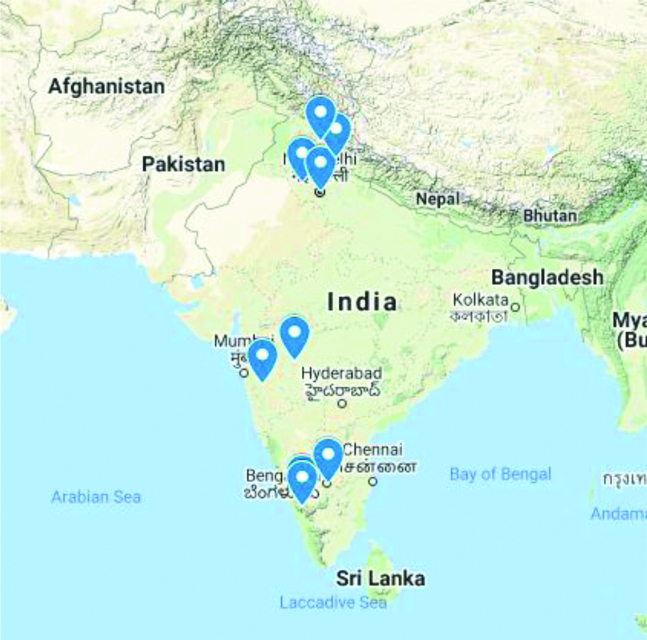
As it is evident from the map in [Table/Fig-2], there is clustering of cases in some states of North India, where the vegetation is mainly grassy, and in wooded areas of South India where human beings live very close to wild animals. Seropostivity was seen in Arunachal Pradesh [3], which suggests that there are as yet unreported cases in that area.
Map showing distribution of cases of Lyme Disease.
Conclusion(s)
Since both the vector and the organism have been found in India, we are possibly sitting on the tip of the iceberg, and conditions are rife for the spread of Lyme disease in India. A high index of suspicion for the disease to treat it early and appropriately is needful so as to prevent development of late complications.
[1]. https://www.cdc.gov/lyme/index.html accessed 24/1/2021 [Google Scholar]
[2]. Patial RK, Kashyap S, Bansal SK, Sood A, Lyme disease in a Shimla boyJ Asso Physicians India 1990 38(7):503-04.10.1016/S0377-1237(08)80140-2 [Google Scholar] [CrossRef]
[3]. Praharaj AK, Jetley S, Kalghatgi AT, Seroprevalence of Borrelia burgdorferi in North Eastern IndiaMed J Armed Forces India 2008 64(1):26-28. [Google Scholar]
[4]. Babu K, Murthy PR, Neuroretinitis as a manifestation of Lyme disease in South India: A case reportOcul Immunol Inflamm 2010 18(2):97-98.10.3109/0927394090335973320370334 [Google Scholar] [CrossRef] [PubMed]
[5]. Jairath V, Sehrawal M, Jindal N, Jain VK, Aggarwal P, Lyme disease in Haryana, IndiaInd J of Dermatol Venereol Leprology 2014 80(4):320-23.10.4103/0378-6323.13689425035356 [Google Scholar] [CrossRef] [PubMed]
[6]. Shenthar J, Shetty SB, Krishnamurthy D, Diagnosis not to be missed: Lyme carditis, rare but reversible cause of complete atrioventricular blockIndian Heart J 2014 66(6):723-26.10.1016/j.ihj.2014.11.00425634416 [Google Scholar] [CrossRef] [PubMed]
[7]. https://timesofindia.indiatimes.com/city/kozhikode/Lyme-disease-outbreak-in-Wayanad/articleshow/18758675.cms Accessed 24/1/21 [Google Scholar]
[8]. Bhatt M, Sehgal R, Gupta R, Mohapatra JN, Sharma S, Aggarwal KC, Two brothers with multiple cranial nerve palsiesIndian J Pediatr 2015 82(4):383-84.10.1007/s12098-014-1605-225408268 [Google Scholar] [CrossRef] [PubMed]
[9]. Sharma A, Guleria S, Sharma R, Sharma A, Lyme disease: A case report with typical and atypical lesionsIndian Dermatol Online J 2017 8(2):124-27.10.4103/2229-5178.20227128405553 [Google Scholar] [CrossRef] [PubMed]
[10]. Sadanandane C, Elango A, Marja N, Sasidharan PV, Raju KHK, Jambulingam P, An outbreak of Kyasanur forest disease in the Wayanad and Malappuram districts of Kerala, IndiaTicks Tick Borne Dis 2017 8(1):25-30.10.1016/j.ttbdis.2016.09.01027692988 [Google Scholar] [CrossRef] [PubMed]
[11]. Guliani BP, Kumar S, Chawla N, Mehta A, Neuroretinitis as presenting and the only presentation of Lyme disease: Diagnosis and managementIndian J Ophthalmol 2017 65(3):250-52.10.4103/ijo.IJO_151_1728440258 [Google Scholar] [CrossRef] [PubMed]
[12]. Tevatia P, Ahmad S, Gupta N, Shirazi N, Lyme disease in north India: A case for concernTrop Doct 2018 48(4):352-55.10.1177/004947551878955230124129 [Google Scholar] [CrossRef] [PubMed]
[13]. Baveja S, Oberoi B, Vashisht D, Das P, Lyme Diseae- A report of Atypical Cutaneous SequelaeIndian Dermatol Online J 2019 10(3):336-37.10.4103/idoj.IDOJ_294_1831149589 [Google Scholar] [CrossRef] [PubMed]
[14]. Farmania R, Jauhari P, Chakrabarty B, Gulati S, Chronic meningitis with persistent hypoglycorrhachia: An unusual presentation of Lyme’s diseaseNeurol India 2019 67(2):563-65.10.4103/0028-3886.25803931085878 [Google Scholar] [CrossRef] [PubMed]
[15]. Sinha P, Oberoi B, Sirohi YS, Sood A, Bhattacharjee S, A case report of early disseminated lyme diseaseNeurol India 2020 68(4):916-18.10.4103/0028-3886.29347632859843 [Google Scholar] [CrossRef] [PubMed]
[16]. Ronghang B, Roy B, Status of tick infections among semi-wild cattle in Arunachal Pradesh, IndiaAnn Parasitol 2016 62(2):131-38. [Google Scholar]
[17]. Sadanandane C, Gokhale MD, Elango A, Yadav P, Mourya DT, Jambulingam P, Prevalence and spatial distribution of Ixodid tick population in the forest fringes of Western Ghats reported with human cases of Kyasanur forest disease and monkey deaths in South IndiaExp Appl Acarol 2018 75(1):135-42.10.1007/s10493-018-0223-529594846 [Google Scholar] [CrossRef] [PubMed]
[18]. Geevarghese G, Fernandes S, Kulkarni SM, A checklist of India ticks (Acari: Ixodoidea)Ind J Animal Sci 1997 67:17-25. [Google Scholar]