Sealants have proved to be one of the easiest methods of caries prevention in young permanent teeth, the placement of which is very technique sensitive. The preventive function of pit and fissure sealant is accomplished mainly by the adhesion of the material to the enamel surface. In an attempt to improve and increase adhesion, acid etching has been introduced by Buonocore MG [1]. However, Hobson RS et al., found that the surface quality of enamel etching with phosphoric acid (H3PO4) was not encountered over the total adhesion surface [2]. Also, phosphoric acid works on the mineralised portion of the enamel only and does not remove the organic content that is less than 1%, but can affect the etching pattern of the enamel. For this reason, various invasive and non-invasive techniques as sodium hypochlorite deproteinisation and application of intermediate bonding agent have been suggested.
Sodium hypochlorite solution has been used in endodontics for its antimicrobial properties and ability to dissolve the organic material from the root canal space without damaging intact dentinal tissues [3]. Rishika et al., reported that the non-invasive method of treatment of the enamel surface by deproteinising with 5.25% sodium hypochlorite for 60 seconds prior to etching has proved to be a promising method for improving the retention [4]. The use of the bonding agent may change the viscosity of the material making it to flow better into the fissures and acid etched surface [5]. According to Askarizadeh N et al., the use of dentin bonding agents between the tooth and fissure sealant can be beneficial for reducing microleakage when there is contamination of the enamel [6]. Thus, this study was aimed to assess the retention and microleakage in pit and fissure sealants following enamel pre-etching with sodium hypochlorite deproteinisation and bonding agent.
Materials and Methods
This in vitro experimental study, approved by Institutional Review Board (IRC Ref no: IRC/18/19), was conducted in Department of Paediatric and Preventive Dentistry at Annoor Dental College, Muvattupuzha, Kerala and Nanotechnology lab at Amrita Institute of Medical Science from 6th November 2019 to 2nd March 2020.
Inclusion criteria: Freshly extracted (orthodontic purpose), intact, caries free permanent maxillary and mandibular 20 premolars and 20 third molars were collected.
Exclusion criteria: Teeth with fractures or cracks, occlusal caries, malformations, enamel hypoplasia, or erosions were excluded from the study.
The selected teeth were cleaned with soft brush and distilled water to remove debris and blood stains and preserved in a glass container containing distilled water at room temperature (37°C) until study procedure [7].
Sample size calculation: Sample Size was calculated using the formula [8]:
The calculated Sample size is:
Input: Effect size f = 1.0511021
α err prob = 0.05
Power (1-β err prob) = 0.95
Number of groups = 4
Total sample size = 20
Actual power = 0.9500000
Sample Preparation
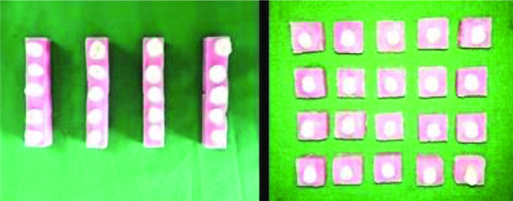
All teeth were mounted on cubic acrylic blocks after thorough washing [Table/Fig-1]. Prepared 20 third molars (a) and 20 premolar teeth (b) were divided into 4 groups containing 5 teeth in each group by random sampling method. Premolars were taken for assessing microleakage and molars were taken for assessing the rate of retention.
Shows molar and premolar samples mounted on acrylic blocks.
Retention subgroups: Group 1a, Group 2a, Group 3a, Group 4a
Microleakage subgroups: Group 1b, Group 2b, Group 3b, Group 4b
Sealant Placement
Pits and fissures of all samples were cleaned with a brush and pumice/water using a low-speed hand piece for approximately 10 seconds. After this, following procedures were carried out.
Group 1: Application of 37% Phosphoric acid and pit and fissure sealant
The enamel was etched with 37% phosphoric acid gel (d tech R) for 15 seconds, washed and thoroughly dried.
Group 2: Application of 5.25% Sodium hypochlorite, 37% Phosphoric acid and pit and fissure sealant
A 5.25% NaOCl was applied on the occlusal surface using sterile cotton for 60 seconds, washed with sterile distilled water for 10 seconds, and completely dried with oil-free compressed air spray. The enamel was then etched with 37% phosphoric acid gel (d tech R) for 15 seconds, rinsed and thoroughly dried.
Group 3: Application of 37% Phosphoric acid, bonding agent and pit and fissure sealant
The enamel was etched with 37% phosphoric acid gel (d tech R) for 15 seconds, washed and thoroughly dried followed by application of bonding agent by using applicator tip.
Group 4: Application of 5.25% Sodium hypochlorite, 37% Phosphoric acid, bonding agent and pit and fissure sealant
A 5.25% NaOCl was rubbed on the occlusal surface using sterile cotton for 60 seconds, washed with sterile distilled water for 10 seconds, and completely dried with oil-free compressed air spray. Then, the enamel was etched with 37% phosphoric acid gel for 15 seconds, washed and thoroughly dried followed by application of bonding agent (Tetric n bond, ivoclar, vivadent).
All the samples were sealed with a sealant (Clinpro TM 3M ESPE, Mexico) using a syringe style tip in compliance with the manufacturer’s instructions. The sealant was placed into the etched fissures by using the tip of the probe and obvious air voids were simultaneously removed. The sealant was then light cured for 20 seconds by placing the tip of the curing light (GUILINS WOODPECKER) perpendicularly to the sealant surface and 1 mm away.
Samples were then stored in sterile distilled water for 24 hours at room temperature. Afterwards, to simulate the oral environment the samples were subjected to thermocycling 500 times in artificial saliva filled baths at 4°C, 37°C, and 57°C with an exposure time of 30 seconds each. Pieces of each molar were then carefully coated with two layers of acid-resistant nail varnish over the axial surfaces of each molar, leaving 1 mm free around the sealant borders. Specimens were dipped for 24 hours in Rhodamine B (Sigma- Aldrich, Mexico) dye solution (0.02 mg in 200 mL of sterile distilled water) and were thoroughly rinsed under tap water for 1 minute to remove excess dye from the enamel surface. The varnish was then scraped, and each specimen was sectioned longitudinally in a buccolingual direction through the sealant using a water-cooled diamond disc (Isomet 5000, Buehler, Unear Precision Saw). The sections were kept dry until microscopic examination [7].
Outcome Measurement
For retention
After thermocycling, retention was assessed by one pre calibrated blinded examiner by passing a probe of diameter 0.5 mm along the margins of the sealant to verify integrity, loss of continuity or failure based on Simonsen’s criteria-1989 [9] which Indicates complete retention, partial loss and complete loss.
For Microleakage
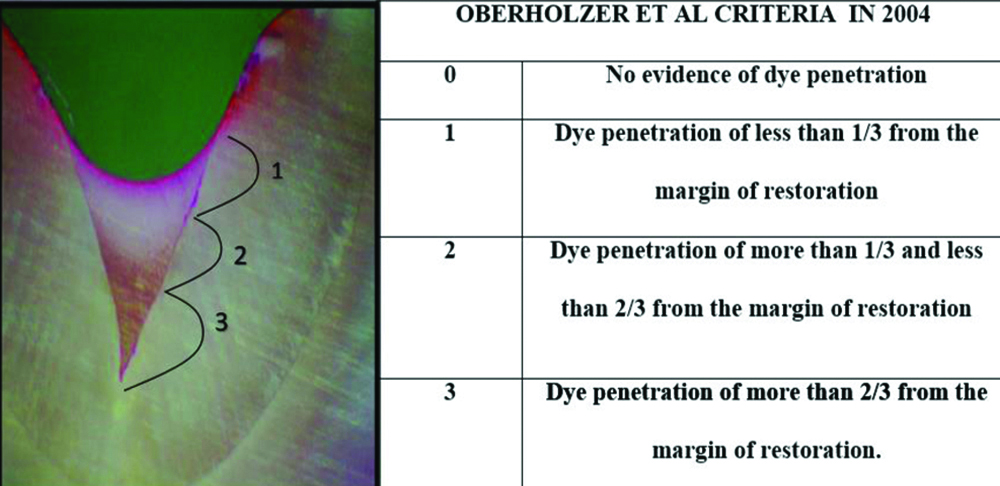
Stereomicroscope (Leica® DMI 4000B, Germany) was used to examine the prepared sections from each specimen. The degree of penetration of Rhodamine B on photographs (10x magnification; λex 553 nm, λem 627 nm) was observed to determine the rate of microleakage. Microleakage was assessed using the criteria by Oberholzer TG et al., in 2004 [10] [Table/Fig-2].
Shows penetration of dye into various levels of fissure [10].
Statistical Analysis
Data was analysed using Statistical Package for the Social Science (SPSS) Windows version 24.0. Mann-Whitney U test and Chi-square test were used to compare the values of microleakage and retention, respectively. The p-value <0.05 considered as significant.
Results
The results showed 100% retention for teeth in Group 2a, Group 3a, Group 4a, while 3 teeth from Group 1a showed partial loss of sealant [Table/Fig-3]. The differences between the groups for retention was statistically significant (p=0.014).
Rate of Retention in each group (Group 1a: acid etching only, Group 2a-deproteinised prior to acid etching, Group 3a-acid etching followed by bonding and Group 4a-deproteinisation followed by etching and bonding).
| Study groups | Retention | Chi-square test |
|---|
| Complete retention | Partial loss | Total loss |
|---|
| Group 1a | 2 | 3 | 0 | χ2=10.558p=0.014(significant) |
| Group 2a | 5 | 0 | 0 |
| Group 3a | 5 | 0 | 0 |
| Group 4a | 5 | 0 | 0 |
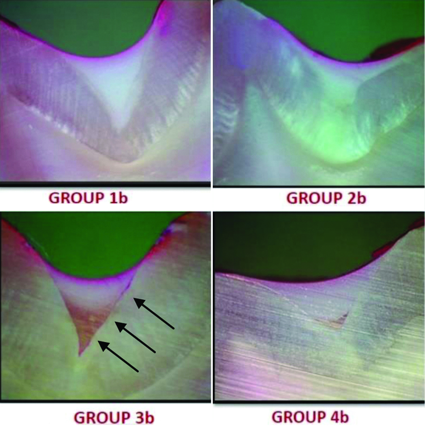
When microleakage was compared among groups, Group 3b showed most microleakage and group 4b showed the least microleakage, which was statistically significant (p=0.009). Statistically significant difference was found in microleakage between group 2b (deproteinisation prior to acid etching) and group 3b (acid etching followed by bonding) with p=0.013 [Table/Fig-4,5].
Rate of microleakage in each group (Group 1b: acid etching only, Group 2b-deproteinised prior to acid etching, Group 3b-acid etching followed by bonding and Group 4b-deproteinisation followed by etching and bonding) (n=20).
| Study groups | Microleakage | Mann-Whitney U test |
|---|
| Grade 0 | Grade 1 | Grade 2 | Grade 3 |
|---|
| Group 1b | 1 | 4 | 0 | 0 | 1b-2b: p=0.2211b-3b: p=0.0181b-4b: p=0.0722b-3b: p=0.0132b-4b: p=0.5133b-4b: p=0.009 |
| Group 2b | 3 | 2 | 0 | 0 |
| Group 3b | 0 | 1 | 2 | 2 |
| Group 4b | 4 | 1 | 0 | 0 |
p-value <0.05 considered significant
Shows a representative image of the microleakage in different groups.
Discussion
Although, placement of pit and fissure sealants causes reduction in occlusal caries, but the effectiveness of sealant may be associated with certain technical issues during its placement that includes tissue management and salivary contamination [11,12]. Problems in the application of sealant can cause microleakage or limited or complete loss, which results in the unsuccessful function of sealant with the rate of 5% to 10% a year [13]. Success of the sealant depends on optimal conditions under which the sealant is applied. The efficacy of sealant in the prevention of caries has been attributed with the degree and duration of the retention of sealant [1].
In 1955, Buonocore MG introduced a new technique of acid etching to enhance the adhesion of restorative material to the enamel [1]. Dental enamel composed of 96% of inorganic matter and organic matter is less than 1%, out of which less than half part contains protein [14]. Phosphoric acid acts mainly on the mineralised part i.e., inorganic portion of the enamel surface but it does not remove the organic material. Due to this outer organic layer, phosphoric acid is unable to etch the surface of enamel efficiently, that results in variable pattern and a distorted surface of enamel for bonding. So, it is necessary to remove the organic part from the enamel surface to refine the quality of pattern of etching, which gave rise to the enamel deproteinisation concept by Espinosa R et al., [15].
In the present study, molar teeth were used for assessing retention of sealant and premolar teeth for assessing the rate of microleakage after sealant placement based on availability of teeth since only teeth with deep pits and fissure chosen for the study. Clinpro sealant which is a hydrophobic sealant was chosen in this study. A study conducted by Mohanraj M et al., reported that the retention of hydrophobic (Clinpro) sealant was superior to hydrophilic sealants [16]. All samples were subjected to thermocycling after sealant application since International Organisation for Standardisation (2003) considered thermocycling as the best process for mimicking thermal changes in the oral environment during in vitro studies [7].
The changes in the materials and methods (such as the use of varieties of dye agents, including methylene blue, basic fuchsin, or rhodamine B as in the present study used by different studies to evaluate microleakage may explain the difference between results. In the present study, Group 1a in which teeth were subjected to acid etching alone showed least retention when compared to other groups where the enamel was pretreated with deproteinisation alone and deproteinisation followed by bonding agent. These findings are in accordance with other studies by Rishika et al., [4], Venezie RD et al., [17], Sarogil I et al., [18] where deproteinisation could enhance the retention of sealants and by Fiegel RJ et al., [13], Singh S et al., [19], where application of bonding agents improved the retention of sealants. Thus, the improved retention could be either due to deproteinisation or intermediate bonding layer.
Pertaining to evaluation of microleakage, Groups 1b and 3b where enamel surface was treated with acid etching alone and acid etching followed by bonding agent respectively showed increased microleakage. In Group 3b, dye had penetrated up to the base of fissure [Table/Fig-5] when compared to other groups, which means the rate of microleakage under sealant margins was more in samples treated with etchant followed by bonding agent and significantly less in groups with pre etching deproteinisation and pre-etching deproteinisation followed by bonding agent application. Thus, deproteinisation played an important role in reducing microleakage than bonding agent in the present study. These findings are in accordance with another study by Garrocho-Rangel A et al., and Borem LM and Feigal RJ, [7,20].
Group 4b, where enamel was pretreated with sodium hypochlorite and bonding agent, showed significantly less microleakage when compared to Group 3b where enamel surface was treated with acid etching alone followed by bonding agent [Table/Fig-5]. This might be due to the fact that etching and deproteinising the enamel, was efficient in removing the smear layer, demineralising the inorganic and organic enamel surface, creating microporosities for a patent and mechanical bond as stated by Erickson RL et al., [21].
Abdelmegid FY, noticed an improved surface roughness of enamel when enamel was pretreated with sodium hypochlorite before and after acid etching than pre-etching with phosphoric acid alone [Table/Fig-6] [22]. Nirwan M et al., concluded that use of bonding agent under sealant does not improve the retention of conventional fissure sealants [23]. This was not in accordance with findings of this study. In contrast, Ramakrishna Y et al., and Ahuja B et al., concluded that use of 37% phosphoric acid alone is still the best method for pretreatment of the enamel and there is no need of enamel deproteinisation [24,25]. As paediatric dentistry is concerned, it is better to reduce the number of treatment steps for the convenience of patient as well as dentist.
List of authors and their observations.
| Author and year | Place | Observations |
|---|
| Baca P et al., in 2007 [26] | Southern Spain | Dentin bonding system used as a sealant does not improve the retention of conventional fissure sealants. |
| Ahuja B et al., [25] in 2010 | Uttar Pradesh, India | Enamel deproteinisation did not grossly alter the surface topographic features of enamel before acid etching in this study. The use of 37% phosphoric acid for 15 seconds still remains the best method for pretreatment of enamel. |
| Garrocho-Rangel A et al., [7] in 2015 | Mexico | Deproteinisation method can be recommended prior to enamel acid etching to obtain better clinical results with sealants. |
| Abdelmegid FY [22] in 2018 | Saudi Arabia | Deproteinising the enamel of immature permanent teeth with 2.5% NaOCl before and after acid etching with 32% H3PO4 increased surface roughness compared to the application of H3PO4 alone. |
| Rishika et al., [4] in 2018 | Uttar Pradesh, India | Enamel deproteinisation along with the use of intermediate bonding layer significantly enhances the retention of pit and fissure sealants in terms of enhanced marginal integrity, decreased marginal discoloration and preserving the anatomical form. |
| Roopa KB et al., [27] in 2019 | Karnataka, India | Deproteinisation does not have an added advantage in the retention of pit and fissure sealant over routine acid etching method. Deproteinisation after etching is better compared to deproteinisation before etching. |
| Mowiena OM et al., [28] in 2019 | Egypt | Deproteinisation prior to acid etching slightly improved shear bond strength and showed a significant reduction in sealant nanoleakage when compared to acid etching only. |
| Bayrak GD et al., [29] in 2020 | Turkey | Enamel deproteinisation before acid etching increases the shear bond strength of the fissure sealant material. The application of deproteinisation agents prior to etching did not decrease the microleakage of the fissure sealant. |
| Present study | Kerala, India | Deproteinisation and bonding agent application both increases retention of sealant as compared to acid etching alone with the difference among them being statistically significant (p-value=0.014).Deproteinisation was a more effective method to control microleakage when compared to application of bonding agent with the difference among them being statistically significant (p-value=0.009). |
Limitation(s)
The main limitation of the study is that it was conducted in an in vitro condition with small sample size. Further studies in in vivo condition with large sample size are recommended by including other factors that are responsible for increasing the retention and reducing microleakage of pit and fissure sealants.
Conclusion(s)
In the present study, retention was enhanced by deproteinisation and bonding agent application. Microleakage under sealant margins were significantly less in groups with pre-etching deproteinisation and pre-etching deproteinisation followed by bonding agent application. However, Microleakage was more when only bonding agent was used without prior deproteinisation. Hence, it can be concluded that deproteinisation can be considered as an effective method for pretreating enamel surface prior to acid etching for sealant placement compared to bonding agent application.
p-value <0.05 considered significant