Anaesthetic Management of Patient with Atrial Septal Defect Posted for Abdominal Hysterectomy
Sara Mary Thomas1, Pranav Kanabar2, Dinesh Chauhan3, Malini Mehta4
1 Associate Professor, Department of Anaesthesia, Smt. BK Shah Medical Institute and Research Centre, Sumandeep Vidyapeeth (Deemed to be University), Piparia, Vadodara, Gujarat, India.
2 Resident, Department of Anaesthesia, Smt. BK Shah Medical Institute and Research Centre, Sumandeep Vidyapeeth (Deemed to be University), Piparia, Vadodara, Gujarat, India.
3 Professor and Head, Department of Anaesthesia, Smt. BK Shah Medical Institute and Research Centre, Sumandeep Vidyapeeth (Deemed to be University), Piparia, Vadodara, Gujarat, India.
4 Professor, Department of Anaesthesia, Smt. BK Shah Medical Institute and Research Centre, Sumandeep Vidyapeeth (Deemed to be University), Piparia, Vadodara, Gujarat, India.
NAME, ADDRESS, E-MAIL ID OF THE CORRESPONDING AUTHOR: Sara Mary Thomas, B 17, Saket Society, Behind Essar Petrol Pump, Sussen-Tarsali Road, Vadodara, Gujarat, India.
E-mail: sara.cinosh@gmail.com
Incidence of Atrial Septal Defect (ASD) in acyanotic congenital heart disease is about 10%. This condition is commonly diagnosed in childhood, although in some cases it is detected in later stages of life. Patients with ASD and Pulmonary Hypertension (PHT) pose a great challenge to anaesthetic management. Authors are presenting a case of general anaesthetic management of 39-year-old lady having ASD with mild to moderate PHT undergoing abdominal hysterectomy, a noncardiac surgery. The patient was induced with Inj. Propofol slow Intravenous (IV) and intubation was achieved with succinylcholine and maintenance was done with atracurium and isoflurane. The primary goal of general anaesthesia was to avoid increase in PHT and to maintain systemic vascular resistance.
Acyanotic congenital heart disease, General anaesthesia, Noncardiac surgery, Pulmonary hypertension
Case Report
A 39-year-old female presented with history of increased bleeding during menstruation since six months. She also had history of generalised weakness, intermittent palpitations and had breathlessness on more than routine work (NYHA Grade II) since last one year. She was diagnosed to have a large uterine fibroid, based on appropriate investigative findings, and was posted for abdominal hysterectomy.
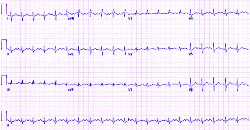
On general examination, patient had mild pallor and weighed 50 kg. For preanesthetic assessment her vitals were recorded, pulse rate was 88 beats per minute and blood pressure was 132/90 mmHg. Her airway assessment showed mouth opening of two and a half fingers, Mallampati grade II and neck movements were normal. Chest auscultation showed normal vesicular breath sound with bilaterally equal air entry. Cardiovascular examination revealed a systolic murmur in the tricuspid area. Electrocardiogram (ECG) showed incomplete Right Bundle Branch Block (RBBB) and right axis deviation as shown in [Table/Fig-1]. 2D Echo reported large osteum secundum ASD (size 24 mm) with left to right shunt, dilated right atrium and right ventricle with mild to moderate pulmonary artery hypertension, moderate Tricuspid regurgitation and good left ventricular function. Cardiologist opinion was taken and she was started on Tab. Torsemide 5 mg once daily seven days before surgery. Her haemoglobin was 10 gm % and other blood reports were within normal limits.
Showing ECG with incomplete RBBB and right axis deviation.
General anaesthesia was planned for the surgery and written informed high-risk consent was taken after proper counselling. Patient was kept fasting for six hours preoperatively. In the operation theatre, 18G vein flow was secured, intravenous line was deaired and Injection (Inj) Ringer Lactate was started. Standard non-invasive monitors including End-Tidal Carbon Dioxide (EtCO2) and temperature monitors were attached. Inj Glycopyrrolate 0.2 mg, Inj Midazolam 1 mg, Inj Ondansetron 4 mg and Inj Fentanyl 75 mcg were given intravenously (IV). Preoxygenation was done with 100% Oxygen for three minutes. Inj Propofol 100 mg was given slow IV as induction agent and airway was secured with portex cuffed endotracheal tube no 7.5 after giving loading dose of muscle relaxant, Inj Atracurium 25 mg IV with Inj Xylocard 80 mg IV Tube was fixed appropriately after checking bilateral equal air entry. Maintenance of anaesthesia was done with Oxygen and Isoflurane. Intermittent doses of Inj. Atracurium 0.1 mg/kg iv was given for muscle relaxation. Intraoperatively, her Mean Arterial Pressure (MAP) was maintained between 65-70 mmHg, heart rate remained in range of 70-80 per minute, oxygen saturation was 98% and EtCO2 was 33-36 mmHg. Intravenous fluid was given judiciously with regular monitoring of urine output and blood loss. At the end of surgery, Inj Xylocard 80 mg IV was given and after adequate reversal of neuromuscular blockade patient was extubated smoothly. Postextubation vitals were normal and patient was monitored in Intensive Care Unit (ICU) for one day. Postoperatively, adequate analgesia was ensured with Inj Paracetamol and Inj Diclofenac.
Patient started her feeds orally on second postoperative day, on return of bowel function and was discharged on fifth postoperative day. She had her first follow-up after seven days and had no new complaints and was referred to cardiology OPD for further management of ASD.
Discussion
Atrial Septal Defect (ASD) is one of the more frequently documented acyanotic congenital cardiac anomalies (about 25% of all adult Congenital Heart Diseases) [1]. ASD is defined by a defect in the interatrial septum permitting pulmonary venous return in left atrium to pass directly to the right atrium. ASD can be categorised based on the defect location in relation to fossa ovalis, the size of the defect and volume of the shunt and can be associated with anomalies resulting into a wide range of clinical manifestations from asymptomatic cardiac sequelae to right heart failure, pulmonary arterial hypertension, and rarely atrial arrhythmias [2]. Osteum Secundum is the most prevalent type of ASD and it amounts to 70% of cases with ratio of 1:2 for males to females, respectively. It involves the fossa ovalis and is located in the mid-septal region [3].
Large ASD, >20 mm leads to significant shunt and can have substantial haemodynamic effects in form of increased pulmonary perfusion and resulting increase in pulmonary vascular resistance, which if chronic, leads to development of PHT [4,5]. Echocardiography is the choice of investigation to confirm the size and type of ASD, volume of shunt and also the presence and grade of PHT. PHT can be graded as mild (36-49 mmHg), moderate (50-59 mmHg) and Severe (>60 mmHg) [6-8]. Ventricular hypertrophy, myocardial ischaemia and arrhythmias and even heart blocks are reported adverse events for long standing PHT and therefore, these can be anticipated during general anaesthesia along with air embolism in cases of ASD [9]. In Eisenmenger syndrome, Pulmonary Vascular Resistance (PVR) is very high and is characterised by irreversible pulmonary vascular disease with reversed or bidirectional shunt flow [5]. Perioperative mortality is more in patients with Eisenmenger syndrome [10].
In our case, a 39-year-old female had a large Osteum Secondum defect with mild to moderate PHT undergoing total abdominal hystrectomy. Usually, the preferred anaesthetic technique for total abdominal hysterectomy is central neuraxial block. However, Authors decided to administer general anaesthesia, as in patients with nonrestrictive intracardiac shunts, central neuraxial anaesthesia sometimes lead to unpredictable reduction in systemic vascular resistance, which could worsen right to left shunt and may result in acute heart failure and hypoxemia. Better maintenance of ventilation and systemic vascular resistance is possible in general anaesthesia [4]. Moreover, mechanical ventilation of lungs by their stretching effect stimulates release of nitric oxide and prostaglandins which are pulmonary vasodilator [11]. One of the important precautions that have to be taken is to avoid systemic air embolisation [5], so the IV lines were cautiously deaired. The patient was induced by administering Inj. Propofol in small incremental doses of 30 mg till the patient lost consciousness. According to Lovell AT the rate and dose of the IV-induction agent are more important than the actual drug itself [5]. N2O was not used for maintenance of anaesthesia so as to decrease the risk of paradoxical air embolism. The intention of anaesthetic management in such patients is to reduce the increase in pulmonary vascular resistance, to stabilise systemic vascular resistance, to safeguard tissue oxygen delivery. Inadvertent rise in PVR leads to acute right heart failure followed by oxygen desaturation and reduced cardiac output [4].
Factors that cause increase in PVR are hypercapnia, hypoxemia, hypothermia, acidaemia, pain. Inadequate anaesthesia and stimulation of sympathetic nervous system can result in increase of systemic vascular resistance and increase in PVR which cumulatively will decrease cardiac output resulting in hypotension leading to decrease in pulmonary blood flow causing desaturation [4,5]. Hence, it was aimed to maintain normotension, euthermia, adequate pain relief and depth of anaesthesia. Patient was optimally oxygenated and ventilated to avoid hypercapnia and desaturation. Intravenous fluid was given judiciously in view of PHT. Inj Xylocard was given intravenously before intubation and extubation to decrease the sympathetic response associated with these procedures. Postoperatively, patient was monitored in ICU and was ensured adequate pain relief. Course in ICU remained uneventful.
Conclusion(s)
Patients having large ASD with PHT can successfully undergo general anaesthesia for noncardiac surgeries like abdominal hysterectomy. This can be achieved by optimal preoperative preparation with meticulous intraoperative management, which aims at avoiding increase in PHT, maintaining systemic vascular resistance, optimal oxygen delivery along with ensuring good postoperative analgesia.
Author Declaration:
Financial or Other Competing Interests: No
Was informed consent obtained from the subjects involved in the study? Yes
For any images presented appropriate consent has been obtained from the subjects. Yes
Plagiarism Checking Methods: [Jain H et al.]
Plagiarism X-checker: Oct 30, 2020
Manual Googling: Nov 21, 2020
iThenticate Software: Dec 13, 2020 (11%)
[1]. Meissner I, Whisnant JP, Khandheria BK, Spittell PC, O’Fallon WM, Pascoe RD, Prevalence of potential risk factors for stroke assessed by transesophageal echocardiography and carotid ultrasonography: The SPARC study. Stroke Prevention: Assessment of Risk in a CommunityMayo Clin Proc 1999 74(9):862-69.10.4065/74.9.86210488786 [Google Scholar] [CrossRef] [PubMed]
[2]. Khairy P, Landzberg MJ, Adult congenital heart disease: Toward prospective risk assessment of a multisystemic conditionCirculation 2008 117:2311-12.10.1161/CIRCULATIONAHA.108.77059418458179 [Google Scholar] [CrossRef] [PubMed]
[3]. Feldt RH, Avasthey P, Yoshimasu F, Kurland LT, Titus JL, Incidence of congenital heart disease in children born to residents of Olmsted County, Minnesota, 1950-1969Mayo Clin Proc 1971 46(12):794-99. [Google Scholar]
[4]. Cannesson M, Earing MG, Collange V, Kersten JR, Anesthesia for noncardiac surgery in adults with congenital heart diseaseAnesthesiology 2009 111(2):432-40.10.1097/ALN.0b013e3181ae51a619602959 [Google Scholar] [CrossRef] [PubMed]
[5]. Lovell AT, Anaesthetic implications of grown-up congenital heart diseaseBr J Anaesth 2004 93(1):129-39.10.1093/bja/aeh17215192002 [Google Scholar] [CrossRef] [PubMed]
[6]. Lee MG, Ko JS, Yoon HJ, Kim KH, Ahn Y, An unusual presentation of an atrial septal defectJ Cardiovasc Ultrasound 2009 17(4):151-52.10.4250/jcu.2009.17.4.15120661343 [Google Scholar] [CrossRef] [PubMed]
[7]. Hines RL, Marschall KE, Stoelting’s anesthesia and co-existing disease. (6th edn). In: Livingstone C (Ed.) 2008 IndiaElsevier:60 [Google Scholar]
[8]. Valdes Cruz LM, Cayre RO, Anomalies of right ventricular outflow tract and pulmonary arteries. In: Valdes-Cruz LM & Cayre RO (Eds.) 1999 Philadelphia, USALippincott-Raven:325-48. [Google Scholar]
[9]. Park YS, Kim JY, Anesthetic management of a patient with large atrial septal defect undergoing laparoscopic cholecystectomy: A case reportSaudi J Anaesth 2020 14(2):249-52.10.4103/sja.SJA_638_1932317887 [Google Scholar] [CrossRef] [PubMed]
[10]. Ammash NM, Connolly HM, Abel MD, Warnes CA, Noncardiac surgery in Eisenmenger syndromeJ Am Coll Cardiol 1999 33:222-27.10.1016/S0735-1097(98)00554-3 [Google Scholar] [CrossRef]
[11]. Young MJ, Gorlin AW, Modest VE, Quraishi SA, Clinical implications of the transversus abdominis plane block in adultsAnesthesiol Res Pract 2012 2012:73164510.1155/2012/73164522312327 [Google Scholar] [CrossRef] [PubMed]