Concomitant Mucormycosis with Aspergillosis in Patients with Uncontrolled Diabetes Mellitus: A Case Series
Arpana Singh1, Aroop Mohanty2, Shweta Jha3, Pratima Gupta4, Neelam Kaistha5
1 Senior Resident, Department of Microbiology, All India Institute of Medical Sciences Rishikesh, Rishikesh, Uttarakhand, India.
2 Assistant Professor, Department of Microbiology, All India Institute of Medical Sciences Gorakhpur, Gorakhpur, Uttar Pradesh, India.
3 Junior Resident, Department of Microbiology, All India Institute of Medical Sciences Rishikesh, Rishikesh, Uttarakhand, India.
4 Professor, Department of Microbiology, All India Institute of Medical Sciences Rishikesh, Rishikesh, Uttarakhand, India.
5 Professor, Department of Microbiology, All India Institute of Medical Sciences Rishikesh, Rishikesh, Uttarakhand, India.
NAME, ADDRESS, E-MAIL ID OF THE CORRESPONDING AUTHOR: Dr. Aroop Mohanty, Assistant Professor, Department of Microbiology, AIIMS Gorakhpur, Gorakhpur-273008, Uttar Pradesh, India.
E-mail: aroopmohanty7785@yahoo.com
Fungal infections are life threatening especially in presence of immunosuppression or uncontrolled diabetes mellitus mainly due to their invasive potential. Mucormycosis of the oculo-rhino-cerebral region is an opportunistic, aggressive, fatal and rapidly spreading infection caused by organisms belonging to Mucorales order and class Zygomycetes. The organisms associated are ubiquitous. Aspergillosis is a common clinical condition caused by the Aspergillus species, most often by Aspergillus fumigatus (A. fumigatus). Both fungi have a predilection for the immunosuppressive conditions, with uncontrolled diabetes and malignancy being the most common among them. Mucormycosis is caused by environmental spores which get access into the body through the lungs and cause various systemic manifestations like rhino-cerebral mucormycosis. Here, a case series of such concomitant infections of Aspergillus and Mucor spp from Rishikesh, Uttarakhand, India is reported.
Diabetes, Fungal infection, Invasive mycoses, Rhinosinusitis
Introduction
Mucorales are the universally distributed saprophytes causing aggressive and opportunistic infection. They are angio-invasive in nature. Aspergillosis is the clinical condition caused by the Aspergillus species most often A. fumigatus [1]. It proves to be fatal, if it infects secondarily to the brain. There are various risk factors like uncontrolled diabetes, immunodeficiency states, solid organ transplantation, underlying malignancy and cirrhosis but they are also associated with immunocompetent people [1]. Despite advances in treatment, mortality is very high. So, early diagnosis and prompt treatment of the condition plays a pivotal role in saving the patient.
Case Series
Case 1
A 50-year-old diabetic female patient presented to the Ear, Nose and Throat Out Patient Department (ENT OPD) with complaints of postnasal discharge on the left side since one year and left-sided facial pain for the last three months. She had undergone septoplasty one year back in a private hospital, where she was given nasal spray-xylometazoline and augmentin 625 mg. There was no other significant past history. On physical examination, patient was afebrile; heart rate was 80 beats/min, respiratory rate was 22/min; blood pressure was 80/60 mm Hg; and oxygen saturation was 88% while breathing room air. Patient’s Random Blood Sugar (RBS) was 207 mg/dl and HbA1C was 11.0% at the time of admission. A presumptive diagnosis of bilateral acute, invasive fungal rhinosinusitis was made. Bilateral Functional Endoscopic Sinus Surgery (FESS) was done. Tissue debridement was done and sent for fungal examination to the Mycology section of the Department of Microbiology. Direct 10% KOH (Potassium hydroxide) wet mount of the tissue demonstrated mixture of thin, septate and few distorted right-angle branching hyaline, broad aseptate hyphae. Patient was started on Liposomal Amphotericin B in a dose of 100 mg/day; the dose was increased up to 200 mg/day over the next one week with regular monitoring of blood parameters for toxicity to the same. She was discharged on the same along with Tab voriconazole orally. Patient successfully completed treatment and on follow-up after three weeks of conservative treatment, repeat KOH examination from nasal swab demonstrated no fungal hyphae.
Case 2
A 60-year-old female patient presented with complaints of fever for three days and ptosis for one day. She was a known case of Type 2 Diabetes Mellitus (T2DM) and hypertension for the past seven years but was on irregular drug metformin and amlodipine. No other significant past history was recorded. Extensive nasal crusts, black eschar in the nasal cavity and septal perforation was showed in nasal examination. On physical examination, patient was febrile; heart rate was 100 beats/min, respiratory rate was 26/min; blood pressure was 88/68 mm Hg; and oxygen saturation was 98% while breathing room air. Eye examination revealed periorbital swelling along with swollen left eye and diminished extraocular movements in all directions. She was diagnosed as case of rhino-ocular mucormycosis with uncontrolled T2DM. FESS was done. KOH examination revealed mixture of thin, septate and broad aseptate hyphae. Treatment was started with Liposomal Amphotericin B, a cumulative dose of 2000 mg was given, followed by Itraconazole 200 mg OD, initially for seven days and then continued for four weeks. During the conservative therapy, she developed hypokalemia for which syrup potassium hydrochloride was administered and anaemia was treated by transfusing one unit of packed cells. A repeat KOH mount was done on follow-up after four weeks of treatment. No hyphae were noted in the KOH mount.
Case 3
A 35-year-old male patient presented to the emergency with complaints of fever for last 20 days and facial swelling on the left side for 15 days. He also had history of T2DM but was on irregular antidiabetic medication-metformin. No other significant history was recorded. Laboratory investigations showed leukocytosis-WBC count was 15×103/μL. Clinical examination showed slight asymmetry in the mouth region and complete closure of eye with no discomfort. These features suggested grade II facial palsy. Diagnostic nasal endoscopy revealed dead necrotic tissue, black eschar and discharge. Debridement was done endoscopically. Debrided tissue was sent for mycological examination. KOH revealed plenty broad aseptate hyphae along with septate hyphae. Finally, the patient was diagnosed as case of left sinonasal mucormycosis with left grade II facial palsy. Patient was started on Liposomal Amphotericin B for 24 days and discharged on step down treatment with tab voriconazole. During follow-up visits after three weeks, KOH was negative for fungal elements. Patient improved significantly.
Microbiological Diagnosis:
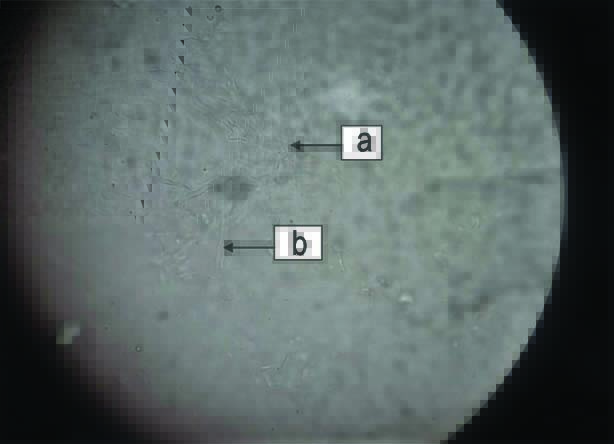
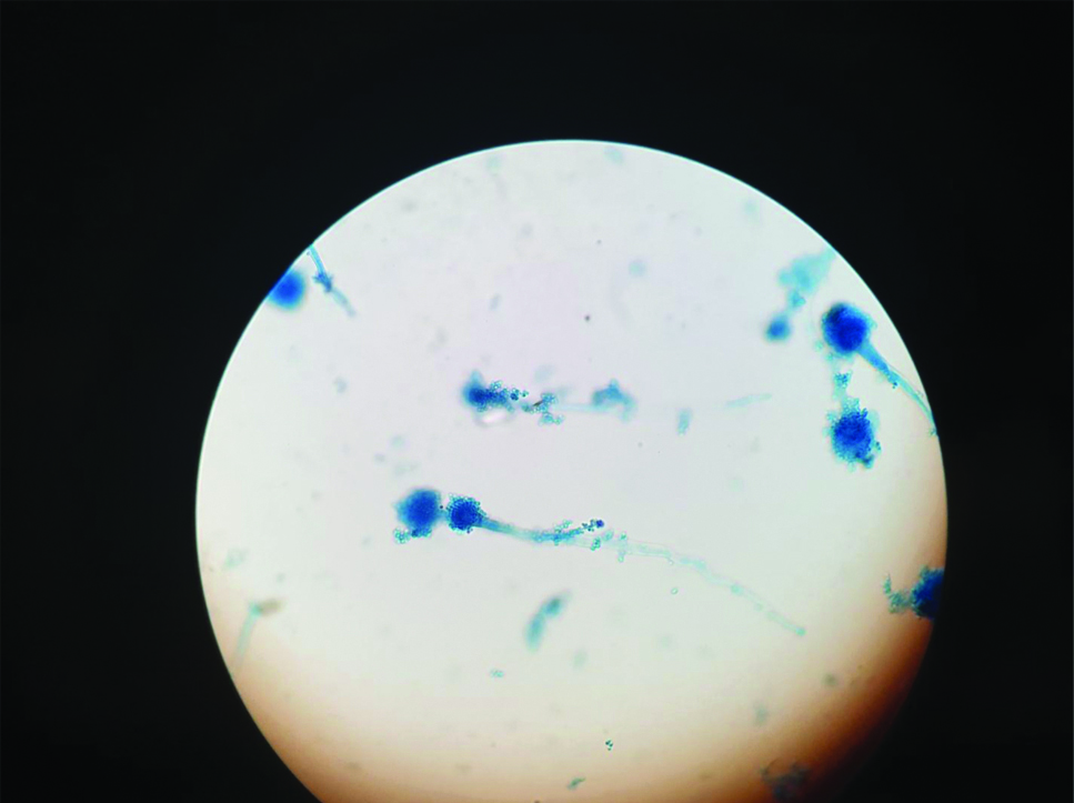
Tissue sample after taking biopsy from nasal mucosa was sent in all the above cases for KOH mount and fungal culture to the mycology laboratory of the Department of Microbiology of our hospital. Direct KOH wet mount revealed mixture of broad aseptate hyaline hyphae along with thin septate hyphae in above three cases [Table/Fig-1]. Fungal culture was done on Sabouraud’s Dextrose Agar (SDA) and was inoculated and incubated at 37°C and 25°C, respectively. It showed a mixture of cottony growth. Lactophenol Cotton Blue (LPCB) mount of the culture showed presence of Mucor spp and Aspergillus spp, [Table/Fig-2,3]. Matrix-Assisted Laser Desorption/Ionization Time of Flight-Mass Spectrometry (MALDI-TOF MS) was also done and Aspergillus spp was identified as Aspergillus fumigatus with a Confidence Interval (CI) of 2.1. Mucor could not be identified up to species level. Patients consent was obtained in all the three cases. The clinical and microbiological data of all three cases is shown in [Table/Fig-4].
Direct KOH wet mount showing broad aseptate hyphae; (a) and thin septate hyphae; (b) (Magnification:400X).
Lactophenol Cotton Blue (LPCB) stain showing Mucor spp, 400X).

Lactophenol Cotton Blue (LPCB) stain showing Aspergillus spp, 400X.
Clinical and microbiological data of patients.
| Age (years) | Sex | Presenting complaints | Underlying disease | KOH | Treatment | Outcome |
|---|
| 50 | Female | Facial pain x 3 monthsPost nasal discharge x 1 year | T2DM | Positive | Inj. Liposomal Amphotericin B Tab.Voriconazole 100 mg BD | Improved |
| 60 | Female | Fever x 3 daysPtosis x 1 day | T2DM | Positive | Inj. Amphotericin B Cap. ItraconazoleNasal douching | Improved |
| 35 | Male | Fever x 20 daysFacial swelling x 15 days | T2DM | Positive | Tab.Voriconazole 100 mg BDInj.Amphotericin | Improved |
Discussion
Invasive mycoses are caused by many pathogens especially in debilitated patients but the frequency of infection and spectrum of the disease is increasingly changing with time. India is the most affected country in the world by Mucormycosis, with about 44.3% of the total number of cases being reported worldwide [1]. Diabetes is a well-known risk factor for invasive fungal infections as the host’s immunological response is altered. Glucose intolerance along with hyperglycemia provides a perfect niche for these fungi to proliferate rapidly. Along with this, decreased granulocyte mediated phagocytosis has been observed in such patients. Various genera which are responsible for infection are Rhizopus (most common), Rhizomucor and Absidia [2]. Due to their angioinvasive nature and predilection for vessels, infarction of the infected tissues is the hallmark of this invasive disease [3] of mucormycosis. The most common among all mucormycosis form is the rhino-orbital cerebral form [4] which can also extends to orbits and even to brain.
Smith HW and Kirchner JA gave a criterion for the clinical diagnosis of mucormycosis which includes the following [5]:
Blood mixed nasal discharge and facial pain, both on the same side.
Drooping of the eyelid, proptosis of the globe and total ophthalmoplegia.
Soft peri-nasal or peri-orbital swelling which progresses to induration and occlusion of blood vessels.
Multiple unrelated weaknesses of the cranial nerves.
Black, necrotic tissues in turbinates that are easily mistaken for dried, crusted blood.
Invasive aspergillosis of the sino-orbital area may simulate features of mucormycosis and malignancy due to the presence of facial mass and local tissue destruction [6-8]. There is an increased incidence of mucormycosis in patients treated with voriconazole for suspected aspergillosis so, the clinical distinction between aspergillosis and mucormycosis is crucial [9].
Pandey D et al., have also described a rare case with mixed infection of mucormycosis, Aspergillus and non-albicans Candida. Others have reported similar co-infections with Mucorales and Aspergillus spp in known diabetics. Rit K et al., documented the co-infection in a 46-year-old with uncontrolled diabetes and squamous cell carcinoma and the patient initially did not respond to voriconazole and then started amphotericin on detecting Mucor but succumbed to death. Nagarkar NM et al., reported the similar case of co-infection of Mucor and Aspergillus spp in a patient of uncontrolled diabetic patient [1,2,4,10,11].
Thus, in case of diabetic patients with fungal rhinosinusitis, more than one fungus can be isolated as seen in this unique case series. Mixed fungal infection with Aspergillus, Mucor and even Candida should be suspected in diabetics in case of invasive mycosis especially in rhino-cerebral mycosis. Fungal culture and histopathology will help the clinicians in guiding the therapy. Early diagnosis and antifungal treatment, early control of disease and patient’s general condition plays fundamental role for survival and morbidity. In cases of mixed infections, more aggressive and focused approach will be required as compared to infection with a single fungal agent. Definitive surgical treatment must be performed only after these requirements have been met and helpful for the fungal eradication. The survival rate in cerebral mucormycosis has improved to more than 50% at present due to early diagnosis, timely and appropriate treatment [12].
Conclusion(s)
Although a very few cases of concomitant infection have been seen and reported in diabetic population, the clinicians should always keep a high index of suspicion so that such cases can be effectively managed and lives be saved.
Author Declaration:
Financial or Other Competing Interests: None
Was informed consent obtained from the subjects involved in the study? Yes
For any images presented appropriate consent has been obtained from the subjects. NA
Plagiarism Checking Methods: [Jain H et al.]
Plagiarism X-checker: Dec 05, 2020
Manual Googling: Jan 04, 2021
iThenticate Software: Dec 24, 2020 (9%)
[1]. Pandey D, Agarwal M, Chadha S, Agarwal D, Mixed opportunistic infection with Mucor, Aspergillus and Candida in oculo-rhinocerebral mycosis: An uncommon caseJ Acad Clin Microbiol 2019 21(1):47-49.10.4103/jacm.jacm_2_19 [Google Scholar] [CrossRef]
[2]. Kumari M, Malhotra P, Bhardwaj M, Co-infection of mucormycosis and aspergillus in a diabetic patient: A rare entityInt J Med Microbiol Trop Dis 2019 5(4):241-43.10.18231/j.ijmmtd.2019.055 [Google Scholar] [CrossRef]
[3]. Greenberg RN, Scott LJ, Vaughn HH, Ribes JA, Zygomycotic (mucormycosis): Emerging clinical importance and new treatmentsCurr Opin Infect Dis 2004 17(6):517-25.10.1097/00001432-200412000-0000315640705 [Google Scholar] [CrossRef] [PubMed]
[4]. Rit K, Saha R, Dey R, Barik G, Rhino-oculo-cerebral aspergillus and mucor co-infections in an immunocompromised patient with type 2 diabetes mellitusMed J DY Patil Univ 2014 7(4):486-88.10.4103/0975-2870.135278 [Google Scholar] [CrossRef]
[5]. Smith HW, Kirchner JA, Cerebral mucormycosis: A report of three casesAMA Arch Otolaryngol 1958 68(6):715-26.10.1001/archotol.1958.0073002073901013593957 [Google Scholar] [CrossRef] [PubMed]
[6]. Martino P, Raccah R, Gentile G, Venditti M, Girmenia C, Mandelli F, Aspergillus colonization of the nose and pulmonary aspergillosis in neutropenic patients: A retrospective studyHaematologica 1989 74(3):263-65. [Google Scholar]
[7]. Sivak-Callcott JA, Livesley N, Nugent RA, Rasmussen SL, Saeed P, Rootman J, Localised invasive sino-orbital aspergillosis: Characteristic featuresBr J Ophthalmol 2004 88(5):681-87.10.1136/bjo.2003.02172515090423 [Google Scholar] [CrossRef] [PubMed]
[8]. Adulkar NG, Radhakrishnan S, Vidhya N, Kim U, Invasive sino-orbital fungal infections in immune competent patients: A clinic-pathological studySpringer Nature 2019 33(6):988-94.10.1038/s41433-019-0358-630765886 [Google Scholar] [CrossRef] [PubMed]
[9]. Hofman V, Dhouibi A, Butori C, Padovani B, Gari-Toussaint M, Garcia-Hermoso D, Usefulness of molecular biology performed with formaldehyde-fixed paraffin embedded tissue for the diagnosis of combined pulmonary invasive mucormycosis and aspergillosis in an immune competent patientDiagn Pathol 2010 5(1):110.1186/1746-1596-5-120205795 [Google Scholar] [CrossRef] [PubMed]
[10]. Nagarkar NM, Verma H, Punia RPS, Co-existing mucormycosis with aspergillosis in a patient with diabetes mellitus- First case reportOtolaryngol Online J 2014 4(4):257-64. [Google Scholar]
[11]. Alfano C, Chiummariello S, Dessy LA, Bistoni G, Scuderi N, Combined mucormycosis and aspergillosis of rhinocerebral regionIn vivo 2006 20(2):311-15. [Google Scholar]
[12]. Benachinmardi KK, Rajalakshmi P, Veenakumari HB, Bharath RD, Vikas V, Mahadevan A, Successful treatment of primary cerebral mucormycosis: Role of microbiologistIndian J Med Microbiol 2016 34(4):550-53.10.4103/0255-0857.19537327934843 [Google Scholar] [CrossRef] [PubMed]