A three-dimensional obturation eliminates leakage pathways from the coronal and apical directions and entombs remaining bacteria in the root canal system [1]. An ideal root canal sealer should meet the general requirements of American National Standard Institute/American Dental Association [2]. A wide variety of root canal sealers are available [3] and AH series was the first-generation epoxy resin used because of its lasting dimensional stability and satisfactory apical sealing ability [3]. Advantages of Roekoseal, which is a recent formulation has been its ability to mechanically adhere to the walls of the root canal due to expansion, retreatment can be easily carried out, if required and the material does have eugenol in its composition and is helpful for radiographic evaluation as the material is well appreciated on radiographs as claimed by the manufacturer [4]. Adseal is another epoxy-based resin sealer reported in literature with acceptable physical properties and good radiopacity [5]. Microleakage testing has been used to determine the possible clinical performance of obturation materials using various methods in-vitro [6]. Dye penetration is by far more accepted method which can be tested by active as well as passive methods. Although the precincts of traditional dye leakage are well-established, this method has still been utilised in some recent studies [7-9]. Dye extraction method might provide more reliable results because it quantitatively measures all of the dye taken up by the sample. Camp J and Pashley D have observed and stated that dye extraction method has yielded equivalent results as fluid transport experimental setup, while saving laboratory time [10]. There are only few studies in the literature where in dye extraction method has been used to evaluate apical microleakage of obturated teeth [8,11].
Thus, the present study was designed to evaluate the apical sealing ability of two resins and one silicon-based root canal sealer and gutta-percha obturated root canals using dye extraction method.
Materials and Methods
The present study was an experimental in-vitro study, carried out at MGM Dental College and Hospital, Navi Mumbai, Maharashtra, India. The study was carried out over a period of 10 months (from August 2013 to May 2014). Ethical approval was obtained from the Institutional Review Board before commencement of the study vide No. MGM/DCH/IEC/05/2012.
Inclusion citeria: Eighty single rooted freshly extracted human permanent maxillary central incisors for periodontal reasons, with single root and single straight patent canal, curvature less than 10 degrees assessed using the Schneider’s method [12] with fully formed apex and a single portal of exit were selected for the study.
Exclusion criteria: The teeth were checked for absence of caries, cracks, and structural defects and if found were excluded from the study.
Study Procedure
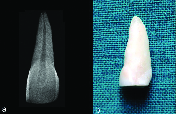
The selected teeth were stored in 0.9% normal saline (Althea Pharma Private Ltd., India) until use and in between the procedures. calculus and soft tissue debris were removed with a universal hand scaler (GDC Fine Crafted Dental Pvt., Ltd., Hoshiarpur, India) and teeth were immersed in 5% Sodium Hypochlorite (NaOCl) solution (Prime Dental Products, India) for one hour to remove any organic component from the root surface. Pre-operative radiographs were taken to assess inclusion and exclusion criteria using Radiovisuography after extraction of the tooth [Table/Fig-1a]. The crowns of the teeth were sectioned at/below cementoenamel junction [Table/Fig-1b] using a diamond disk (Mani Inc, Tachigiken, Japan) with water coolant at a slow speed, and the length was standardised at 15 mm with help of vernier caliper (HM & Company, Mumbai).
a) Pre-operative Intraoral periapical radiograph of the permanent maxillary central incisor with a single, straight root canal. b) Sectioned permanent maxillary central incisor.
Access cavity was refined using #2 stainless steel round bur (Mani Inc, Tachigiken, Japan) by holding the roots in saline moistened 2×2 gauze. Working Length (WL) was established by introducing a size #10 K-file (Mani Inc, Tachigiken, Japan) until tip of instrument was visible at the apical foramen. This file was then retracted by 1 mm and WL was established at this length. Biomechanical preparation of the root canals was done using Step-Back technique with hand K-files. Irrigation was done between each instrument with 2 mL, 5% NaOCl and recapitulation with previous smaller instrument carried to full WL. The smear layer was removed using 2 mL, of 17% Ethylene Diamine Tetra-acetic Acid (EDTA) (Dentwash, Prime Dental Products, India) wash for one minute followed by saline irrigation and then with 2 mL, of 5% NaOCl. Finally, the canals were irrigated with 5 mL of normal saline. All canals were then dried with absorbent points (Mani, Japan) and randomly divided into five groups and obturated subsequently using gutta-percha points (Dentsply Maillefer, China) and the three sealers AH Plus, Roekoseal, Adseal manipulated as per manufacturer’s instructions. Flowable composite resin material (Tetric N-Flow, Ivoclar Vivadent) was placed in the coronal cavity. The obturated roots were then incubated at 37°C for seven days in an incubator before subjecting it to dye penetration study.
The roots were divided into five groups of 16 teeth each (n=16); three test groups, one positive and one negative group, which were all subjected to passive dye penetration and dye extraction method as follow:
Group I- Guttapercha/AH Plus: Cold lateral compaction of Guttapercha with AH Plus sealer (Dentsply Maillefer USA). The lateral surfaces of the roots were coated with three layers of nail varnish (Revlon, India) except on the apical 2 mm, to allow dye penetration into the accessory and lateral canals.
Group II- Guttapercha/Roekoseal: Cold lateral compaction of Guttapercha with Roekoseal sealer (Roeko, Germany). The lateral surfaces of the roots were coated with three layers of nail varnish except on the apical 2 mm.
Group III- Guttapercha/Adseal: Cold lateral compaction of Guttapercha with Adseal sealer (MetaBiomed Cheongju South Korea). The lateral surfaces of the roots were coated with three layers of nail varnish except on the apical 2 mm.
Group IV- Positive Control: In this group root canals were not obturated. The lateral surfaces of the roots were coated with three layers of nail varnish except on the apical 2 mm.
Group V- Negative control: In this group root canals were filled with adhesive wax and all root surfaces were coated with three layers of nail varnish.
For the Passive dye penetration test, roots were suspended in stationary glass vials (Himedia, India) containing 2% methylene blue (Himedia, India) such that only apical 2 mm of the tooth root was immersed in the dye, for seven days. After removal from the dye, the roots were washed in running water for one hour. Then the roots were subjected to dye extraction test [8] by placing them in eppendorf tubes (Himedia, India) containing 1 mL, 65% wt concentration of nitric acid (Qualigens Fischer Scientific, India) and left sealed for 72 hours. Centrifugation was carried out at 14,000 rpm in a high-speed centrifuge machine (Remi cooling microfuge CM-12). At the end of the procedure, 500 μL of supernatant solution obtained post-dye extraction was analysed using an Ultra-Violet spectrophotometer (Thermo scientific, Evolution 201) which measured the absorbance of monochromatic beam of light passing through the supernatant solution containing the dye comparing this with the blank solution which was concentrated nitric acid and thus, giving a digital read out of the absorbance by the dye present in the supernatant solution [8].
Statistical Analysis
Statistical analysis was performed using IBM SPSS statistical software version 20 (Armonk, New York, United States). Descriptive analysis was done to find mean and standard deviation. Normality of data was assessed by using Shapiro-Wilk test. The data was found to be normally distributed, thus one-way ANOVA followed by Tukey’s post-hoc test was performed. Significance was set at 5%. Null Hypothesis stated that there was no difference in apical sealing ability of the two-resin and one silicon-based sealer and gutta percha obturated root canal using dye extraction method.
Results
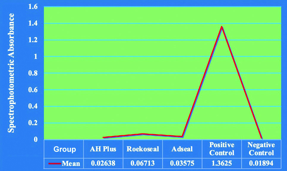
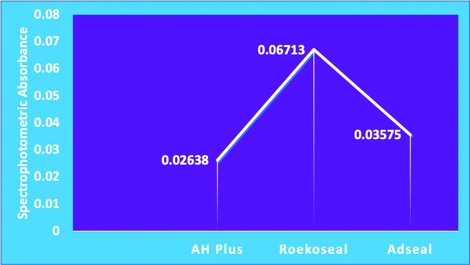
Among the experimental groups, the dye absorbance value was least in the AH Plus group with a mean of 0.02638±0.007320 and highest in Roekoseal group with a mean of 0.06713±0.010372; the dye absorbance value for Adseal was higher than AH Plus but lower than Roekoseal with a mean of 0.03575±0.007946. The Dye absorbance value for positive control group was highest 1.36250±0.171289. The negative control group showed least mean absorbance value of dye of 0.01894±0.008282 [Table/Fig-2].
Table showing mean, standard deviation, Standard Error of the Mean (SEM), lower bound and upper bound values for confidence interval.
| Group | n | Mean | SD | SEM | 95% Confidence interval for Mean |
|---|
| Lower bound | Upper bound |
|---|
| AH Plus | 16 | 0.02638 | 0.007320 | 0.001830 | 0.02247 | 0.03028 |
| Roekoseal | 16 | 0.06713 | 0.010372 | 0.002593 | 0.06160 | 0.07265 |
| Adseal | 16 | 0.03575 | 0.007946 | 0.001986 | 0.03152 | 0.03998 |
| Positive control | 16 | 1.36250 | 0.171289 | 0.042822 | 1.27123 | 1.45377 |
| Negative control | 16 | 0.01894 | 0.008282 | 0.002071 | 0.01452 | 0.02335 |
The comparative analysis of the mean values of dye absorbance of all the five groups is given in [Table/Fig-3a] and that of experimental groups are presented in [Table/Fig-3b].
Graphical representation of the comparative analysis of dye absorbance of all the five groups.
Graphical representation of the comparative analysis of dye absorbance of experimental groups.
The graph represents spectrophotometric absorbance of dye for each group. The normality of data in each group was tested using Shapiro-Wilk test and the data was found to be normally distributed, since all the p-values were p>0.05 for the above tests [Table/Fig-4].
Test of Normality using Shapiro-Wilk Test.
| Groups | Statistic | df | Sig. |
|---|
| AH plus | 0.975 | 16 | 0.913 |
| Roekoseal | 0.982 | 16 | 0.980 |
| Adseal | 0.880 | 16 | 0.393 |
| Positive control | 0.944 | 16 | 0.403 |
| Negative control | 0.954 | 16 | 0.557 |
df: Degree of freedom; p-value <0.05 considered statistically significant
The mean absorbance values of the five groups were compared using one-way ANOVA. [Table/Fig-5] shows p<0.001 which means there was a significant difference between the dye absorbance values of all the groups.
One-way analysis of variance table.
| Parameter | Sum of squares | df | Mean square | F | Sig. |
|---|
| Between groups | 22.509 | 4 | 5.627 | 949.491 | <0.001 |
| Within groups | 0.444 | 75 | 0.006 | | |
| Total | 22.953 | 79 | | | |
df: Degree of freedom; p-value <0.05 consiedred statistically significant
Further to compare the level of absorbance of dye among various groups, Tukey’s post-hoc test was applied showing mean difference [Table/Fig-6]. When AH Plus was compared with Roekoseal the p-value was 0.568 and 0.997 with Adseal whereas when Roekoseal was compared with Adseal the p-value was 0.778. There was no statistically significant difference amongst different experimental groups.
Multiple Comparisons using Tukey’s post-hoc test.
| (I) Sealer | (J) Sealer | Mean difference (I-J) | Std. Error | Sig. | 95% Confidence interval |
|---|
| Lower bound | Upper bound |
|---|
| AH plus | Roekoseal | -0.040750 | 0.027218 | 0.568 | -0.11683 | 0.03533 |
| Adseal | -0.009375 | 0.027218 | 0.997 | -0.08546 | 0.06671 |
| Positive control | -1.336125* | 0.027218 | <0.001 | -1.41221 | -1.26004 |
| Negative control | 0.007438 | 0.027218 | 0.999 | -0.06864 | 0.08352 |
| Roekoseal | Adseal | 0.031375 | 0.027218 | 0.778 | -0.04471 | 0.10746 |
| Positive control | -1.295375* | 0.027218 | <0.001 | -1.37146 | -1.21929 |
| Negative control | 0.048188 | 0.027218 | 0.398 | -0.02789 | 0.12427 |
| Adseal | Positive control | -1.326750* | 0.027218 | <0.001 | -1.40283 | -1.25067 |
| Negative control | 0.016813 | 0.027218 | 0.972 | -0.05927 | 0.09289 |
| Positive control | Negative control | 1.343563* | 0.027218 | <0.001 | 1.26748 | 1.41964 |
*The mean difference is significant at the 0.05 level
The results of the present study were in agreement with the null hypothesis which stated that there was no difference in the apical sealing ability of resin and silicon-based sealer guttapercha filled root canals using dye extraction method. Though the p-value did not show any statistically significant difference amongst experimental groups, the mean dye absorbance value of AH Plus was found to be least and that of Roekoseal was highest in the experimental groups and Adseal had intermediate values.
Discussion
In spite of thorough chemo-mechanical preparation of the root canal, it has been observed that microorganisms still remain active in the dentinal tubules [1]. AH Plus has good physical and chemical properties with good sealing ability and hence is considered as a gold standard to which other sealers are compared [13]. In the present study, roots in group I were obturated using AH Plus sealer and Guttapercha. AH Plus sealer being a two-component paste material is based on a slow polymerisation reaction of epoxy resin amines, where the conversion of monomers into polymers occurs gradually occurs [14], where the diepoxide compounds and polyamines paste are mixed together during material manipulation, and each amine group reacts with an epoxide group to form a covalent bond. The resulting polymer thus obtained shows heavy cross-linkages and is rigid and strong [15], which may offer possible explanation for its low solubility and high dimensional stability [16,17].
According to Tay FR et al., the critical area of fillings is located at the sealer/dentin interface [18]. Chemical reaction of exposed amino groups in collagen of the root canal dentin forms a covalent bond between the resin and the collagen upon opening of the epoxide ring, leading to a superior adaptation of the epoxy resin to bond to root dentin [19]. As stated by manufacturers, AH Plus has a slower setting time (37°C, 8 hours) than Adseal (37°C, 70 min) and Roekoseal (37°C, 45 to 50 min). In the study by Kokkas AB et al., AH Plus showed significant deeper maximum penetration depth, 59 μm which was enhanced by the removal of smear layer [20], Thus, in the current study 17% EDTA was used to remove the smear layer along with NaOCl. When AH Plus, Epiphany and EndoREZ were evaluated for apical leakage in a study by Dultra F et al., AH Plus, Epiphany and EndoREZ did not differ statistically to each other (p>0.05). EndoFill zinc oxide and eugenol sealer presented the highest dye penetration mean and was statistically different from the other groups (p<0.01) [21]. In a study by Reddy A et al., Resilon Epiphany group showed more leakage as compared to guttapercha/AH Plus sealer with a significant p-value of 0.028 [22], all in agreement with the results of this study. On the contrary, Kqiku L et al., compared apical sealing ability of laterally condensed gutta percha/AH plus and Resilon/Epiphany using the active versus passive dye microleakage and observed that laterally condensed guttapercha/AH Plus showed more leakage than Resilon/Epiphany (p<0.05) [23]. In group II, of the present study, the root canals were obturated using Roekoseal which is a highly biocompatible polydimethylsiloxane-based root canal sealer. Working time (15-30 min.) of Roekoseal is shortened by use of heat and about 45-50 minutes are required for the curing to take place. The sealer expands (0.2% by volume) on setting and results in mechanical adhesion to the root canal walls. Under pressure the sealer becomes less viscous and flows into the dentinal tubule, the reason being the thixotropic nature of the sealer [24].
In the current study, dye absorbance value was highest with Roekoseal group, indicating maximum dye penetration. Yigit DH and Gencoglua N observed that the mean value of flow measurement for Roekoseal (6.31±0.27) was less than that of AH Plus (9.50±0.20) [25]. Wu MK et al., used fluid transport model to measure leakage along Single Cone (SC) fillings with guttapercha and Roekoseal sealer at one week and at one year and observed that SC fillings with Roekoseal sealer in wide and straight canals prevented fluid transport for one year [26]. Kazemi RB et al., observed dimensional changes of endodontic sealers, followed over a period of 180 days; AH26 and a silicon-based sealer were significantly more stable than zinc-oxide eugenol-based sealers. Furthermore, the silicon-based sealer showed 1% initial expansion [27]. Cobankara FK et al., at the end of one week noted that root fillings with Roekoseal were leaking more than AH Plus, Ketac Endo and Sultan, but after 21 days, the situation was reversed where Roekoseal showed best sealing when compared to other sealer tested (p>0.05) [28]. In group III, Adseal was used where the dye absorbance values for Adseal were higher than AH Plus but lower than Roekoseal. Properties such as solubility, radiopacity, film thickness, flow, setting time, and adaptation to the root canal walls of three epoxy resin based sealers: AH Plus, Acroseal, and Adseal were evaluated by Marciano MA et al., and they observed similar root canal adaptation, solubility, flow, and film thickness of all sealers. In the study, it was observed that, Adseal had a setting time of 70 minutes as compared to AH Plus 711.33 minutes. The different percentages of hardeners found in the Adseal and AH Plus might explain the differences in the setting time [29]. The seal that was provided by Adseal sealer was better than Proroot MTA and MTA Fillapex correlation (p-value <0.01) between all groups [30].
Dye penetration technique is the most commonly used technique, yet has the drawback that it is a qualitative test and yields a high level of variation [10]. Dye extraction represents an improvement in comparison with the dye penetration technique alone that underestimates the extent of the dye in the root canal because of simple linear measurements after longitudinal splitting, cross-sectioning or clearing of the specimens [31]. The quantitative nature of the dye extraction technique involves spectrophotometric analysis of the volume of tracer [10]. Disadvantage of the dye extraction method is the inability to measure microleakage without destroying the root specimens during treatment with concentrated nitric acid. Therefore, repeated observation of the same specimens over time to reveal changes in sealing ability is not possible [32]. Methylene blue dye penetration method was utilised in the current study for the evaluation of apical microleakage because it is a relatively inexpensive and reliable method [33]. If a small molecule such as a dye cannot penetrate the root canal filling in in-vitro studies, clinically it implies that root canal filling material will prevent bacteria and their by-products from entering the root canal [34].
In the present study, AH Plus has shown to give minimum dye absorbance values, inferring to indicate minimum dye leakage, influencing the strategic advantage of the sealer over the other sealers.
Limitation(s)
The present study was carried out using a sample size of 80 roots and each root could be used for one reading only as the root gets completely dissolved in 65% wt concentration of nitric acid used in the dye extraction process and further readings cannot be obtained, compared to the passive dye penetration where the readings can be verified, whenever required. Studies using different techniques of obturation can be carried out to compare different sealer performance while using two different apical leakage measurement techniques like fluid filtration and dye extraction method.
Conclusion(s)
Under the limitations of this in-vitro experimental study, it was concluded that all test groups showed apical microleakage. Though the p-values did not show any statistically significant difference amongst experimental groups, AH Plus gave best results with least dye extracted, inferring least dye penetration and minimum microleakage. Roekoseal allowed maximum dye to penetrate. Thus, AH Plus which is considered as a gold standard root canal sealer, defended its pivotal position in success of endodontic treatment. Adseal, also showed promising results and can be further evaluated for its value as a competent sealer in endodontics. Further clinical studies are necessary to evaluate microleakage.
df: Degree of freedom; p-value <0.05 considered statistically significant
df: Degree of freedom; p-value <0.05 consiedred statistically significant
*The mean difference is significant at the 0.05 level