Video Laryngoscope Assisted Awake Nasal Intubation with Restricted Mouth Opening in an Operated Radical Neck Dissection Patient
Tejash Hariduttbhai Sharma1, Avani Prafulchandra Vayeda2, Malini K Mehta3, Dinesh K Chauhan4
1 Associate Professor, Department of Anaesthesia, Smt. B.K. Shah Medical Institute and Research Centre, Sumandeep Vidyapeeth Deemed University, Piparia, Waghodia, Vadodara, Gujarat, India.
2 Third Year Resident, Department of Anaesthesia, Smt. B.K. Shah Medical Institute and Research Centre, Sumandeep Vidyapeeth Deemed University, Piparia, Waghodia, Vadodara, Gujarat, India.
3 Professor, Department of Anaesthesia, Smt. B.K. Shah Medical Institute and Research Centre, Sumandeep Vidyapeeth Deemed University, Piparia, Waghodia, Vadodara, Gujarat, India.
4 Professor and Head, Department of Anaesthesia, Smt. B.K. Shah Medical Institute and Research Centre, Sumandeep Vidyapeeth Deemed University, Piparia, Waghodia, Vadodara, Gujarat, India.
NAME, ADDRESS, E-MAIL ID OF THE CORRESPONDING AUTHOR: Dr. Tejash Hariduttbhai Sharma, Shyam Residency, Nr. Fire Station, Ganesh Chokdi, Induchacha Lane, Tp 13, Chaanijakat Naka, Vadodara-390024, Gujarat, India.
E-mail: drtejash@mail.com
Naso-Endotracheal Intubation (NEI) for anaesthesia in oral and maxillofacial surgeries are difficult in case of oral mass, micrognathia, restricted mouth opening, pathological anatomy of upper airway and restricted neck extension. This case report presents the clinical and anaesthetic management for a 46-year-old male patient recently operated for left buccal mucosa squamous cell carcinoma with restricted mouth opening of 1.5 cm, a big rent of 4×5 cm on left cheek and restricted neck extension and flexion scheduled for forehead flap surgery. Patient was intubated with non-channeled Video Laryngoscope (VLS) assisted awake nasal intubation.
Airway blocks, Bougie, King vision video laryngoscope, Non-channeled blade
Case Report
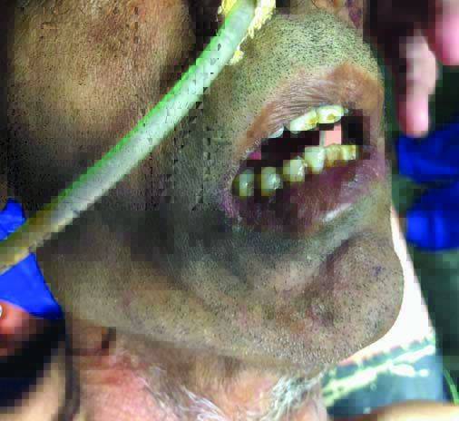
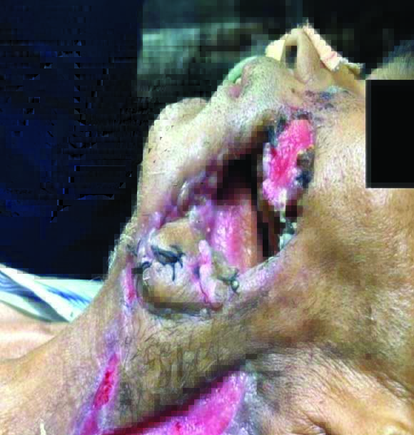
A 46-year-old male patient was posted for left pectoralis major myocutaneous flap surgery under general anaesthesia. He was diagnosed for left buccal mucosa squamous cell carcinoma and radical neck dissection with deltoid flap was performed. He presented with restricted mouth opening of 1.5 cm [Table/Fig-1], extension and flexion of neck was restricted and a big rent of 4×5 cm [Table/Fig-2] on left cheek was there owing to failed graft.
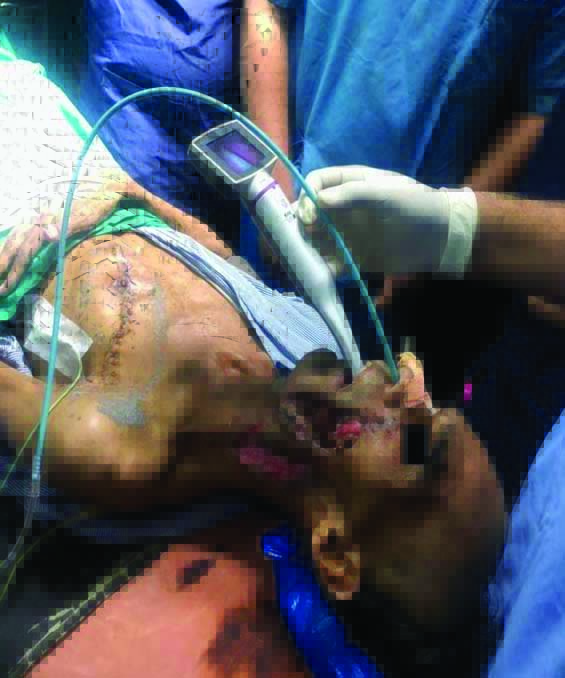
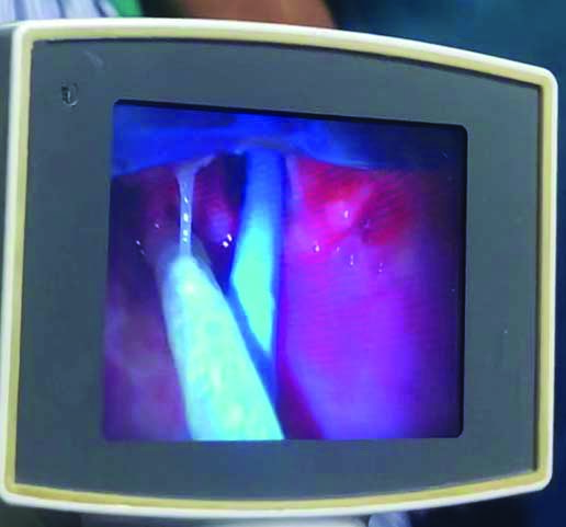
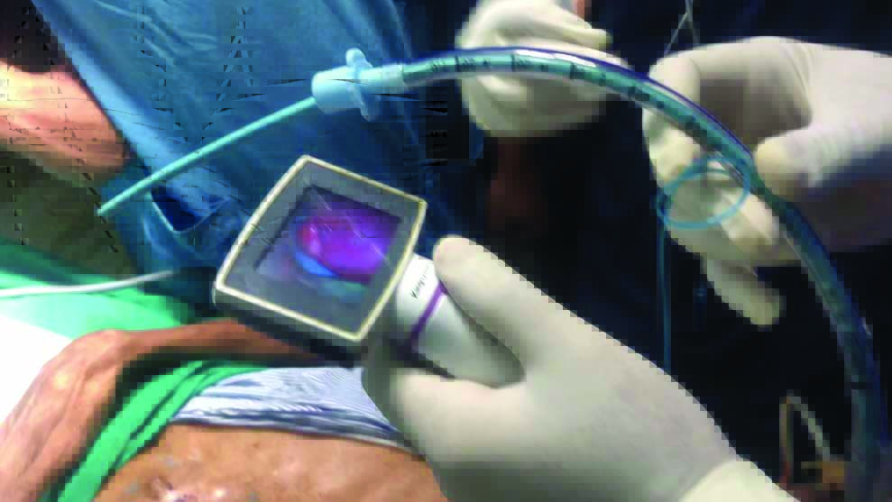
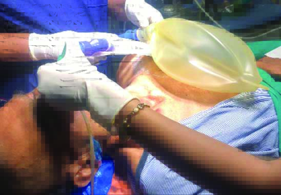
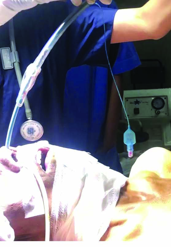
After explaining the procedure, an informed written consent was obtained. Tab. alprazolam 0.5 mg was given the night before surgery. Patient was kept nill per orally overnight. On the day of surgery, pre-operative check-up was performed. A multipara monitor was attached to the patient and vitals were monitored. Pre-medication with inj. glycopyrrolate 0.4 mg, inj. ondansetron 4 mg and inj. midazolam 0.5 mg i.v., inj. ranitidine 50 mg i.v. through 18 G vein flown and inj. Ringer’s lactate 500 mL were started. Patient was given oxygen through nasal prongs. Bilaterally superior laryngeal nerve block was given with 2 mL 1% inj. lignocaine and transtracheal recurrent laryngeal nerve block was given with 2% inj. lignocaine and topical nasal and oral spray was done with 10% inj. lignocaine. A non-channeled VLS was introduced [Table/Fig-3] in oral cavity to visualise vocal cords. Lignocaine 2% jelly was squeezed intranasally and also applied over bougie. Bougie was travelled through the nasopharynx into trachea via vocal cords under direct visualisation of VLS [Table/Fig-4]. A 7 mm internal diameter portex Endotracheal Tube (ETT) was railroaded over bougie, thus ETT was advanced further [Table/Fig-5]. Bilateral air entry was confirmed [Table/Fig-6]. Inj. propofol 120 mg, inj. atracurium 30 mg and inj. tramadol 50 mg i.v. was given and then ETT cuff was inflated. Patient was extubated after complete reversal of muscle relaxant keeping ventilating bougie in situ [Table/Fig-7].
Insertion of VLS and Bougie.
Passage of bougie through vocal cords.
Passage of ETT over bougie.
Confirmation of bilateral air entry.
Extubation with ventilating bougie in situ.
Discussion
In patients with head and neck carcinoma, the three axis (oral-pharynx-tracheal) alignment is very difficult. Given the condition of limited mouth opening and non-availability of fibreoptic bronchoscope, a VLS is a better alternative. VLS gives good supraglottic view and allows better condition for intubation under direct visualisation of vocal cords. Most modern VLS require a mouth opening of >2.5 cm [1]. An interincisor distance of 20 mm is required to insert the narrow blades. A channeled blade is more suitable for bougie guided oral intubation with an interincisor distance of more than 2 cm, as the channeled blade’s diameter is 18 mm2.
The King Vision has an anterior-posterior diameter of 13 mm in non-channeled blade. A King Vision VLS with number 3 non-channeled blade was used in the index case, for awake nasal endotracheal intubation. It was difficult to mask ventilate the patient due to a large rent on the cheek and given the restricted mouth opening it was a difficult intubation scenario, thus awake intubation was opted for.
A complete airway block was provided with smooth video laryngoscopy during the awake state. It is necessary that the patient understands and co-operate well during the procedure. VLSs are easier to use and need relatively less expertise in comparison to fibreoptic scope by inexperienced anaesthesiologists for awake intubation [2-5]. The indirect view of upper airway improves glottis visualisation, including in suspected or encountered difficult intubation [6].
A case report by Ali QE et al., also described the use of King Vision VLS in a restricted mouth opening patient of ankylosing spondylitis successfully with channeled blade while in the present case, non-channeled blade was used owing to less mouth opening with nasal guided intubation [7].
The range of extension was limited owing to the previous surgery therefore, VLS provided better visualisation without manipulating the head neck or the laryngoscope blade, which is also described in the case reported by Ali QE et al., [8]. A non-channeled blade facilitated nasotracheal intubation in comparison to McGrath MAC video laryngoscope in a randomised controlled trial for predicted difficult intubations [9]. Non-channeled blades are generally thinner, easier to insert and provide a good quality view to the vocal cords even in the case of significantly limited mouth opening [10].
Conclusion(s)
It is possible to intubate a patient via nasal route with the help of non-channeled VLS and bougie in restricted mouth opening of less than 1.5 cm and neck movement with good airway preparation in settings where flexible fibreoptic scope is not available.
Author Declaration:
Financial or Other Competing Interests: None
Was informed consent obtained from the subjects involved in the study? Yes
For any images presented appropriate consent has been obtained from the subjects. Yes
Plagiarism Checking Methods: [Jain H et al.]
Plagiarism X-checker: Oct 30, 2020
Manual Googling: Dec 08, 2020
iThenticate Software: Dec 22, 2020 (5%)
[1]. Niforopoulou P, Pantazopoulos I, Demestiha T, Koudouna E, Xanthos T, Video-laryngoscopes in the adult airway management: A topical review of the literatureActa Anaesthesiol Scand 2010 54(9):1050-61.10.1111/j.1399-6576.2010.02285.x20887406 [Google Scholar] [CrossRef] [PubMed]
[2]. de Pinho Martins M, Bastos Alexandre M, Fontes Cardoso L, Pinto Calçada, Adolfo BC, Case report: Tracheal intubation with King Vision in a patient with oral opening <1 cmEuropean Journal of Anaesthesiology 2014 31:27010.1097/00003643-201406001-00778 [Google Scholar] [CrossRef]
[3]. Rai MR, Parry TM, Dombrovskis A, Warner OJ, Remifentanil target-controlled infusion vs propofol target-controlled infusion for conscious sedation for awake fibreoptic intubation: A double-blinded randomised controlled trialBr J Anaesth 2008 100:125-30.10.1093/bja/aem27918037667 [Google Scholar] [CrossRef] [PubMed]
[4]. Sukhupragarn W, Churnchongkolkul W, Glidescope intubation after failed fiberoptic intubationPaediatr Anaesth 2010 20:901-02.10.1111/j.1460-9592.2010.03373.x20716090 [Google Scholar] [CrossRef] [PubMed]
[5]. Jeyadoss J, Nanjappa N, Nemeth D, Awake intubation using Pentax-AWS video laryngoscope after failed fibreoptic intubation in a morbidly obese patient with a massive thyroid tumour and tracheal compressionAnaesth Intensive Care 2011 39:311-12.https://www.aaic.net.au/Document/?D=20101107 [Google Scholar]
[6]. Chemsian RV, Bhananker S, Ramaiah R, VideolaryngoscopyInt J Crit Illn Inj Sci 2014 4:35-41.10.4103/2229-5151.12801124741496 [Google Scholar] [CrossRef] [PubMed]
[7]. Ali QE, Amir SH, Siddiqui OA, Pal K, King vision video laryngoscope: A suitable device for severe ankylosing spondylitisEgyptian Journal of Anaesthesia 2017 33(1):129-31.10.1016/j.egja.2016.08.001 [Google Scholar] [CrossRef]
[8]. Ali QE, Ahmed SO, Hussain AS, Sarfaraz A, Shaista J, King Vision video laryngoscope for severe post burn contracture neck: an encouraging experienceRev. Bras. Anestesiol 2017 67(6):641-43.10.1016/j.bjan.2016.08.00627662773 [Google Scholar] [CrossRef] [PubMed]
[9]. Zhu H, Liu J, Suo L, Zhou C, Sun Y, Jiang H, A randomised controlled comparison of non-channeled king vision, McGrath MAC video laryngoscope and Macintosh direct laryngoscope for nasotracheal intubation in patients with predicted difficult intubationsBMC Anaesthesiol 2019 19:16610.1186/s12871-019-0838-z31470814 [Google Scholar] [CrossRef] [PubMed]
[10]. Votruba J, Brozek T, Blaha J, Henlin T, Vymazal T, Donaldson W, Video laryngoscopic intubation using the king visionTM laryngoscope in a simulated cervical spine trauma: A comparison between non-channeled and channeled disposable bladesDiagnostics 2020 10:13910.3390/diagnostics1003013932138162 [Google Scholar] [CrossRef] [PubMed]